

 PDF
PDF ePub
ePub Citation
 Print
Print


Introduction
Integrins comprise a family of allosteric, heterodimeric, transmembrane glycoproteins that regulate cell-cell, cell-extracellular matrix, and sometimes cell-pathogen interactions [1415]. All known integrins are noncovalently combined heterodimers composed of two subunits, α and β, at the cell surface that display activated and non-activated conformations. Nineteen diverse integrin α subunits and eight β subunits exist in multicellular animals, forming at least 25 αβ heterodimers, which may make integrins the most structurally and functionally diverse and complex family of cell adhesion molecules [412162128]. Among the 25 αβ integrins identified to date, αvβ3 integrin is probably the most extensively studied [56]. When the function of αvβ3 integrin is constrained, vascular endothelial cells become apoptotic, tumor growth is suppressed, and the tumor may even disappear [8]. αvβ3 is the most active integrin in that it binds to many different extracellular matrix ligand proteins with an exposed arginine-glycine-aspartic (RGD) tripeptide sequence, including fibronectin (Fn), fibrinogen (Fg), vitronectin (Vn), lamin, collagen, von Willibrand factor, and osteopontin [222232425]. Integrin αvβ3 is usually expressed at low levels on quiescent endothelial cells in vivo, but its expression is highly upregulated during wound angiogenesis [11]. Therefore, RGD radiotracers that specifically bind to αvβ3 integrin can be widely applied in other angiogenesis-related abnormalities. These properties make αvβ3 integrin an attractive target for visualization of neovasculature [1]. Integrin αvβ3 is one of the key regulators of pathological angiogenesis and endothelial functions in general.
Integrin αvβ3 is also involved in many signal transduction pathways, in both directions across the cell membrane. It has been demonstrated that αvβ3 integrin can transmit apoptotic signals [13]. Stupack et al. [26] demonstrated that cells adherent within a three-dimensional extracellular matrix undergo apoptosis when unligated integrins are expressed, which recruits caspase 8 to the membrane, identifying an unexpected role of αvβ3 integrin in the regulation of apoptosis. Zhao and Ross showed that unoccupied αvβ3 integrin regulates osteoclast apoptosis via a component of integrin by transmitting a positive death signal [31]. Apart from its role in signal transduction, αvβ3 integrin can adhere to the underlying basement membrane, acting as a cell-adhesion transmembrane receptor, while it also mediates the adsorption and invasion of a variety of viruses into susceptible cells. For example, αvβ3 integrin is a functional receptor for Foot-and-mouth disease virus (FMDV) that plays a vital role in the infection process. FMDV is a member of the genus Aphthovirus of the family Picornaviridae that enters cells via a receptor-mediated endocytotic pathway, causing foot-and-mouth disease, a highly infectious and economically important disease that impacts domestic cloven-hoofed animals [9].
Although there have been many studies of αvβ3 integrin, we understand relatively little about the roles it plays in FMDV infection. Specifically, the role of αvβ3 integrin in tissue tropism and pathogenesis of viruses is still unclear. In this study, we cloned full-length cDNA of suckling mouse integrin subunits αv and β3, and established a CHO-677 cell line stably expressing αvβ3. This will be a useful cell model for examination of the effects of a single αvβ3 integrin receptor in mediation of FMDV infection. We then evaluated the susceptibility of this cell line to FMDV type O/ZK/93 (FMDV/O/ZK/93).
Materials and Methods
Cells, virus and antibodies
293T cells and heparan sulfate-deficient Chinese hamster ovary (CHO-677 or pgsD-677, ATCC, CRL-2244) cells were cultured in Ham'F-12 (HyClone, USA) medium supplemented with 10% fetal bovine serum (FBS; HyClone, USA), 1% streptomycin (0.2 mg/mL) and penicillin (200 U/mL). Baby hamster kidney (BHK-21) cells were maintained in Eagle's minimal essential medium (Invitrogen, USA) containing 10% FBS. All cells were incubated at 37℃ with 5% CO2.
FMDV/O/ZK/93 was isolated from a naturally infected pig in the City of ZhouKou, HeNan Province, China, during the 1993 outbreak and propagated in BHK-21cells. FMDV/O/ZK/93 was used for viral challenge, and is the major and optimal candidate for an FMD vaccine.
Guinea pig anti-FMDV serum and rabbit polyclonal antiserum directed against mouse integrin subunit αv and β3 were obtained from the Lanzhou Veterinary Research Institute. Fluorescein isothiocyanate (FITC)-conjugated goat anti-guinea-pig IgG antibody and FITC-conjugated anti-rabbit IgG antibody were purchased from Sigma-Aldrich (USA).
Cloning and sequencing the integrin subunit αv and β3 genes
Genomic RNA was extracted from the tongue or lung tissues of suckling mice using an RNeasy Mini kit, as previously described [29]. The use of all animals in this study was approved by the Review Board of Lanzhou Veterinary Research Institute, Chinese Academy of Agricultural Sciences. cDNA was synthesized from the extracted RNA with AMV reverse transcriptase (Takara Bio, Japan) using random primers (20 pmol/mL) and used as the template for amplification of the αv and β3 transcripts with PCR. The PCR primer pairs, αvF/αvR and β3R/β3F, respectively, are shown in Table 1. The PCR reaction was performed in a volume of 40 µL. The cycling parameters were as follows: 40 cycles of denaturation at 95℃ for 3 min, annealing at 58℃ for 30 sec, and elongation at 72℃ for 4 min, followed by a final elongation step at 72℃ for 10 min before the reaction was cooled to 4℃ for further processing. The amplicons were purified with a QIAquick Gel Extraction Kit (Takara Bio) and cloned separately into the pGEM-T Easy vector (Promega, USA). The PCR products were verified by electrophoresis and sequenced in both directions by GeneWiz (China).
Identifying integrin subunit αv and β3 homology in model organisms
The sequence data were assembled and analyzed with the Lasergene software (DNASTAR). The complete genomic sequence was analyzed using BLASTN at the National Center for Biotechnology Information (NCBI) website. Multiple sequence alignments were conducted using ClustalW. A phylogenetic tree was constructed with the MEGA5 software using the neighbor-joining method [27]. The reliability of the neighbor-joining tree was estimated by bootstrap analysis using 1,000 replicates.
Construction of the recombinant lentiviral plasmid
The recombinant plasmid was constructed as previously described [29]. Briefly, pOK12 and the internal ribosome entry site (IRES) fragment were amplified from pOK12 and the pIRES2-EGFP plasmid (stored in our laboratory), respectively, using the corresponding primer pairs (Table 1). The IRES fragment was then cloned into pOK12 to generate pOK-IRES. The αv PCR product was digested and cloned into the pOK-IRES plasmid to generate the recombinant plasmid pOK-αv-IRES, which was digested to obtain the αv-IRES fragment. This fragment was subsequently cloned into the pLVX-Tight-Puro vector (Clontech, USA) to generate the recombinant plasmid, pLVX-αv-IRES. The amplified β3 fragment was cloned into pLVX-αv-IRES to generate the recombinant lentiviral plasmid pLVX-αv-IRES-β3. All the products were sequenced using GENWIZ (USA).
Establishing a CHO-677 cell line continuously expressing murine αvβ3 integrin
Using the Lenti-X Tet-Off Lentiviral Advanced Induced Expression System (Takara Bio, USA), we transduced 293T packaging cells with the lentiviral vector pLVX-αv-IRES-β3 to express the lentivirus. The virus was then used to infect the CHO-677 target cells according to the lentiviral expression system user manual. To establish a stable αvβ3-transgenic CHO-677 cell line, a single clone was cultured under selection with 500 µg/mL G418 and 2 µg/mL puromycin. After approximately 14 days, cell cloning islands were observed. After 20 rounds of continuous cloning, a stable αvβ3-transgenic CHO-677 cell line was obtained and designated as CHO-677-mαvβ3.
Characterization of the CHO-677-mαvβ3 cell line
To determine whether the αv and β3 genes were stably integrated into the CHO-677 cell genome, we analyzed expression of the αv and β3 transcripts and proteins in CHO-677-mαvβ3 cells using PCR and IFA, respectively. The αv and β3 mRNA was detected at the 20th passage by PCR and the corresponding primer pairs (Table 1). The PCR products were analyzed by electrophoresis. IFA was conducted as described previously [17]. Briefly, CHO-677-mαvβ3 cells were seeded on 35 mm diameter plates and grown to approximately 80% confluence. The cells were then fixed with 4% paraformaldehyde for 30 min, permeabilized with 0.1% Triton X-100 for 25 min, and blocked with 5% bovine serum albumin for 1 h at room temperature. Next, cells were incubated with rabbit polyclonal antiserum directed against mouse integrin subunit β3 (1:50 dilution) or αv (1:50 dilution) for 1 h at 37℃, then reacted with FITC-conjugated anti-rabbit IgG secondary antibody (1:400 dilution) in a dark room. The cell nuclei were subsequently stained with 4',6-diamidino-2-phenylindole (DAPI; Invitrogen) for 15 min, after which photographs were taken under an Olympus BX40 fluorescence microscope (Olympus, Japan).
Infectivity assays
We infected the CHO-677-mαvβ3 cell line with FMDV/O/ZK/93 virus to study the functional features of the cells, conducted a plaque assay, and analyzed the growth kinetics of the virus. In addition, we used quantitative real-time reverse transcription-PCR (qRT-PCR) and IFA to analyze the growth characteristics of FMDV/O/ZK/93 in the CHO-677-mαvβ3 cell line.
Plaque assay
CHO-677-mαvβ3 and the parental CHO-677 cells were seeded in six-well cell culture plates 48 h before infection. Dilutions (10-fold) of the virus were prepared in Ham's F-12 medium using an inoculum volume of 200 µL per well. One hour after adsorption, the inoculum was removed and the cells were overlain with 50% gum tragacanth and 50% 2 × minimal essential medium supplemented with 2% fetal bovine serum. The plates were then incubated for 48 h, fixed with acetone and methanol (1:1), and stained with crystal violet (Histochoice; Amresco, USA).
Growth kinetics of the virus
The growth kinetics of the virus were analyzed in BHK-21 cells. Briefly, CHO-677-mαvβ3 cells and the parental CHO-677 cells were infected with FMDV/O/ZK/93 at the same multiplicity of infection (MOI = 1), after which the virus was allowed to adsorb for 1 h at 37℃. The medium was replaced with fresh medium twice during the incubation period. At 12, 24, 36, 48, and 60 h post-infection (hpi), the plates were subjected to three rounds of freeze/thaw, after which the viral titers in cell culture media were determined by TCID50 on BHK-21 cells. Three independent experiments were performed.
qRT-PCR
To determine the level of viral replication, both cell types were prepared in triplicate as described above and the viral supernatants were harvested at the indicated times after infection. Total RNA was extracted from the cells using a QIAxtractor kit (Qiagen, Germany) according to the manufacturer's instructions. qRT-PCR was performed with the StrataGene Mx3000P Real-Time PCR System (Agilent, USA) using a SYBR Premix Ex Taq Kit (Takara Bio), as previously described [10].
IFA
Viral proteins were detected with IFA. CHO-677-mαvβ3 and CHO-677 cells were seeded on 35 mm diameter plates, grown to approximately 80% confluence, and then infected with FMDV/O/ZK/93 at MOI = 0.4. At 22 hpi, the cells were fixed, permeabilized, and blocked as described above. The cells were then incubated with guinea pig anti-FMDV serum (1:100 dilution) at 37℃ for 1 h, then reacted with FITC-conjugated goat anti-guinea-pig IgG antibody (1:400 dilution) in a dark room. The cell nuclei were stained and the cells photographed.
Antibody blockade assay
We established a CHO-677-mαvβ3 cell line that only expressed αvβ3-integrin using the lentviral-mediated gene transfer technique. This integrin was shown to function as a receptor for virus attachment. However, it was not known which domain of the αvβ3 integrin played a key role in mediating virus infection; therefore, we performed an antibody blockade assay. Briefly, CHO-677-mαvβ3 cells were seeded in 35-mm-diameter dishes and incubated for 48 h at 37℃ under 5% CO2. Confluent cell monolayers were washed with PBS (pH 7.5) containing 2 mM CaCl2 and 1 mM MgCl2, after which the cells were divided into two groups, one that was incubated with PBS (800 µL/well) (mock treatment) and another that was incubated with a 400-fold dilution of anti-β3 serum (800 µL/well). After 1.5 h, the supernatants were removed and the cells were infected with 500 µL FMDV/O/ZK/93 (MOI = 2) at 37℃. At 24 h postinfection, the titer of infectious virus in the cell culture media was determined by TCID50 on BHK cells.
Results
Cloning and sequencing the integrin subunit αv and β3 genes
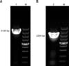
In the past 20 years, αvβ3 integrin has been extensively investigated; however, most of these studies were performed using human integrin. To investigate the role of αvβ3 integrin in animals, we amplified the integrin subunit αv and β3 genes from the tongue or lung tissues of suckling mice (panels A and B in Fig. 1). The sequencing results showed an αv PCR fragment of ~3135 nucleotides (accession no. KP296148) and a β3 PCR fragment of ~2364 nucleotides (accession no. KP296149), as expected.
Identification of homologues of murine integrin subunits αv and β3 in model organisms
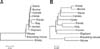
We compared the αv nucleotide sequence of the suckling mouse with its homologues in other species by a BLAST search of the NCBI database. The suckling mouse αv sequence was highly homologous to the mouse gene (99.2%). The amino acid sequence of suckling mouse αv was also highly homologous to the corresponding human (94.2%), swine (92.1%), bovine (92.2%), elephant (92.5%), horse (91.7%), and camel (92%) genes (Table 2). Similarly, the nucleotide and deduced amino acid sequences of β3 in suckling mouse were much more similar to those of horse, sheep, and camel β3 than to elephant or panda β3 (Table 2). These results were confirmed by phylogenetic analysis (panels A and B in Fig. 2).
Characterization of the recombinant plasmid
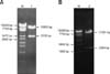
To confirm that the lentiviral recombinant plasmid pLVX-αv-IRES-β3 was built successfully, double enzyme digestion experiments were performed. The pLVX-αv-IRES-β3 plasmid was digested by BamHI and NheI to identify the αv fragment, then digested by NotI and MluI to identify the β3 fragment. As shown in Fig. 3, the results of the double enzyme were verified by electrophoresis.
Characterization of the CHO-677-mαvβ3 cell line
PCR and IFA were used to detect the integrin subunits αv and β3 at the gene and protein levels in the CHO-677-mαvβ3 cell line. The αv and β3 transcripts were amplified by PCR at the 20th passage (panel A in Fig. 4). The sequencing results showed that both genes were correct. We then detected the expression of the αvβ3 heterodimer in the CHO-677-mαvβ3 cell line using IFA. The CHO-677-mαvβ3 cells were immunostained for αvβ3 integrin, whereas the parental CHO-677 cells were not (panel B in Fig. 4), implying that the αv and β3 genes were successfully introduced and stably expressed in the recombinant cells.
Infectivity assays
To investigate the functional features of the CHO-677-mαvβ3 cell line, we infected the cells with FMDV/O/ZK/93 virus, then analyzed the growth characteristics of the virus and the susceptibility to infection of the cells. Plaque assays were performed to compare the plaque sizes and quantities produced in CHO-677-αvβ3 and CHO-677 cells, and the plaque phenotypes and FMDV yields were characterized in both cells. As shown in panel A in Fig. 5, the virus produced larger and more plaques in the CHO-677-mαvβ3 cells than in the CHO-677 cells.
The growth kinetics of the virus were determined on BHK-21 cells using viral titers to compare the in vitro growth characteristics of FMDV/O/ZK/93 in the CHO-677-mαvβ3 cell line and the parental cells. Samples were collected at specific times after infection and TCID50 was titrated on BHK-21 cells. The peak titers of FMDV/O/ZK/93 were 105.3 TCID50/0.1 mL in CHO-677-mαvβ3 cells at 36 hpi and 103.9 TCID50/0.1 mL in the parental cells at 48 hpi (panel B in Fig. 5). We next determined the expression of viral proteins with IFA. Intracellular cytoplasmic fluorescence was detected in the infected CHO-677-mαvβ3 cell line, whereas no fluorescence was detected in the CHO-677 cells (panel C in Fig. 5). These findings indicated that the utilization efficiency of FMDV/O/ZK/93 for CHO-677-mαvβ3 cells was relatively high.
To analyze the replication capacity of the virus, we determined the copy numbers of FMDV/O/ZK/93 viral RNAs in both cells by qRT-PCR using the supernatants collected at different time points. As shown in panel D in Fig. 5, the parental cells clearly had lower levels of RNA replication than the CHO-677-mαvβ3 cell line. These results indicate that the CHO-677-mαvβ3 cells are susceptible to FMDV, and therefore confirm that the CHO-677-mαvβ3 cell line was successfully constructed.
Antibody blockade assay
Preliminary studies using antibody blockade analysis showed that treatment of cells with anti-β3 serum reduced the virus titer in cell culture media compared to that of mock-treated control (Fig. 6), indicating that the ability of integrin αvβ3 to mediate virus infection is dependent on the presence of the β3 subunit domain.
Discussion
Although αvβ3 integrin has been studied extensively and in-depth in the field of medicine, few studies of animal αvβ3 integrin have been conducted, and those that have focused on using the transient expression of integrin subunits to study receptors and FMDV infection. Clark et al. [6] colleagues studied the transient functional expression of αvβ3 integrin on vascular cells during wound repair, while Neff et al. [20] examined the high-efficiency utilization of bovine αvβ3 integrin as a receptor for FMDV by transiently expressing αv and β3 subunit cDNAs. However, the transient expression of αvβ3 cannot be controlled well, and its repeatability is not high because it is affected by many factors. Therefore, we established a stable CHO-677-mαvβ3 cell line using a highly efficient lentivirus-based inducible expression system. Recombinant lentiviral vectors are powerful and efficient tools for transferring heritable genetic material into the genome of virtually any cell type [37]. Lentiviruses are also perhaps the most versatile retroviruses because they can infect, transduce, and sustain their expression in almost any mammalian cell. The αv and β3 subunits were also connected by the IRES sequence, so that both subunits were expressed within the same cellular environment under control of the same promoter, which may have facilitated formation of the αvβ3 heterodimer. The integrin subunit αv and β3 genes were extracted from the tongue or lung tissues of suckling mice. Although αv and β molecules have been studied in other papers, murine αv and β molecules have not been reported. Suckling mice have been shown to be a good animal model for studying viruses and are often used in experiments due to their simple and convenient application. For example, suckling mice can be used to propagate the virus and the 50% lethal dose (LD50) test is determined in suckling mice. Additionally, commercial murine antibodies can be more easily obtained, which will facilitate studies of FMDV using this model. More importantly, suckling mouse αv and β3 genes show high sequence homology to their human, swine, bovine and camel counterparts.
FMDV utilizes four integrins (αvβ1, αvβ3, αvβ6, and αvβ8) and heparan sulfate (HS) as cell receptors to initiate viral infection. Chinese hamster ovaries lack β1, β3, β6 and β8 integrin genes, and CHO-677 cell is a heparan sulfate-deficient Chinese hamster ovary cell. Specifically, CHO-677 cells do not express αvβ1, αvβ3, αvβ6, αvβ8 and HS, while the constructed CHO-677-αvβ3 cell line can stably express murine αvβ3 integrin. Expression of the integrin subunits αv and β3 at the gene and protein levels in the CHO-677-mαvβ3 cell line showed that the αv and β3 genes were successfully introduced into the CHO-677 cells and expressed stably.
αvβ3 integrin has been shown to act in many cells as a receptor for the internalization of FMDV. Entry of FMDV into host cells is a complex process initiated by virus binding to a specific cell surface receptor. Although receptors for FMDV have been identified, there is little information regarding the precise mechanisms by which these receptors promote entry. Therefore, we used FMDV to infect the CHO-677-mαvβ3 cell line to verify the mediating effects of αvβ3 on infection. The effects of FMDV replication on the CHO-677-mαvβ3 cell line suggested that FMDV RNA levels were higher in the CHO-677-mαvβ3 cell line than in CHO-677 cells. After peak replication at 36 h and 48 h, respectively, the viral RNA levels began to decline in both cells. The level of FMDV RNA in the CHO-677-mαvβ3 cell line was significantly influenced by αvβ3 integrin, which is consistent with the results observed for the viral growth curves. A standard plaque assay was also used to characterize the pathogenicity of FMDV in CHO-677-mαvβ3 and CHO-677 cells. FMDV produced more plaques in CHO-677-mαvβ3 cells than in CHO-677 cells. The expression of viral proteins differed significantly in FMDV/O/ZK/93-infected CHO-677-mαvβ3 and CHO-677 cells according to IFA, indicating that the CHO-677-mαvβ3 cell line was constructed successfully and susceptible to FMDV infection.
Many of the intracellular signaling pathways activated by integrins are dependent on the β-subunit cytoplasmic domain, which indicates a further possible function of the β3 cytoplasmic domain in facilitating virus entry. Previous studies have demonstrated that the ability of integrin αvβ3 to function as a receptor for FMDV is not dependent on the presence of complete subunit cytoplasmic domains [19]. In this study, we used an antibody blockade assay to study the role of the β3 subunit in mediating infection. Our results demonstrated that the β3 subunit played a critical role in post attachment events. Miller et al. [18] studied the role of the β6 cytoplasmic domain in infection and found that αvβ6 did not merely pass the virus onto a second receptor for internalization, but also played an active role in events subsequent to attachment. Zhang et al. [30] showed that the integrin β6-1 subunit could induce partial protection against FMDV in guinea pigs. Although the αv and β6 subunits have been widely investigated, there have been few investigations of the role of αvβ3 integrin in infection. Therefore, this cell line, which stably expresses murine αvβ3 integrin, could be a useful cell model to research the mechanism of a single αvβ3-receptor on virus infection, and should also be a good vehicle for enhancing viral titers.
In conclusion, we cloned the full-length cDNA of the integrin subunits αv and β3 from suckling mice and established a CHO-677-mαvβ3 cell line stably expressing suckling mouse αvβ3 integrin that had increased susceptibility to FMDV. Further studies should investigate the functions and mechanisms of αv and β3 subunits using this newly developed CHO-677-mαvβ3 cell line, including its involvement in signaling pathways, cell apoptosis, and inflammatory response in animals.

 XML Download