

 PDF
PDF ePub
ePub Citation
 Print
Print


Introduction
Mammary carcinoma is one of the most important diseases leading to increased morbidity and mortality in female dogs [23]. Recent stem cell research in veterinary medicine suggests that mammary carcinoma may be driven by cancer stem cells (CSCs) [21926]. This subset of cancer cells has been identified in some types of canine tumors [41528] including mammary tumors. In canine mammary tumors (CMTs), the existence of CSCs has been confirmed by expression of several well-known molecular stem cell markers, CD44, CD24 [13], octamer-binding transcription factor(Oct)-3/4 [27], and aldehyde dehydrogenase (ALDH) 1 [14], that serve as key factors for cell differentiation and maintenance of pluripotency [910]. This subpopulation can also be isolated and enriched in tumorsphere (TS) models for CSC research [422].
To date, a series of cell-based markers and intrinsic signal pathways have been explored to characterize CSCs for the development of innovative cancer treatments by targeting CSCs. In the past few decades, the cyclooxygenase (COX)-prostaglandin E2 (PGE2) pathway has been widely studied and emerged as an important regulator of inflammation-induced or non-inflammatory carcinoma [8]. Aberrant COX-2 expression has also been observed in CMTs and is thought to be a negative prognostic factor [6923]. Recently, it was reported that COX-2-derived PGE2 protects embryonic stem cells from apoptosis [12] and is considered an important mediator in the stem cell network and stem cell-carcinoma niche [11]. However, few studies on the possible role of COX-2 in CSC-mediated tumor initiation for primary CMTs have been performed. Therefore, we conducted the present investigation to clarify the potential role of COX-2 in sustaining CSCs during tumor progression by measuring COX-2 expression and examining its correlation with CSC biomarkers (CD44, Oct-3/4, and epidermal growth factor receptor [EGFR]) in CMTs and CSC-enriched TSs.
Materials and Methods
Retrospective samples and histopathology
CMT tissues were collected from dogs that underwent surgical resection at the Veterinary Teaching Hospital of China Agricultural University (China). Mammary tissues were histologically classified by pathologists according to previously described protocols [36]. The size, histological subtype, histological malignancy, Ki-67 proliferative index, and lymphatic invasion were assessed for each tumor. Cases of malignant mammary tumors (MMTs) were divided according to histological grade. Four normal mammary gland (NMG) tissue samples were included as control samples.
Cell culture and TS formation
A total of 24 mammary tumor tissue samples from 79 malignant cases were aseptically collected and processed. Briefly, the tissues were minced into small pieces of about 1 mm, then transferred and digested for 4~16 h at 37℃ with 200 U/mL collagenase (Sigma-Aldrich, USA) and 100 U/mL hyaluronidase (Sigma-Aldrich) whilst being stirred. Digestion was terminated and the solution was centrifuged. The resulting cell pellets were suspended and passed through a 40-µm strainer. The single cells were then collected and resuspended for TS culture in serum-free Dulbecco's modified Eagle medium/Ham's F-12 medium (DMEM/F12) supplemented with B27 (Gibco-BRL, USA), epidermal growth factor (EGF; 20 ng/mL; PeproTech, USA), basic fibroblast growth factor (bFGF; 20 ng/mL; PeproTech), and 0.4% bovine serum albumin (BSA; Sigma-Aldrich). After 10~14 days of culturing, the spheres were pelleted and fixed in 10% neutral buffered formalin for 30 min before paraffin embedding.
Immunohistochemistry (IHC)
De-paraffinized tissue and cell block sections were subjected to heat-induced or pepsin digestion antigen retrieval and incubation in 3% hydrogen peroxide. Next, the sections were incubated overnight with primary antibodies specific for COX-2 (diluted 1 : 100; Vector Laboratories, USA), CD44 (diluted 1 : 100; Santa Cruz Biotechnology, USA), POU5F1B transcription factor (Oct-3/4, diluted 1 : 80; Santa Cruz Biotechnology), EGFR (diluted 1 : 100; Zymed Laboratories, USA), and Ki-67 (diluted 1 : 150; BD Pharmingen, USA) at 4℃ using a two-step Polymer Detection System (GBI Labs, USA). Antibody binding was visualized with 3,3'-diaminobenzidine tetrahydrochloride and the sections were counterstained with Mayer's hematoxylin. For the negative controls, the primary antibody was replaced with phosphate-buffered saline (pH 7.2).
Immunohistochemical scoring (IHS)
IHS was performed by estimating the percent of positive cells and labeling intensity. The percentages of positive cells were scored as 0 (0% positive cells), 1 (< 10% positive cells), 2 (10~50% positive cells), 3 (51~80% positive cells), or 4 (> 80% positive cells). Staining intensity was scored as 0 (negative), 1 (weak), 2 (moderate), or 3 (strong). Scores for the percentage of positive cells and staining intensity were multiplied to calculate an IHS value from 0 to 12. IHS values ≤ 6 were considered to represent low expression while values 7~12 were considered high. The Ki-67 proliferation index was determined based on the percentage of positive cells: 0 (negative), 1+ (≤ 10% of cells labeled), 2+ (11~50% of cells labeled), or 3+ (51~100% of cells labeled). Each section was examined using a microscope (Nikon, Japan) by two pathologists who were blind to the clinical characteristics and prognosis.
Statistical analysis
Statistical analyses were performed using SPSS software (ver. 16.0; SPSS, USA) and GraphPad Prism 5.0 (GraphPad Software, USA). Categorical data are expressed as frequencies and percentages. A chi-square test and Fisher's exact test were used to evaluate categorical variables. P values < 0.05 were considered significant.
Results
Clinicopathological findings
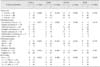
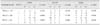
Histological examination was performed for seven mammary hyperplasia (MH) samples, 79 MMTs including different histological subtypes, and four normal mammary gland (NMG) tissue samples as a control. Histological classification of the MMTs showed that these cases included three carcinoma in situ, 10 complex carcinoma, nine carcinosarcoma, 16 ductal carcinoma, one inflammatory carcinoma, 15 simple carcinoma, and 16 ductal carcinoma. Well-(WD), moderately (MD), and poorly differentiated (PD) tumors accounted for 17 (21.5%), 47 (59.5%), and 15 (20.0 %) of the MMTs, respectively (Table 1).
IHC analysis of COX-2, CD44, Oct-3/4, and EGFR expression
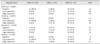
Among the MMTs, high COX-2 expression was observed in 36 cases (45.6% of the total cases), and low COX-2 expression was found in 25 cases (31.6%). Similarly, the high and low expression rates were 51.9% (41/79) and 35.4% (28/79) for CD44, 22.9% (18/79) and 15.2% (12/79) for Oct-3/4, and 49.4% (39/79) and 40.5% (32/79) for EGFR, respectively. All the unlisted cases were negative for each labeled protein. All the NMGs were negative or had low expression levels for these proteins, and expression varied in the MHs (Table 2).
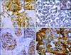
COX-2 expression was observed in the cytoplasm, cell membrane, and nucleus of the neoplastic epithelial cells with weak to moderate immunoreactivity in some stromal cells. Highly aggressive tumor tissues and neoplastic cells had intense staining in a heterogeneous pattern compared to noninvasive tissues (panel A in Fig. 1). COX-2 expression was significantly higher in the MMTs than other types of specimens (p = 0.001). Oct-3/4 was widely detected in malignant neoplastic cells with strong and diffuse nuclear staining patterns (panel B in Fig. 1). Normal and hyperplastic tissues were mildly positive for Oct-3/4 while strong expression of this factor was observed predominantly at the invasive edge of malignant tissues (p = 0.016). CD44-specific staining was primarily found in the cell membrane of epithelial and myoepithelial cells (panel C in Fig. 1), but was absent from the apical epithelial surface, especially in normal and hyperplasic mammary tissue. CD44 was more extensively expressed in invasive components of the MMTs compared to NMGs although this difference was not statistically significant (p = 0.079). EGFR expression patterns were similar to those of CD44, and its membrane and cytoplasmic localization was consistently observed in epithelial cells of MMTs regardless of histological subtype (panel D in Fig. 1). Malignant tumors also had a higher positive ratio for EGFR than other types of specimens (p = 0.103; Table 2).
Association of clinicopathological parameters with COX-2, CD44, Oct-3/4, and EGFR
Elevated COX-2 expression was significantly associated with tumor size (p = 0.006), histological subtype (p = 0.017), histological grade (p = 0.032), lymphatic invasion (p = 0.033), and Ki-67 index (p = 0.022). Oct-3/4 expression strongly correlated with the Ki-67 index (p = 0.018). CD44 expression was statistically associated with lymphatic invasion (p = 0.005). EGFR expression was significantly related to histological subtype (p = 0.019) and Ki-67 index (p = 0.020). Significant associations between protein expression and selected variables are summarized in Table 1.
Correlation between COX-2 and CSC markers
In moderately and poorly differentiated MMTs, COX-2 expression was strongly associated with CD44 (r = 0.402, p = 0.000; r = 0.810, p = 0.005) and EGFR (r = 0.367, p = 0.04; r = 0.700, p = 0.02) expression. In contrast, no statistical correlation was found between COX-2 and Oct-3/4 expression (Table 3).
IHC phenotype of TSs
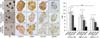
Cell blocks from 24 CMTs with different histological grades (seven WD tumors including one simple carcinoma and six simple carcinomas, nine MD tumors including three ductal carcinomas and six solid carcinomas, and eight PD tumors consisting of four solid carcinomas, three complex carcinomas, and one carcinosarcomas) were analyzed to evaluate COX-2, CD44, Oct-3/4, and EGFR immunolabeling. Representative TSs derived from tubular carcinomas (WDs), tubulopapillary carcinomas (MDs), and solid carcinomas (PDs) were assessed. The TSs had inter-sphere heterogeneity and a compact morphology. Enhanced COX-2 expression was consistently observed in the cytoplasm with sporadic nuclear staining in three TSs. COX-2 expression was stronger in TSs derived from more malignant (more poorly differentiated) tumors. CD44 staining was enriched in cellular membranes. Nuclear localization of Oct-3/4 was extensive in solid carcinomagenerated spheres compared to the other two samples. EGFR was located in the cytoplasm with negative or weak immunoreactivity. Comparative analysis showed that there was a significant difference in COX-2 (PD 10.0 ± 2.1, MD 7.6 ± 1.9, WD 10.0 ± 2.1; p = 0.006), CD44 (PD 10.9 ± 1.9, MD 8.9 ± 2.5, WD 6.5 ± 1.4; p = 0.002), and Oct-3/4 (PD 9.4 ± 2.5, MD 6.0 ± 1.4, WD 5.5 ± 1.4; p = 0.001) expression among the three histological grades (p < 0.01). This was not observed for EGFR expression (PD 1.7 ± 1.4, MD 2.2 ± 2.1, WD 2.0 ± 1.4; p = 0.856). Staining specific for CD44 and Oct-3/4 had a similar level of intensity along with elevated COX-2 expression in the TSs (Fig. 2).
Discussion
CSCs have the ability to self-renew and generate the diverse types of cells that form tumors. This makes CSCs a potential therapeutic target in human and veterinary oncology. Oncoprotein COX-2 is generally characterized as a pivotal regulator of growth and anti-apoptosis signals used by malignant tumor cells [724], and may also have an impact on CSCs [161826]. In the current study, the possible role of COX-2 in tumor initiation was explored by assessing the expression patterns of COX-2 and CSC-related markers.
COX-2 plays different roles in tumor initiation. According to our data, enhanced COX-2 expression and a heterogeneous pattern were commonly observed in pre-malignant and malignant tumor tissues, and were strongly associated with malignant features such as large tumor size, complex histological type, poor differentiation, and an aggressive phenotype as previously reported in the literature [5821]. Absent or low levels of COX-2 expression were found in normal and hyperplastic mammary tissues while expression was dramatically increased in tumor tissues. These results also imply that inducible COX-2 expression may promote canine mammary tumorigenesis. Moreover, differential stromal COX-2 staining in fibroblasts, endothelial, and infiltrating inflammatory cells may also suggest that stroma-derived COX-2 contributes to the acquired proliferative and invasive properties of tumor cells through tumor-stromal interaction during mammary tumorigenesis [1]. For cases of human breast cancer, it has been reported that proliferative tumors (COX-2+ or Ki-67+) possess more unfavorable characteristics and have a poorer prognosis than non-proliferative tumors (COX-2+ or Ki-67+) [20]. In our study, we found that MTs with rapid growth (Ki-67+) as well as moderate and poorly differentiated ones showed elevated COX-2 expression, which may suggest a dynamic switch of functional COX-2 during tumor proliferation and differentiation. It has been reported that down-regulated COX-2 expression suppresses the malignant phenotype, delays the onset of tumors formed by poorly differentiated cancer cells [24], and reverses the epithelial-to-mesenchymal transition [18].
In order to better understand the function of COX-2 in CSC-mediated tumor initiation, we evaluated the distribution of CSCs in mammary tissues by examining CD44, Oct-3/4, and EGFR expression patterns, and their respective associations with clinicopathological parameters. Aberrant expression of these factors was frequently observed in aggressive components of malignant tumors characterized by poor differentiation and rapid proliferation. Nevertheless, normal and hyperplastic tissues also had lower levels of expression. These findings indicate that CSCs maintain their malignant properties and can form a recurrent tumor that is more malignant. In contrast to COX-2, no significant correlation between CSC marker expression and poor prognostic variables was found. We therefore propose that these markers are not suitable independent prognostic factors. However, we further examined the co-expression of COX-2, CD44, Oct-3/4, and EGFR in MMTs and found significant associations among COX-2, CD44, and EGFR. These results show that COX-2 activation in CSCs may be CD44- or EGFR-dependent [1729]. Although we did not observe a relationship between COX-2 and Oct-3/4 in primary CMTs, it has been previously hypothesized that Oct-3/4 expression is associated with nuclear COX-2 activation [25] and COX-2high or Oct-3/4+ cells may acquire stem cell-like features in cases of human cancer [22].
The present study has provided new insight into the potential role of COX-2 in CSC-mediated tumor initiation by evaluating the expression patterns of COX-2 and CSC markers in TSs, which consist primarily of partially differentiated progenitor cells with few CSCs. The robust expression of COX-2 in heterogeneous patterns and at higher levels in more aggressive tumor-derived TSs suggests that COX-2 may contribute to the self-renewing ability and maintenance of CSCs. We compared COX-2, CD44, Oct-3/4, and EGFR expression in TSs according to tumor differentiation. The results confirmed that COX-2 expression accompanied enhanced CD44 and Oct-3/4 expression to a similar degree, and COX-2 up-regulation was associated with CSC enrichment in in vitro models. Therefore, it is reasonable to believe that COX-2 expression is necessary for CSC-mediated tumor initiation.
Taken together, our data highlight the potential role of COX-2 in CSC-mediated tumor initiation. These findings suggest that CSCs may be more sensitive to direct COX-2 inhibition than the general cell population. Further studies should be conducted to explore COX-2-dependant elimination of CSCs as well as the development of novel therapeutic approaches for CMTs.
 XML Download