

 PDF
PDF ePub
ePub Citation
 Print
Print


Introduction
Mycoplasma (M.) hyosynoviae and M. hyorhinis are ubiquitous pathogens of swine known to cause arthritis and polyserositis, respectively, in pigs post-weaning [73033]. Transmission of both pathogens presumably occurs either vertically or horizontally after initial exposure [141935]. M. hyorhinis is more likely to be found in pigs immediately following weaning [2135]. After initial colonization of the upper respiratory tract, bacteremia may occur leading to the development of polyarthritis, polyserositis, and potentially pneumonia [101726]. M. hyosynoviae primarily colonizes the tonsils of weaned pigs but clinical arthritis only develops around 3 to 6 months of age [132529].
Despite our understanding of the clinical disease, the true prevalence, incidence, and overall dynamics of infection by these mycoplasmas in swine populations is relatively unknown. Given the re-emergence of Mycoplasma-associated clinical arthritis that impacts both animal well-being and the profitability of clinically-affected populations [11], more epidemiological data will be needed to effectively implement disease control strategies. Due to the difficulty with culturing these species of mycoplasma, development of rapid, accurate, and culture-independent methods for quantifying these pathogens in multiple sample matrices is crucial. Therefore, the current study was conducted to establish quantitative real-time PCR (qPCR) assays and test the ability of these methods to detect both M. hyosynoviae and M. hyorhinis in pen-based oral fluid samples along with nasal and tonsillar fluids as proxies for diagnostic specimens commonly used in swine herd screening. In addition, this study also verified the effects of an automated and a manual method of DNA extraction commonly performed in veterinary diagnostic laboratories on test result interpretation.
Materials and Methods
Genomic DNA extraction protocols
As a part of the diagnostic test validation, two distinct methods for total DNA extraction from pen-based oral, nasal, and tonsillar fluids were compared: magnetic beads (MB, automatic) and spin column (SC, manual). The MB (MagMAX Total Nucleic Acid Isolation Kit; Applied Biosystems, USA) and SC (High Pure PCR Template Preparation Kit; Roche, USA) DNA extraction protocols were performed according to the manufacturers' instructions. For the MB procedure, DNA was extracted using a semi-automated nucleic acid purification system (KingFisher 96 magnetic particle processor; Thermo Fisher Scientific, USA). All DNA samples were frozen at -20℃ until qPCR analysis.
Primer selection and design
The M. hyosynoviae-specific primer pair, herein called "Lauerman qPCR", was used for validation. This primer set was originally presented by Lauerman [18], but its validation and diagnostic test performance were never formally published. These primers were used in the current study for validation and verification of diagnostic performance without discrediting the initial primer pair development. A new primer set was produced to detect M. hyorhinis. All primers used for this study targeted sequences within the hypervariable region of the 16S ribosomal DNA (rDNA) of the bacteria. For initial design and confirmation of specificity across all swine mycoplasmas, the 16S partial sequences for swine mycoplasmas were aligned. DNA sequence alignment was performed using a ClustalW algorithm available with commercially available software (DNASTAR Lasergene software ver. 8.0; DNASTAR, USA). The Lauerman qPCR primer sequences were 5'-CAGT TGAGGAAATGCAACTG-3' (forward) and 5'-TAGCTGCG TCAGTGATTGG-3' (reverse). The M. hyorhinis qPCR primer sequences were 5'-GCATGTTGAACGGGATGTAGCAAT-3' (forward) and 5'-TGAAGCTGTGAAGCTCCTTTCTATTA CTC-3' (reverse). Specificity of the two primer pairs was subsequently re-confirmed in silico by checking them against a bacterial ribosomal database [4] and a probeCheck platform [23].
qPCR
qPCR specific for M. hyosynoviae and M. hyorhinis was performed using a fluorescence-based assay consisted of 2×commercial master mix (QuantiTect SYBR Green PCR Master Mix; Qiagen, USA) at a final concentration of 1×, each forward and reverse primer (0.4 µM final concentration), 2.5 µL template DNA, and nuclease-free water (up to 25 µL of the total reaction volume). All qPCR assays were performed with a commercial platform (ABI 7500 Fast Real-time PCR system; Life Technologies, USA). The assays were run under the following conditions: 15 min at 95℃ followed by 45 cycles of 15 sec of denaturing at 94℃, 30 sec of annealing at 63℃ (M. hyosynoviae) or 59℃ (M. hyorhinis), and 30 sec of extension at 72℃; and a final melt curve from 95℃ to 59℃. Assays for M. hyosynoviae and M. hyorhinis were performed in separate reactions given the difference in annealing temperature. All samples were tested in duplicate. In the case of discordant results (i.e., one positive and one negative), the sample was re-tested twice to achieve a consensus.
qPCR data interpretation
Analysis of the cycle threshold (Ct) value was performed using on-board qPCR software (ABI 7500 Fast software; Life Technologies) by setting the threshold manually at 0.04 with the baseline set from cycles 3~15. A sample was considered positive for M. hyosynoviae if the amplification curve did not exceed a Ct value of 35 and had a melting temperature (Tm) of 81.5 ± 0.5℃. Samples were positive for M. hyorhinis only if the limit of detection did not exceed a Ct value of 38 and had a Tm of 75.9℃ ± 0.5℃. Samples collected after an experimental challenge with all four major swine mycoplasmas, including the two tested in the present study, were used as in-house positive and negative controls for pen-based oral, nasal, and tonsillar fluids [12].
Assay specificity
To measure the specificity of each primer pair, a collection of bacterial isolates commonly found in swine that had been previously identified was utilized [34]. The collection included both mycoplasmas and non-mycoplasma species present in the swine respiratory tract, joints, or peritoneal cavities. This panel was specifically chosen due to its representativeness of known swine pathogens that could possibly be a confounding factor for development of the assays. More importantly, the panel had also been previously used to develop M. hyopneumoniae-specific PCR primers, therefore indicating its validity.
Analytical sensitivity
Two plasmids were synthesized (Integrated DNA Technologies, USA) as quantitative standards. One contained the 160-base pair target region of M. hyorhinis 16S rDNA and the other contained the 392-base pair target region of M. hyosynoviae 16S rDNA. Insert sequences were synthesized into pIDTSMART plasmids containing restriction sites flanking each end of the insert sequences. Both plasmids were linearized and prepared in ten 10-fold serial dilutions ranging from 1 × 10⁹ to 1 copy for sensitivity testing and standard curve analysis. Standard curves were generated in triplicate to determine the limit of detection and efficiency of each reaction. In order to estimate the total number of bacterial genome copies (genome equivalents), one copy of the 16S rDNA gene was used for either mycoplasma by taking into consideration data available from the ribosomal RNA database [20]. DNA isolated from serially diluted cultures of either mycoplasma was used to confirm results predicted by the plasmid standard curves. Mycoplasma culture procedures were performed as previously described [31]. Ultimately, the plasmid standard curves were used throughout the study for consistent quantification of bacterial load in the diagnostic specimens.
Diagnostic specimens for qPCR performance testing
For diagnostic performance verification using field samples that can serve as proxies for swine herd screening, five commercial finishing sites that received pigs from a common sow herd were selected for sampling. Each site had 2,000 to 4,000 18- to 24-week-old pigs housed in one or two barns. Sites were selected based on the clinical history reported by the consultant veterinarian of Mycoplasma-associated arthritis in previous groups of pigs produced from the same groups of sows in the herd. The clinical history was confirmed by the Iowa State Veterinary diagnostic laboratory and used as a defining criterion for the presence of both pathogens in piglets produced on that farm. No vaccines or specific antibiotic treatments known to be effective against either M. hyosynoviae or M. hyorhinis had been administered before or during sampling. Animal care and management protocols remained unchanged compared to routine practices. No indication of clinical disease was observed at the time of sampling. Diagnostic specimens were collected within one day at each of the five sites. A total of 340 specimens were analyzed in the study, including 60 pen-based oral fluid samples, 81 nasal swabs, and 49 tonsil scrapings. At each site, pen-based oral fluid samples were collected as previously described by leaving a rope for approximately 25 min in place to maximize the number of pigs coming into contact with it and yielding 30 mL of sample [27].
Pigs for individual sampling (nasal swabs and tonsil scrapings) were selected from the same pens from which oral fluids were collected. Nasal swabs were obtained from each naris and placed into sterile 5-mL polystyrene round bottom snap-cap tubes (BD Biosciences Discovery Labware, USA) containing 2 mL of sterile 1× phosphate buffer solution (PBS; Gibco, USA). Tonsil scrapings were collected and placed into sterile 5-mL tubes containing 3 mL of sterile 1× PBS (Gibco) as previously described [35]. All samples were stored at -20℃ until assayed by qPCR.
Field longitudinal study
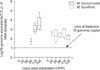
A longitudinal study was ultimately carried out in one wean-to-finish barn with pigs placed at approximately 4~5 weeks of age. The barn housed 220 animals in each of the 11 pens with a total of 2,420 animals. The pig unit was selected as part of the same production complex from where samples were collected to evaluate the initial diagnostic test performance. During placement, five pens were randomly selected, and pen-based oral fluid samples were collected by caretakers and the herd veterinarian at six time points: 0, 28, 58, 88, 118, and 148 days post-placement (DPP). The average age of the animals at the start of the study was 28 days.
Animals and animal care
All animal experimental protocols were approved by the Iowa State University Institutional Animal Care and Use Committee (no. 7-11-7181-S).
Statistical analysis
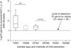
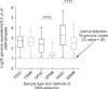
Statistical analysis and graphic depiction of data were performed with GraphPad Prism 6 (GraphPad Software, USA) along with its online software for calculating Kappa Cohen's coefficient (QuickCalcs; GraphPad Software) and McNemar's test (QuickCalcs; GraphPad Software). qPCR test results are presented as the number and percentage of positives in addition to the log10 of bacterial load shown in a box-and-whisker plot. Kappa Cohen's coefficient and McNemar's test (p < 0.05) were used to determine the agreement among qPCR test results when using different methods of genomic DNA extraction across multiple diagnostic specimens. An unpaired T-test (p < 0.05) using Welch's correction for different variance was used to compare the total bacterial yield per specimen between the two methods of genomic DNA extraction.
Results
Analytical sensitivity and specificity
M. hyosynoviae and M. hyorhinis primer sets were shown to be species-specific. The analytical sensitivity analysis using the plasmid DNA standard curve revealed linearity with M. hyosynoviae (y = -3.4667 + 38.333/R2 = 0.998) and M. hyorhinis (y = -3.6167 + 41.861/R2 = 0.999). qPCR efficiencies were 94% and 89% for M. hyorhinis and M. hyosynoviae, respectively. The assay limit of detection was found to be 10 genome equivalent copies for both M. hyosynoviae (Ct value of 35) and M. hyorhinis (Ct value of 38).
Comparative analysis of DNA extraction methods for qPCR test performance
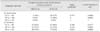
M. hyosynoviae was less likely to be detected in all diagnostic specimens used in this study with a maximum of 37% positive test results for tonsillar fluids and 28% for pen-based oral fluids. None of the nasal fluid samples were positive for M. hyosynoviae. In contrast, M. hyorhinis was detected in all specimens with a maximum of 96%, 75%, and 77% for tonsillar fluids, pen-based oral fluids, and nasal fluids, respectively (Table 1). Overall, the spin column method of DNA extraction increased test sensitivity for both M. hyosynoviae and M. hyorhinis (Table 1, Figs. 1 and 2).
Longitudinal study
Results of the longitudinal study revealed distinct patterns of detection for M. hyosynoviae and M. hyorhinis in serially collected pen-based oral fluids. M. hyosynoviae was not found until 88 DPP when only 40% of the pens were positive (2 of 5) with all samples testing positive thereafter. M. hyorhinis was initially detected in 60% of the samples on day 28 DPP (3 of 5) and subsequently in all five pens. Quantification of mycoplasma load in oral fluids showed that the overall bacterial load was low over time (Fig. 3).
Discussion
M. hyosynoviae and M. hyorhinis can be isolated from the tonsils and nasal cavity of experimentally inoculated and naturally exposed animals using standard culture procedures [9151624]. However, standard culture techniques are often complicated by overgrowth of other fast-growing bacteria [82235]. Therefore, qPCR assays were developed and validated in the current study to be used as high-throughput and rapid diagnostic alternatives for detecting and quantifying M. hyosynoviae and M. hyorhinis in specimens used as proxies for field samples routinely used in swine population screening.
Since M. hyosynoviae and M. hyorhinis are presumably long-term colonizers of the upper respiratory tract of pigs, it was hypothesized that pen-based oral, tonsillar, and nasal fluids would yield positive test results provided that these organisms were known to be circulating in the population [3131732]. This criterion was established while taking into consideration that a herd with a clinical history of Mycoplasma-associated arthritis was selected for sampling. As shown in the present investigation, both M. hyosynoviae and M. hyorhinis could be detected in tonsillar and pen-based oral fluids whereas only M. hyorhinis was found in nasal fluids. To date, it is still unclear whether or not M. hyosynoviae has a differential tropism for tonsils compared to the nasal cavity, or if an ecological barrier exists that determines the inhabitance and ecology of these structures, or niches. It appears that M. hyorhinis is less likely to be influenced by anatomical location. While these results are intriguing, careful interpretation is warranted since confounding factors such as sampling, number of animals, true prevalence, dynamics of infection, and host immunity may alter data interpretation.
Comparison between two different genomic DNA extraction protocols performed in the current investigation was made based on a previous report in which significant differences were observed in the rate of porcine reproductive and respiratory syndrome virus detection in oral fluids using different methods [2]. Additionally, many veterinary diagnostic laboratories use automatic genomic DNA extraction procedures to expedite test results, thus increasing the relevance of the comparison here. In this study, the manual procedure using a spin column was shown to increase test sensitivity for both M. hyosynoviae and M. hyorhinis. As recently suggested [3], this step can affect detection of these mycoplasmas in pen-based oral fluids. As demonstrated here, this may be true for other specimens as well. Results of the present investigation suggest that the number of false negatives can be affected by the genomic DNA extraction protocol used depending on the study design and nature of sampling. Importantly, these findings should increase the awareness of veterinarians and diagnosticians to not only interpret the qPCR test results as is (e.g., cycle threshold values as positive or negative), but to incorporate the clinical history of the farm, specifically the expected prevalence, to determine the positive and negative predictive values of the test.
While previous research has demonstrated that a variety of other swine pathogens may be found in porcine oral fluids [135628], the current study is the first to show that M. hyosynoviae can also be detected by qPCR in pen-based oral fluid samples. In our longitudinal study, delayed detection of either mycoplasma may have been caused by multiple factors such as protective circulating maternal antibodies, low rate of transmission, few infected animals at the time of weaning, or unknown host factors that confer temporary immunity. This is assuming that group-based sampling reflects the individual status of pen mates, which is yet to be proven. In summary, this study provided a new molecular approach to be used in veterinary diagnostics for specific quantification of two re-emerging swine pathogens. Our findings can serve as a foundation for future studies to understand the true prevalence, incidence, and impact of production interventions for controlling M. hyosynoviae and M. hyorhinis infection in pig populations.
 XML Download