Abstract
Tea contains polyphenols and is one of the most popular beverages consumed worldwide. Because most tyrosinase inhibitors that regulate melanogenesis are phenol/catechol derivatives, this study investigated the inhibitory effects of Camellia sinensis water extracts (CSWEs), including black tea, green tea, and white tea extracts, on melanogenesis using immortalized melanocytes. CSWEs inhibited melanin accumulation and melanin synthesis along with tyrosinase activity in a concentration-dependent manner. These inhibitory effects were superior to those of arbutin, a well-known depigmenting agent. The anti-melanogenic activity of black (fermented) tea was higher than that of a predominant tea catecholamine, epigallocatechin gallate. CSWEs, especially black tea extract, decreased tyrosinase protein levels in a concentration-dependent manner. These results suggest that the anti-melanogenic effect of CSWEs is mediated by a decrease in both tyrosinase activity and protein expression, and may be augmented by fermentation. Thus, CSWEs could be useful skin-whitening agents in the cosmetic industry.
Increased production and accumulation of melanin is a characteristic of many skin diseases, including acquired hyperpigmentation, melasma, postinflammatory melanoderma, and solar lentigo [835]. Melanin is one of the most widely distributed pigments, and is found in bacteria, fungi, plants, and animals. This compound is mostly produced in melanosomes of melanocytes that are dendritic cells found in the basal layer of the epidermis [16]. Abnormal accumulation of melanin in specific parts of the skin resulting in over-pigmented patches (e.g., melasma, freckles, ephelide, and senile lentigines) can be aesthetically undesirable [31]. Because acquired hyperpigmentation usually occurs in light-exposed areas of the skin, this condition has psychosocial and cosmetic relevance.
In human skin, melanin exists in two basic forms, black/brown eumelanin and yellow/red pheomelanin, that are synthesized from tyrosine via a common biosynthetic pathway [13]. The key enzyme in melanin biosynthesis is copper-dependent tyrosinase that catalyzes the initial rate-limiting step for conversion of tyrosine to 3,4-dihydroxyphenilalanine (DOPA), and is controlled by tyrosinase-related protein (TRP)-1 and TRP-2 [20]. Several depigmenting agents modulate skin pigmentation by regulating the transcription and activity of tyrosinase as well as that of TRP-1 and TRP-2. Tyrosinase inhibitors such as hydroquinone, kojic acid (KA), and arbutin (AT) have been used to treat hyperpigmention disorders but have recently been found to be unsafe for use in humans [101824]. Consequently, there is increasing interest in plant-derived extracts that can induce depigmentation without negative side effects on human health.
Tea is one of the most popular beverages worldwide. Fresh tea leaves contain polyphenols, primarily the catechins epigallocatechin (EGC), epicatechin (EC), epigallocatechin gallate (EGCG), and epicatechin gallate (ECG). These four catechins, of which EGCG is the most abundant, account for up to 30% of tea leaf dry weight [12]. Green tea (GT) is made from rolled and dried leaves that are steamed and/or pan-fried to inactivate endogenous polyphenol oxidase (PPO). White tea (WT) is unfermented tea produced from new growth buds and young leaves. The buds may be shielded from sunlight during growth to reduce chlorophyll contents, which makes the leaves appear white [2]. Black tea (BT) is produced without heat inactivation. The fermentation/oxidation phase after rolling enables large-scale PPO-catalyzed conversion of simple phenolics into more complex polyphenols such as thearubigins and theaflavins that are responsible for the characteristic BT reddish-brown color and astringency [32]. Most tyrosinase inhibitors are phenol/catechol derivatives, structurally similar to tyrosine or DOPA, and thus act as alternative tyrosinase substrates that are converted without pigment production [21]. Many studies have shown that phenolic compounds have antioxidant activity due to their capacity for scavenging free radicals and chelating metal ions along with an ability to prevent free radical formation [1262934].
In the present study, we investigated the anti-melanogenic effects of Camellia sinensis L. (family Theaceae) water extracts (CSWEs) and the potential mechanisms by which they induce skin depigmentation. The effects of CSWEs on tyrosinase activity, melanin synthesis, and expression of melanogenic enzymes at the protein and mRNA levels were evaluated in melan-A cells. These cells are immortalized mouse melanocytes characterized by high levels of tyrosinase and melanin expression [530].
Melan-A cells were obtained from Dr. Dorothy Bennett (St. George's Hospital, UK). EGC, EC, EGCG, ECG, a theaflavin standard mixture, AT, KA, 3-(4,5-dimethyl-thiazol-2-yl)-2, 5-diphenyl-tetrazolium bromide (MTT), 3,4-dihydroxy-L-phenylalanine (L-DOPA), L-tyrosine, and dimethyl sulfoxide (DMSO) were purchased from Sigma-Aldrich (USA). TRP-1 and TRP-2 were purchased from Santa Cruz (USA). All reagents used in this study were of analytical grade.
BT (China), GT (Korea), and WT (China) leaves were obtained from an oriental medicinal herb market (Korea). The leaves (600 g of each type) were boiled in 6 L of distilled water for 2 h in a heating extractor (COSMOS-660; Kyungseo Machine, Korea), concentrated, and powdered by lyophilization. Yields of the BT, GT, and WT extracts were 10.2, 21.3, and 20.0%, respectively.
Tea extract samples (200 mg) were digested using 10 mL of concentrated HNO3 for 15 min in a microwave acid digestion system (MARS5; CEM, USA). The digests were diluted to 50 mL in a volumetric flask with deionized water. The total lead (Pb), arsenic (As), and cadmium (Cd) concentrations were then determined using an inductively coupled plasma mass spectrometer (ICP-MS; ELAN 9000; PerkinElmer, USA). Total mercury (Hg) levels were measured using 20 mg of CSWE in an automatic mercury analyzer (MA-2; Nippon Instruments Corporation, Japan).
Catechin levels were measured with an HPLC-photodiode array detector (Waters 2996 PDA Detector; Waters, USA) and a liquid chromatography system (Waters e2695; Waters). The extract sample (10 µL) was injected into the HPLC column (5 µm, 250 × 4.6 mm, Shiseido CAPCELL PAK C18 UG 120; Shiseido, Japan). Mobile phase A was composed of 0.1% acetic acid and mobile phase B was composed of 100% acetonitrile. The catechins were eluted with 95% mobile phase A at 0 min, 75% mobile phase A at 20 min, 100% mobile phase B at 21 min, and 95% mobile phase A at 36 min. The flow rate was 1.0 mL/min at 40℃. Peaks were monitored at 280 nm and UV spectra were recorded.
Theaflavins were quantified with an HPLC-photodiode array detector (Waters 2998 PDA Detector; Waters) and a liquid chromatography system (Waters e2695; Waters). The extract sample (10 µL) was injected into the HPLC column as described above. Mobile phase A was composed of 0.1% acetic acid:acetonitrile:tetrahydrofuran (96:2:2), and mobile phase B was 100% acetonitrile. The theaflavins were eluted with 100% mobile phase A at 0 min, 40% mobile phase A at 45 min, and 100% mobile phase A at 47 min. The flow rate was 1.0 mL/min at 40℃. Peaks were monitored at 310 nm and UV spectra were recorded. The individual catechins and theaflavins were identified by comparing the retention times of the analytes with those of reference standards.
Total polyphenol contents of the tea extracts were evaluated using a Folin-Denis assay [11]. Total flavonoid contents were assessed as previously described by Davies et al. [9] with modifications. The test material (100 µL) was transferred into test tubes before 1 mL of di (ethylene glycol) reagent and 100 µL of 1 N NaOH were added. The mixture was shaken vigorously and incubated in hot water at 37℃ for 60 min. Absorbance was then measured at 420 nm.
Melan-A cells were grown in Roswell Park Memorial Institute (RPMI)-1640 medium supplemented with 10% fetal bovine serum (FBS), 1% penicillin/streptomycin (P/S), and 200 nM 12-O-tetradecanoylphorbol-13-acetate (TPA) at 37℃ in a 10% CO2 incubator for 72 h prior to the experiment.
Cell viability was assessed with an MTT assay. Melan-A cells were seeded in a 96-well plate (0.5 × 10⁴ cells/well) for 24 h and then treated with 200 µL of the CSWEs (0~50 µg/mL) in RPMI-1640 medium for 48 h. The medium was then replaced with RPMI-1640 medium containing 0.5 µg/mL MTT and the cells were further incubated for 3 h. After centrifugation at 168 × g for 10 min to pellet the cells, the medium was removed, 200 µL of DMSO were added, and the solution was incubated for 15 min with shaking to resuspend the cells. Cell viability was assessed by measuring absorbance at 540 nm with an ELISA reader (680; Bio-Rad Laboratories, Japan).
Melan-A cells were seeded in a 48-well plate (2 × 10⁴ cells/well) for 24 h and then treated with 500 µL of CSWEs (0~12.5 µg/mL) in RPMI-1640 medium for 72 h. Thereafter, the cells were washed and incubated with fresh tea extract solutions for another 72 h. The cells were observed with a model CKX41SF microscope (Olympus, Japan) and photographed at 200× magnification using a U-TVO.5XC-3 digital camera (Olympus) equipped with DMC e310 software (INS Industry, Korea). Next, the cells were dissolved in 1 N NaOH, and absorbance was measured at 490 nm (OD 490) using an ELISA reader. Melanin content was estimated as the OD 490 value/µg of protein and expressed as a percentage relative to the untreated control value (100%).
For the intracellular tyrosinase activity assay, melan-A cells were seeded in 60-mm cell culture dishes (4 × 10⁵ cells/well) for 24 h and then treated with 5 mL of tea extracts (0~12.5 µg/mL) for 48 h. The cells were washed with phosphate buffered saline, detached with 200 µL of 1% Triton X-100, transferred to Eppendorf tubes, subjected to extraction on ice with agitation six times every 10 min, and centrifuged at 18,620 × g for 20 min at 4℃. Next, 100 µL of L-DOPA were added and the mixture was incubated at 37℃ with 10% CO2 for 1 h. The OD 490 nm was then measured using an ELISA reader. Tyrosinase activity was estimated as the OD 490/µg protein/min and expressed as a percentage of the untreated control value (100%). For tyrosinase activity in the cell extract, the cultured melan-A cells were centrifuged, and 50 µL of supernatant was mixed with 49 µL of 0.1 M phosphate buffer (pH 6.8) and 1 µL of tea extracts (0~12.5 µg/mL). Subsequently, 100 µL of 0.2% L-DOPA was added, the absorbance was measured, and the percentage activation was calculated.
Melan-A cells treated with the CSWEs (6.25, 12.5, and 25.0 µg/mL) for 48 h were lysed by sonication in 0.1 M Tris-HCl (pH 7.2) buffer containing 1% Nonidet P-40, 0.01% SDS, and protease inhibitor cocktail (Roche, Germany). Protein concentration in the cell lysates was measured using a protein assay kit (Pierce, USA) with bovine serum albumin as the standard. Equal amounts of protein (10 µg) were separated by electrophoresis in a 10% polyacrylamide gel, transferred onto nitrocellulose membranes, and incubated with antibodies against tyrosinase/prolyl endoprotease-7 (PEP-7, 1:10,000 dilution), TRP-1/PEP-1 (1:10,000), and TRP-2/PEP-8 (1:10,000) kindly provided by Dr. Vincent J. Hearing (National Institutes of Health [NIH], USA). The membranes were then incubated with horseradish peroxidase-conjugated anti-rabbit IgG (1:1,000; Amersham, UK). Antibody binding was detected by chemiluminescence using electrochemical luminescence (ECL) reagents (Amersham). Band intensity was semi-quantified using the Image J program (NIH); β-actin was used as an internal control.
Total RNA was isolated from cells treated with 6.25 and 12.5 µg/mL tea extracts for 48 h using TRIzol reagent (Life Technologies, USA) according to the manufacturer's instructions. Equal amounts of total RNA (5 µg) were reverse transcribed in a 40-µL reaction mixture containing 8 µL of Molony murine leukemia virus reverse transcriptase (M-MLV RT), 5× buffer, 3 µL of 10 mM dNTPs, 0.45 µL of 40 U/µL RNase inhibitor, 0.3 µL of 250 U/µL M-MLV RT (Promega, USA), and 3.75 µL of 20 µM oligo dT (Bioneer, Korea). Single-stranded cDNA was then used as a template for PCR amplification in a reaction containing 4 µL of 5× green Go Taq Flexi buffer, 0.4 µL of 10 mM dNTPs, 0.1 µL of 5 U/µL Taq polymerase, 1.2 µL of 25 mM MgCl2 (Promega), and 0.4 µL of each primer (20 µM). The primer sequences were as follows: 5'-CAT TTT TGA TTT GAG TGT CT-3' (forward), 5'-TGT GGT AGT CGT CTT TGT CC-3' (reverse) for tyrosinase; 5'-GCT GCA GGA GCC TTC TTT CTC-3' (forward), 5'-AAG ACG CTG CAC TGC TGG TCT-3' (reverse) for TRP-1; 5'-GGA TGA CCG TGA GCA ATG GCC-3' (forward), 5'-CGG TTG TGA CCA ATG GGT GCC-3' (reverse) for TRR-2; and 5'-ACC GTG AAA AGA TGA CCC AG-3' (forward), 5'-TAC GGA TGT CAA CGT CAC AC-3' (reverse) for β-actin. PCR conditions for tyrosinase and TRP-1 were as follows: 28 cycles of denaturation at 94℃ for 60 sec, annealing at 56℃ for 60 sec, and extension at 72℃ for 60 sec. PCR conditions for TRP-2 were 28 cycles of denaturation at 94℃ for 60 sec, annealing at 64℃ for 60 sec, and extension at 72℃ for 60 sec. PCR for β-actin was carried out with 30 cycles of denaturation at 94℃ for 30 sec, annealing at 51℃ for 30 sec, and extension at 72℃ for 60 sec. The PCR products were separated on 1.2% agarose gels. Expected sizes of the amplified tyrosinase, TRP-1, TRP-2, and β-actin fragments were 1192, 268, 1044, and 528 bp, respectively. DNA band density was semi-quantitatively analyzed using a Kodak Gel Logic 100 image analysis system (Eastman Kodak, USA). The gene expression of tyrosinase, TRP-1, and TRP-2 was normalized relative to β-actin as an internal control.
To examine the safety of the CSWEs as an herbal medicine, we evaluated toxicity of the extracts by analyzing their heavy metal contents. Pb, As, Hg, and Cd levels in BT, GT, and WT were below the maximum permissible levels for herbal medicines set by the Ministry of Food and Drug Safety (MFDS). The Hg concentration in all three extracts was below the analytical detection limit (Table 1). These results indicated that the tea extracts could be safely used as herbal medicines and in cosmetics.
To further examine the potential toxicity of BT, GT, and WT, the viability of melan-A cells exposed to the extracts at concentrations between 0 and 50 µg/mL was measured for 48 h. The well-known depigmenting agents AT and KA along with the primary tea catecholamine EGCG were included as positive controls. Cells were viable in the presence of all concentrations of AT, KA, and BT similar to the untreated controls (Fig. 1). At 25 µg/mL, GT and WT also increased cell viability; however, a higher concentration (50 µg/mL) reduced melan-A cell viability to 62% and 76% relative to the untreated control, respectively. EGCG was found to have the greatest cytotoxicity among the tested compounds. At 25 and 50 µg/mL, this reagent decreased cell viability to 38% and 6% relative to the control group, respectively. Thus, BT, KA, and AT had the weakest cytotoxicity (≤ 50 µg/mL), followed by GT and WT (≤ 25 µg/mL), and EGCG (≤ 12.5 µg/mL).
The total polyphenol concentrations of AT, KA, EGCG, BT, GT, and WT were 24, 32, 435, 117, 112, and 123 mg/g, respectively, as determined using the tannic acid standard curve (Table 2). The total flavonoid concentrations of AT, KA, EGCG, BT, GT, and WT were 13, 14, 180, 82, 77, and 85 mg/g, respectively, according to the rutin standard curve (Table 2). The tea extracts were found to contain much higher levels of total polyphenols and flavonoids than AT and KA. Among the teas, WT had the highest concentrations followed by BT and GT.
GT (196.58 ± 0.81 mg/g) contained greater amounts of catechins than WT (137.48 ± 0.79 mg/g) and BT (15.30 ± 0.60 mg/g). In contrast, BT contained 1.49 ± 0.10 mg/g of theaflavins including theaflavin, theaflavin-3 gallate, theaflavin-3' gallate, and theaflavin-3,3' digallate, which were not detected in GT or WT (Table 3).
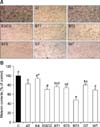
To assess the hypopigmentation effect of the tea extracts, melan-A cells were exposed to the test reagents at concentrations ranging from 0 to 12.5 µg/mL for 72 h and examined by microscopy. The untreated control cells contained abundant black precipitates indicative of melanin synthesis and well-developed dendrites (panel A in Fig. 2). On the other hand, melanin deposition and dendrite formation were conspicuously reduced in the cells treated with BT, GT, and WT.
We also quantitatively analyzed melanin contents of the cells. AT, KA, EGCG, BT, GT, and WT reduced melanin synthesis compared to the control in a concentration-dependent manner (data not shown). At a dose of 12.5 µg/mL, AT, KA, EGCG, BT, GT, and WT decreased melanin contents by 18% (p < 0.00l), 7% (p < 0.05), 29% (p < 0.00l), 52% (p < 0.00l), 20% (p < 0.00l), and 31% (p < 0.00l) compared to that observed in untreated cells, respectively (panel B in Fig. 2). The tea extracts were found to more significantly inhibit melanin synthesis in melan-A cells than AT and KA. Among the teas, BT showed the highest inhibitory effect which was superior even compared to that of EGCG.
Tyrosinase is the rate-limiting enzyme in melanin synthesis. To examine the inhibitory effect of the tea extracts on tyrosinase activity, melan-A cells were exposed to the test materials at concentrations between 0 and 12.5 µg/mL for 48 h. AT was used as a positive control because this compound was found to exert a greater inhibitory effect on melanin synthesis than KA. Compared to the untreated control group, tyrosinase activity in cells treated with AT, EGCG, BT, GT, and WT was reduced in a concentration-dependent manner (data not shown). At 12.5 µg/mL, intracellular tyrosinase activity was decreased by 12%, 61%, 52%, 45%, and 58%, respectively (p < 0.001; Fig. 3), indicating that tyrosinase activity in mealan-a cells was reduced more by the tea extracts than AT. Among the teas, WT had the greatest inhibitory effect, which was similar to that of EGCG. Additionally, treatment with 12.5 µg/mL of AT, EGCG, BT, GT, and WT decreased cell-extracted tyrosinase activity by 13%, 18%, 25%, 15%, and 16%, respectively (p < 0.001; data not shown). Among the extracts, BT had the most potent inhibitory effect, superior even to that of EGCG. These results suggest that the inhibition of melanin synthesis by CSWEs is related to decreased melanocyte tyrosinase activity.
To determine whether the inhibition of tyrosinase activity by CSWEs was associated with tyrosinase expression, melan-A cells were exposed to the extracts at concentrations ranging from 0 to 25 µg/mL for 48 h. Tyrosinase protein expression was then measured by Western blotting (panel A in Fig. 4). Treatment with BT, GT, and WT reduced tyrosinase protein expression in a concentration-dependent manner (panel B in Fig. 4). At 12.5 µg/mL, BT, GT, and WT significantly (p < 0.001) decreased tyrosinase protein levels by 72%, 18%, and 25%, respectively, compared to the control. In contrast, AT at all tested concentrations did not affect the protein expression of tyrosinase. We also measured the protein expression of the melanogenic enzymes TRP-1 and TRP-2, and found that the levels of both proteins were not altered by AT or CSWE treatment (data not shown).
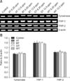
We also performed RT-PCR to examine the mRNA levels of tyrosinase, TRP-1, and TRP-2 in cells exposed to the extracts at concentrations ranging from 0 to 12.5 µg/mL for 48 h (panel A in Fig. 5). No difference in mRNA expression of melanogenic enzymes was observed between the treated and control cells (panel B in Fig. 5). These results suggest that the inhibitory effect of tea extracts on cellular tyrosinase activity is related to decreased tyrosinase protein expression.
Tea is one of the most popular beverages in the world and is also known for its health-promoting effects. Four types of tea are produced from the leaves of Camellia sinensis: WT, GT (both unfermented teas), oolong tea (semi-fermented tea), and BT (fermented tea). In a previous study, oolong tea water extract was reported to inhibit melanogenesis in B16 mouse melanoma cells [4]. However, the melanogenic effects of BT, GT, and WT have not been sufficiently investigated. In the present study, we examined the hypopigmentation effects of BT, GT, and WT (CSWEs) in immortalized melanocytes.
EGCG, a predominant tea catechin, has been reported to more effectively inhibit melanin synthesis than KA in the spontaneously immortalized mouse melanocyte cell line Mel-Ab [15]. Additionally, oolong tea water extract was found to exert a greater inhibitory effect on melanogenesis in B16 melanoma cells than ascorbic acid [4]. In the current study, we discovered that BT and WT had higher levels of anti-melanogenic activity than AT, KA, and EGCG. In addition, BT more effectively reduced melanin deposition and dendrite formation along with total melanin contents than GT and WT. HPLC analysis revealed that BT contained theaflavins (1.49 ± 0.10 mg/g) that were not detected in GT or WT. Theaflavins, a mixture of theaflavin, theaflavin-3-gallate, theaflavin-3'-gallate, and theaflavin-3,3'-digallate, are formed by two catechin monomers via the action of PPO during fermentation. Among these compounds, theaflavin-3,3'-digallate was previously found to prevent melanogenesis more efficiently than EC, ECG, EGC, EGCG, and KA in mouse B16 melanoma cells while the other theaflavins had no significant anti-melanogenic effect [36]. Therefore, it is believed that the depigmentation effects of CSWEs can be augmented by fermentation, and the potential depigmentation component of BT might be theaflavin-3, 3'-digallate.
Tyrosinase, a rate-limiting enzyme in melanogenesis, is a melanocyte-specific copper-containing glycoprotein located within specialized organelles called melanosomes [20]. Tea and its components have been shown to inhibit intracellular and cell-extracted tyrosinase activities. EGCG causes such inhibition in mushrooms [1523], and oolong tea reduces tyrosinase activity in B16 mouse melanoma cells [4]. Tea flavonoids have metal chelation abilities [26]. For example, proanthocyanidins, which are flavonoids from grape seeds, inhibit tyrosinase by chelating tyrosinase-bound copper [34]. BT theaflavins and GT flavonols are also known to exert antioxidant effects by radical scavenging and metal chelation [28]. WT was also reported to have higher levels of antioxidant activity than GT [33].
In the present study, we found that the levels of flavonoids in EGCG, BT, GT, and WT were 180, 82, 77, and 85 mg/g, respectively, which may account for the inhibition of tyrosinase activity. Among the teas, WT exerted the greatest inhibitory effect on intracellular tyrosinase activity followed by BT and GT. In contrast, BT had the highest inhibitory effect on cell-extracted tyrosinase activity followed by WT and GT. Of these two types of tyrosinase activities, cell-extracted one was coincident with melanin synthetic activities of CSWEs in the current study. Although tyrosinase may be inhibited through different mechanisms (e.g., blocking of catalytic or regulatory sites as well as protein degradation) [25] that remain unclear in melan-A cells, our results suggest that CSWEs may inhibit tyrosinase activity via copper chelation. Recently, rhododendrol [4-(4-hydroxyphenyl)-2-butanol], a depigmentation-inducing phenolic compound developed as a lightening/whitening cosmetic by Kanebo Cosmetics Inc. (Japan) was reported to cause depigmentary disorders such as leukoderma and idiopathic vitiligo [1427]. This reagent was also found to be cytotoxic against human melanocytes at high concentrations, suggesting that tyrosinase-dependent accumulation of endoplasmic reticulum stress and/or activation of the apoptotic pathway may contribute to melanocyte cytotoxicity. Although BT had the lowest cytotoxicity among the tested reagents, and WT and GT had lower cytotoxicity than EGCG in the current investigation, further studies should be conducted to evaluate depigmentary disorders associated with CSWEs. Additionally, the relationship between cytotoxicity and molecular events following CSWE-tyrosinase interaction should be clarified.
CSWEs, especially BT, also significantly (p < 0.001) decreased tyrosinase protein expression in a concentration-dependent manner. In contrast, AT did not affect tyrosinase protein levels. This finding is consistent with ones from previous studies showing that AT does not change the expression of tyrosinase, TRP-1, or TRP-2 in B16 melanoma cells [17], melan-A cells [37], and human melanocytes [6]. These data indicate that AT depigmentation activity may be based on post-translational inhibition of tyrosinase activity [619]. It has been reported that oolong tea decreases tyrosinase protein and mRNA levels in B16 mouse melanoma cells, thereby inhibiting melanogenesis at the transcriptional level [4], whereas EGCG reduces tyrosinase expression in Mel-Ab cells at the protein level [15]. In the present study, we found that CSWEs decreased tyrosinase protein expression but did not affect mRNA levels in melan-A cells, suggesting that the teas may decrease the post-transcriptional stability of tyrosinase and accelerate its degradation [3].
Our results are in agreement with those from a previous report showing that danazol inhibits melanogenesis in B16 mouse melanoma cells by downregulating the expression of tyrosinase protein but not mRNA [7]. In addition, ethanol extracts of traditional Chinese herbs (Radix ophiopogonis and Folium perillae) also cause depigmentation in melan-A cells by suppressing tyrosinase activity at the post-transcriptional level [37]. Overall, these results indicate that herbal extracts may have diverse mechanisms of depigmentation due to their complex composition.
In conclusion, our study has demonstrated that CSWEs (BT, GT, and WT) inhibit melanogenesis more efficiently than AT in melan-A cells. Among the different teas, BT (fermented tea) may be the most effective depigmentation agent with the lowest cytotoxicity and highest anti-melanogenic activities that are superior to those of EGCG. The potential mechanism underlying the depigmentation effects of CSWEs is the inhibition of tyrosinase activity. This is due to accelerated tyrosinase degradation and reduced levels of the protein, but not modulation of mRNA transcription. The potential depigmentation reagents in BT are theaflavins. However, this hypothesis should be further investigated by isolating and identifying biologically active components contained in BT.
Figures and Tables
 | Fig. 1Effect of different tea extracts on cell viability. Melan-A cells were treated with the indicated concentrations of the test reagents (0~50 µg/mL) for 48 h. Values represent the mean ± SD of three independent experiments. AT: arbutin, KA: kojic acid, EGCG: epigallocatechin gallate, BT: black tea, GT: green tea, WT: white tea. |
 | Fig. 2Anti-melanogenic effect of the tea extracts. (A) Morphological changes of melan-A cells treated with the extracts. The treatment with 12.5 µg/mL for 72 h reduced the amount of black precipitate and dendrite extension compared to the untreated control. BT at lower concentrations of 3.125 µg/mL (BT1) and 6.25 µg/mL (BT2) also significantly inhibited melanogenesis. (B) Melanin production in cells treated with the test reagents (12.5 µg/mL) for 72 h was measured with an ELISA. BT1 is 3.125 µg/mL, BT2 is 6.25 µg/mL, and BT3 is 12.5 µg/mL. Values represent the mean ± SD of three independent experiments. Statistically significant differences (p < 0.001) identified by an ANOVA and Duncan's multiple range test are indicated by different letters. *p < 0.05 compared to the untreated control. 200× magnification. C: control. |
 | Fig. 3Effect of the tea extracts on tyrosinase activity in melan-A cells. Tyrosinase activity in the cells treated with 12.5 µg/mL of the test reagents for 48 h was measured with an ELISA. BT1 is 3.125 µg/mL, BT2 is 6.25 µg/mL, and BT3 is 12.5 µg/mL. Values represent the mean ± SD of three independent experiments. Statistically significant differences (p < 0.001) identified by an ANOVA and Duncan's multiple range test are indicated by different letters. |
 | Fig. 4Effect of the tea extracts on tyrosinase protein expression in melan-A cells. Tyrosinase protein expression in cells treated with the test reagents (0~25 µg/mL) for 48 h was analyzed by Western blotting. (A) A representative gel. (B) Relative protein expression normalized relative to β-actin levels. Values represent the mean ± SD of three independent experiments. Treatment with BT, GT, or WT decreased tyrosinase protein expression in a concentration-dependent manner compared to the untreated control. Significant differences (p < 0.001) identified by an ANOVA and Duncan's multiple range test are indicated by different letters. **p < 0.01 compared to the untreated control. |
 | Fig. 5Effect of the tea extracts on mRNA expression of tyrosinase, TRP-1, and TRP-2 in melan-A cells. mRNA expression in cells treated with the test reagents (0~12.5 µg/mL) for 48 h was analyzed by RT-PCR. (A) A representative gel. (B) Semi-quantification of mRNA expression at 12.5 µg/mL by normalization to β-actin. Values represent the mean ± SD of three independent measurements. mRNA expression of tyrosinase, TRP-1, and TRP-2 was not affected by treatment with BT, GT, or WT. |
Table 1
Heavy metal contents in the water extracts of black (BT), green (GT), and white (WT) teas

Values represent the mean of duplicate measurements. ND: not detected. Maximum permissible levels for herbal medicines set by the Korea Food and Drug Administration (KFDA): Pb, 5 ppm; As, 3 ppm; Hg, 0.2 ppm; and Cd, 0.3 ppm [22].
References
1. Al-Azzawie HF, Alhamdani MSS. Hypoglycemic and antioxidant effect of oleuropein in alloxan-diabetic rabbits. Life Sci. 2006; 78:1371–1377.

2. Alcázar A, Ballesteros O, Jurado JM, Pablos F, Martín MJ, Vilches JL, Navalón A. Differentiation of green, white, black, oolong, and pu-erh teas according to their free amino acids content. J Agric Food Chem. 2007; 55:5960–5965.

3. Ando H, Kondoh H, Ichihashi M, Hearing VJ. Approaches to identify inhibitors of melanin biosynthesis via the quality control of tyrosinase. J Invest Dermatol. 2007; 127:751–761.

4. Aoki Y, Tanigawa T, Abe H, Fujiwara Y. Melanogenesis inhibition by an oolong tea extract in B16 mouse melanoma cells and UV-induced skin pigmentation in brownish guinea pigs. Biosci Biotechnol Biochem. 2007; 71:1879–1885.

5. Bennett DC, Cooper PJ, Hart IR. A line of non-tumorigenic mouse melanocytes, syngeneic with the B16 melanoma and requiring a tumour promoter for growth. Int J Cancer. 1987; 39:414–418.

6. Chakraborty AK, Funasaka Y, Komoto M, Ichihashi M. Effect of arbutin on melanogenic proteins in human melanocytes. Pigment Cell Res. 1998; 11:206–212.

7. Chang TS, Lin JJ. Inhibitory effect of danazol on melanogenesis in mouse B16 melanoma cells. Arch Pharm Res. 2010; 33:1959–1965.

8. Cullen MK. Genetic epidermal syndromes: disorders characterized by lentigines. In : Nordlund JJ, Boissy RE, Hearing VJ, King RA, Ortonne JP, editors. The Pigmentary System: Physiology and Pathophysiology. New York: Oxford University Press;1998. p. 760–766.
9. Davies R, Massey RC, McWeeny DJ. The catalysis of the N-nitrosation of secondary amines by nitrosophenols. Food Chem. 1980; 6:115–122.

10. Draelos ZD. Skin lightening preparations and the hydroquinone controversy. Dermatol Ther. 2007; 20:308–313.

11. Folin O, Denis W. On phosphotungstic-phosphomolybdic compounds as color reagents. J Biol Chem. 1912; 12:239–243.

12. Graham HN. Green tea composition, consumption, and polyphenol chemistry. Prev Med. 1992; 21:334–350.

13. Hearing VJ. Biogenesis of pigment granules: a sensitive way to regulate melanocyte function. J Dermatol Sci. 2005; 37:3–14.

14. Kasamatsu S, Hachiya A, Nakamura S, Yasuda Y, Fujimori T, Takano K, Moriwaki S, Hase T, Suzuki T, Matsunaga K. Depigmentation caused by application of the active brightening material, rhododendrol, is related to tyrosinase activity at a certain threshold. J Dermatol Sci. 2014; 76:16–24.

15. Kim DS, Park SH, Kwon SB, Li K, Youn SW, Park KC. (-)-Epigallocatechin-3-gallate and hinokitiol reduce melanin synthesis via decreased MITF production. Arch Pharm Res. 2004; 27:334–339.

16. Kim YJ, Uyama H. Tyrosinase inhibitors from natural and synthetic sources: structure, inhibition mechanism and perspective for the future. Cell Mol Life Sci. 2005; 62:1707–1723.

17. Lee YS, Kim HK, Lee KJ, Jeon HW, Cui S, Lee YM, Moon BJ, Kim YH, Lee YS. Inhibitory effect of glyceollin isolated from soybean against melanogenesis in B16 melanoma cells. BMB Rep. 2010; 43:461–467.

18. Lim JT. Treatment of melasma using kojic acid in a gel containing hydroquinone and glycolic acid. Dermatol Surg. 1999; 25:282–284.

19. Maeda K, Fukuda M. Arbutin: mechanism of its depigmenting action in human melanocyte culture. J Pharmacol Exp Ther. 1996; 276:765–769.
20. Mallick S, Singh SK, Sarkar C, Saha B, Bhadra R. Human placental lipid induces melanogenesis by increasing the expression of tyrosinase and its related proteins in vitro. Pigment Cell Res. 2005; 18:25–33.

21. Menter JM, Etemadi AA, Chapman W, Hollins TD, Willis I. In vivo depigmentation by hydroxybenzene derivatives. Melanoma Res. 1993; 3:443–449.
22. Ministry of Food and Drug Safety (KR). Permission levels and experimental methods of heavy metals for herb medicines, etc. Notice 2008-2. 2008. 01. 08.
23. No JK, Soung DY, Kim YJ, Shim KH, Jun YS, Rhee SH, Yokozawa T, Chung HY. Inhibition of tyrosinase by green tea components. Life Sci. 1999; 65:PL241–PL246.

24. Ortonne JP, Nordlund JJ. Mechanisms that cause abnormal skin color. In : Nordlund JJ, Boissy RE, Hearing VJ, King RA, Ortonne JP, editors. The Pigmentary System: Physiology and Pathophysiology. New York: Oxford University Press;1998. p. 489–502.
25. Passi S, Nazzaro-Porro M. Molecular basis of substrate and inhibitory specificity of tyrosinase: phenolic compounds. Br J Dermatol. 1981; 104:659–665.

26. Rice-Evans CA, Miller NJ, Bolwell PG, Bramley PM, Pridham JB. The relative antioxidant activities of plant-derived polyphenolic flavonoids. Free Radic Res. 1995; 22:375–383.

27. Sasaki M, Kondo M, Sato K, Umeda M, Kawabata K, Takahashi Y, Suzuki T, Matsunaga K, Inoue S. Rhododendrol, a depigmentation-inducing phenolic compound, exerts melanocyte cytotoxicity via a tyrosinase-dependent mechanism. Pigment Cell Melanoma Res. 2014; 27:754–763.

28. Serafini M, Ghiselli A, Ferro-Luzzi A. In vivo antioxidant effect of green and black tea in man. Eur J Clin Nutr. 1996; 50:28–32.
29. Seyoum A, Asres K, El-Fiky FK. Structure-radical scavenging activity relationships of flavonoids. Phytochemistry. 2006; 67:2058–2070.

30. Sheffield MV, Yee H, Dorvault CC, Weilbaecher KN, Eltoum IA, Siegal GP, Fisher DE, Chhieng DC. Comparison of five antibodies as markers in the diagnosis of melanoma in cytologic preparations. Am J Clin Pathol. 2002; 118:930–936.

31. Solano F, Briganti S, Picardo M, Ghanem G. Hypopigmenting agents: an updated review on biological, chemical and clinical aspects. Pigment Cell Res. 2006; 19:550–571.

32. Sun CL, Yuan JM, Koh WP, Yu MC. Green tea, black tea and breast cancer risk: a meta-analysis of epidemiological studies. Carcinogenesis. 2006; 27:1310–1315.

33. Thring TS, Hili P, Naughton DP. Anti-collagenase, anti-elastase and anti-oxidant activities of extracts from 21 plants. BMC Complement Altern Med. 2009; 9:27.

34. Uehara S, Mizutani Y, Nakade M, Sakata O, Sasaki I, Arakane K, Suzuki T, Uchida R, Tokutake S. Inhibitory effects of proanthocyanidin-rich extracts from grape seeds on melanogenesis. J Jpn Cosmet Sci Soc. 2003; 27:247–255.
35. Urabe K, Nakayama J, Hori Y. Mixed epidermal and dermal hypermelanoses. In : Nordlund JJ, Boissy RE, Hearing VJ, King RA, Ortonne JP, editors. The Pigmentary System: Physiology and Pathophysiology. New York: Oxford University Press;1998. p. 909–911.


 PDF
PDF ePub
ePub Citation
 Print
Print


 XML Download