

 PDF
PDF ePub
ePub Citation
 Print
Print


Introduction
Sheep production associated with the generation of milk, leather, wool, and mutton constitutes a major proportion of agricultural output in several countries worldwide. Seasonal reproductive physiology is the basis for characterizing various wool sheep breeds, and is influenced by habitat latitude as well as time of the year [3]. The mating period usually occurs during the fall when the number of hours of light per day is reduced [1]; this limits annual flock prolificacy.
The effects of reproductive seasonality can be minimized or reversed by the strategic use of reproductive technologies [5]. Hormone-based protocols have been widely employed to increase the reproductive performance and productivity of sheep. The use of fixed-time artificial insemination (FTAI) allows the synchronization of lambing periods, organization of lambs into batches suitable for meeting market demands, an earlier onset of puberty, and improved conception rates regardless of whether the estrus is observed or not [1]. The estrus cycle can be manipulated by maintaining the luteal phase using progesterone (P4) and analogues (progestagens), and/or by interrupting the luteal phase with prostaglandin F2α or a synthetic analog [16,21]. The follicular phase can also be altered with equine chorionic gonadotropin (eCG) administered around the time that P4 exposure ceases. This increases the occurrence and speed of follicular development and ovulation, thereby improving the fertility rate following insemination [2].
FTAI following estrus synchronization with progestagens has been reported to produce varying conception rates (84.6% [10], 71.4%, 80.4% [21], or 54.5% [7]). Many protocols involve long periods (12 to 14 days) of progestagen administration that provides good synchronization rates in cyclic and acyclic ewes but variable fertility rates [4,11]. It has also been found that long-term exposure to progestagens extends the phase of follicular dominance and leads to the ovulation of aged oocytes; this is less efficient than short-term progestagen treatment [22]. These inconsistencies in the literature and differences of opinions highlight the need for a consistent and efficient standard hormone-based protocol. Therefore, the aim of the current study was to evaluate the effects of norgestomet implants placed for long (14 days), medium (9 days), and short (5 days) periods on the estrus synchronization and conception rates of crossbred ewes undergoing FTAI during the breeding season in the southern hemisphere.
Materials and Methods
Experiment location and animals
This experiment was conducted in Brazil during April and May 2013, corresponding to the natural breeding season with an average of 11 h and 38 min of daylight. The local climate was subtropical, humid, and mesothermal. The study site was located at a longitude of 50°44'28'' west and latitude of 23°43'40'' south at 976 meters above sea level.
The study was performed in accordance with the ethical guidelines of Londrina State University (Brazil) animal welfare committee. Seventy non-pregnant crossbred adult sheep (Santa Inês x Texel) with body condition scores [18] ranging from 2.5 to 3.5 (on a scale of 1~5) were used. The sheep were raised in an extensive farming system, on pasture containing Paspalum notatum and Brachiaria decumbens, and had access to water and mineral salt (Nutristar Ovinos, Londrina, Brazil) ad libitum. During the night, the animals were housed in a pen.
Experimental design
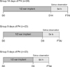
Ewes with similar body condition and age were divided into three groups and subjected to the same procedure for estrus synchronization except that the length of time during which the norgestomet devices (Crestar; Intervet, The Netherlands) were implanted varied. The three groups had the implants for 14 (G14 days of P4, n = 24), 9 (G9 days of P4, n = 23), or 5 (G5 days of P4, n = 23) days.
Protocol for estrus synchronization
A schematic of the treatments administered in this experiment is shown in Fig. 1. The norgestomet implants were cut in half using scissors. One half (containing 1.5 mg norgestomet) was implanted subcutaneously in the ear of each ewe. At the time of implant removal, 400 IU of eCG (Folligon; Intervet) and 22.5 µg D-cloprostenol (Preloban; Intervet) were injected intramuscularly into every ewe. No implant was lost and no ear infections were observed.
Estrus detection and distribution
Four Santa Inês males that were marked with brisket paint (grating paint and mineral oil moisture) were used to detect estrus in the synchronized ewes. Observation for signs of estrus was conducted on the day that the implants were removed from 4:00 to 7:00 pm. The sheep were monitored for 2 days after the devices were withdrawn from 7:00 to 10:00 am and from 4:00 to 7:00 pm. We considered a ewe to be positive for estrus when the female was marked by the ram and was standing while the male mounted her. Heat duration was measured from the first positive sign recorded until the ewe started to reject the male. On the third day after FTAI, a final period of estrus observation was performed from 7:00 to 10:00 am. In total, estrus observation was conducted over a 72-h period. Even the ewes that did not show signs of estrus were inseminated.
Artificial insemination (AI)
A pool of semen containing samples collected from one Texel, one White Dorper, and two Dorper rams (all 2 to 6 years old) was used for AI. Semen samples were collected using an artificial vagina (Walmur, Porto Alegre, Brazil) and combined. Sperm motility and vigor were then assessed with a light microscope (Nikon, Japan). Next, the concentration and percentage of abnormal sperm were determined using a Neubauer hemocytometer slides (AGB Scientific, Ireland) following standard operation procedures [10]. The semen was diluted using Tris-egg yolk extender (Trizma Base; Sigma, USA) to the concentration of 2 × 10⁸ sperm/mL. The sperm used for AI had 80% progressive motility, a vigor score of 3, and 85% were normal sperm cells.
Pregnancy diagnosis
Pregnancy was detected by transrectal ultrasonography (5.0 MHz, Aloka SSD-500; Aloka, Japan) 30 days after FTAI.
Statistical analysis
For this experiment, 70 ewes were randomly divided into three treatment groups. Differences in the rate of estrus onset during the 72-h observation period, the interval between the removal of the progestagen device and estrus onset, estrus duration, and conception rate were analyzed using Fisher's exact test (SigmaStat 3.5; Systat Software, USA). For all of the analyses, p ≤ 0.05 was considered to be significant. Conception rates were analyzed using logistic regression with the "Car" statistical package of "R" software (R development core team 2013, Austria).
Results
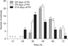
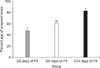
Among the synchronized ewes, estrus was noted in 85.7% (60/70) over the 72-h observation period following the removal of the norgestomet devices. No significant difference was observed in the percentage of ewes exhibiting estrus: 18/24 (75.0%) for G14 days of P4, 22/23 (95.7%) for G9 days of P4, and 20/23 (87.0%) for G5 days of P4. Differences in the dynamics of estrus associated with the various treatments were noted as presented in Fig. 2. The shortest average interval between implant removal and the onset of estrus was observed in the G14 days of P4 group (34.2 ± 8.9 h; p < 0.05). In contrast, this interval was not significantly different between ewes in the G9 days of P4 (41.9 ± 6.1 h) and G5 days of P4 (44.0 ± 6.7 h) groups. The mean duration of estrus did not differ (p > 0.05) between animals from the various groups (28 ± 15.5 h for G14 days of P4, 30 ± 12.1 h for G9 days of P4, and 26 ± 8.3 h for G5 days of P4). Conception rates are shown in Fig. 3. The G14 days of P4 group had a higher pregnancy rate (83.3%) than sheep in the G5 days of P4 group (47.8%; p < 0.05) but not ones in the G9 days of P4 group (60.0%).
Discussion
In the current study, the effect of norgestomet administration for different lengths of time on estrus synchronization and conception rates in ewes was evaluated. The increased conception rates observed with the 14-day treatment period are contrary to the results from a study by Martins et al. [13] using medroxiprogesterone acetate (MAP) for 12 and 6 days. Additionally, our findings conflict with the theory that long-term exposure to progestagen results in the excessive growth and persistence of dominant follicles. According to this hypothesis, oocytes released from these follicles during ovulation are of poor quality [22]. The pregnancy rate of 83.3% obtained in this experiment with long-term (14-day) hormone treatment suggests that the released oocytes retained a satisfactory level of quality and were able to establish pregnancies. Conflicting results from our investigation and those in the literature may be due to differences in the breed of animals or drugs that were used. Another explanation is that compromised fertility may result from long-term protocols involving the use of intravaginal devices due to the vaginal/cervical environment becoming suboptimal for sperm transport. Our protocol would have overcome this drawback because progestagen was administered through a subcutaneous device implanted in the animal's ear. Furthermore, norgestomet implants placed subcutaneously in the ears of ewes are known to increase serum progesterone levels [20].
The similarity of the 14-day protocol with the physiological events of the luteal phase may provide a reasonable explanation for the relatively high conception rates obtained in our study. It is known that corpus luteum regression in ovine females occurs between 13 and 15 days after ovulation, and that the rapid decline in progesterone is essential for the next onset of estrus and subsequent ovulation [6]. This may explain why the G14 days of P4 group had the highest conception rates since a percentage of ewes in the G9 days of P4 and G6 days of P4 groups may have had an active corpus luteum present in the ovary at the time of implant removal. For those animals, follicular development and ovulation events were surely delayed until progesterone secreted from the corpus luteum decreased following prostaglandin-induced luteolysis. Likewise, a percentage of these same animals would have benefited from delaying FTAI to optimize the conception rate.
For births to occur at the beginning of lambing season, estrus synchronization technology can be used to maximize the number of ewes becoming pregnant at the beginning of the breeding season. The interval between implant removal and estrus onset was significantly shorter in the G14 group (34.2 ± 8.9 h; p < 0.05) compared to the G9 days of P4 (41.9 ± 6.1 h) and G5 days of P4 (44.0 ± 6.7 h) groups. This result is similar to findings from studies by Salehi et al. [19], and Martemucci and D'Alessandro [12] in which the synchronization of estrus for 14 days via controlled internal drug release or fluorogestone acetate intravaginal sponges, respectively, promoted the onset of estrus within 36 h or 33.1 ± 4.3 h after removing the device.
In the current study, the percentage of ewes in estrus was similar (p > 0.05) among the three groups. This observation concurred with results obtained by Salehi et al. [19]. In addition, there was no significant difference in the duration of estrus between the groups. Our data indicate the effectiveness of the three protocols to synchronize estrus among the ewes.
In conclusion, treatment with norgestomet for different periods of time resulted in a high percentage of ewes in estrus within the 72-h observation period. Estrus duration among the three groups of ewes was also similar. However significant shortening of the interval between norgestomet implant removal and estrus in the animals treated with progestagen for 14 days (p < 0.05) was accompanied by a higher conception rate following FTAI. Future studies are required to evaluate the effect of inducing luteolysis at least 48 h prior to implant removal along with the impact on estrus dynamics and pregnancy following FTAI.
 XML Download