

 PDF
PDF ePub
ePub Citation
 Print
Print


Introduction
Porcine circovirus-2 (PCV-2) is a small, non-enveloped DNA virus that is a member of the Circoviridae family. To date, four main PCV-2 open reading frames (ORFs) have been identified that encode five viral proteins. Rep and Rep', both encoded by ORF1, are considered essential for viral replication [4]. ORF2 encodes a 27.8 kDa immunogenic capsid protein (Cap) that is the only structural protein of PCV2 [32]. The ORF3 and ORF4 genes produce an 11.9 kDa protein [27] and a 6.5 kDa protein [15], respectively. PCV-2 is horizontally transmitted via the mucosal route [12,33], and is the causative pathogen associated with post-weaning multisystemic wasting syndrome (PMWS) that mainly affects 7- to 15-week-old piglets with a mortality of up to 50% [12]. PMWS was first described in the 1990s and has since been responsible for dramatic economic losses to the swine industry worldwide [1,8].
Mucosa is the major portal of entry for a great majority of pathogens, and thus vaccines capable of eliciting mucosal immunity can fortify disease resistance at the mucosal frontlines [34]. However, most licensed vaccines administered via the parenteral route fail to induce protective mucosal immunity, which may only confer protection from clinical disease, and cannot eliminate infection at local mucosal invasion sites [34]. In this sense, the current vaccination strategy is not optimal since vaccinated animals remain susceptible to infection and serve as carriers of infectious viruses capable of transmission to other animals. In contrast, mucosal vaccines are designed to induce broad protective immunity at the mucosa, which will be more efficacious in providing protection against pathogen entry at mucosal sites. However, targeting all mucosal compartments to generate both local and systemic protective immunity is still a considerable challenge. Efforts should focus on screening for antigens with high immunogenicity, developing efficient mucosa-targeting delivery vectors and routes, and selecting a potent immunostimulatory adjuvant [37].
Adenovirus is a promising vaccine vector even in the presence of preexisting anti-vector immunity [2,5,6,17,38]. Based on previously published epitope mapping of the PCV-2 capsid protein [24], we constructed a recombinant replication-defective adenovirus expressing the major epitopes of the PCV-2 capsid protein (rAd/Cap/518) [11] and evaluated its efficacy in mice. The results showed that rAd/Cap/518 can induce specific mucosal, humoral, and Th1-type cellular immunity following intranasal delivery, which was determined to be the optimal mucosal immunization route [28]. However, mucosal immunity induced by a simple antigen is generally less efficacious and persists for only a short time [26,34,37]. Therefore, delivering an adenoviral vector with mucosal adjuvant might enhance mucosal immunity.
Unmethylated cytosine-phosphate-guanosine oligodeoxynucleotides (CpG ODN) can interact with and stimulate cells that express Toll-like receptor-9 (TLR-9), thereby initiating an immunomodulatory cascade that culminates in the production of Th1 and proinflammatory cytokines or chemokines [16,20,22]. Co-administration of CpG ODN with different types of antigen and vaccine vector along with various delivery routes have been shown to improve humoral and/or cellular immune responses, resulting in enhanced host immunity [3,10,14]. In the current study, CpG ODN was administered together with rAd/Cap/518 using an intranasal immunization protocol to evaluate the potential immune-enhancing effects in mice and provide insights into the development of novel mucosal vaccines against PCV-2.
Materials and Methods
Materials and reagents
rAd/Cap/518 was previously constructed [11] and preserved by Henan Agricultural University (China). Cap protein used for in vitro stimulation during flow cytometry and as a coating for indirect ELISA (iELISA) was produced in the pET-32 plasmid using an Escherichia coli prokaryotic expression system (Sangon Biotech, China). Empty adenovirus vector without the exogenous gene (wild-type rAd) was provided by Dr. Ping Jiang (Nanjing Agricultural University, China). ODN 1826 with unmethylated CpG (CpG ODN): 5-TCCATGACGTTCC TGACGTT-3 and ODN without CpG (non-CpG ODN): 5-TCCAGGACTTTCTCAGGTT-3 [4,15] were synthesized by Sangon Biotech with all bases phosphorothioate-modified.
Immunization and challenge protocols
All animal experiments were approved by the Ethics Committee for Animal Experimentation of the provincial Bureau of Health of Henan (China). BALB/c mice were used as an animal model as previously described [6,7,18,19]. A total of 175 specific pathogen-free (SPF) female BALB/c mice 6 to 8 weeks old were obtained from the Experimental Animal Center of Henan Province (China). The animals were kept under SPF conditions in individually ventilated cages (IVC) with unlimited access to rat food and water, and were housed under controlled temperature (around 22℃) and relative humidity (40%~50%) conditions with a 12 h light/dark cycle. The mice were randomly divided equally into seven groups. The experimental groups and immunization scheme are shown in Table 1.
A total volume of 100 µL rAd/Cap/518 mixed with adjuvant or placebo (Sigma, USA) was delivered to each animal into the nostrils for intranasal immunization (three times, 15~20 µL in each nostril every time). The animals were boosted 2 weeks after priming immunization (Table 1). Fourteen days after boosting immunization, five mice out of each group were challenged intranasally with 3 × 10⁵ tissue culture infective dose 50 (TCID50) of wild-type PCV-2 YY virus (genotype PCV2b), which was preserved by Animal Infectious Diseases Laboratory, Henan Agricultural University.
Sample collection
Serum, saliva, and lung and intestinal lavage samples were collected from five mice in each group to detect PCV-2-specific IgG and IgA 14, 21, 28, and 35 days after priming immunization as previously described [5,17]. Blood was collected through 20-gauge needle (Nipro, Japan) intravenous catheter from the lateral saphenous vein and allowed to clot overnight at 4℃. Serum was recovered from the clotted blood by centrifugation at 15,000 × g for 5 min, 4℃. Salivation was induced by administering 2 mg of carbamylcholine (Sigma) intraperitoneally to collect saliva with swab.
The mice were euthanized with ketamine (93 mg/kg body weight; Sigma) before lung and intestinal lavage. The airways were washed with 1 mL of phosphate-buffered saline (PBS) to harvest lung lavage fluids. The small intestines of the mice were removed and clamped with string at each end. Next, 1 mL of PBS containing 1% fetal bovine serum (Gibco; Life Technologies, USA) was injected into the intestines using a 27-gauge needle (Nipro) and vigorously vortexed to harvest the intestinal fluids. The lung and intestinal lavage fluids were clarified by centrifugation at 4℃ 12,000 × g for 5 min. The supernatants were then collected and stored at -70℃. Splenic lymphocytes were harvested to evaluate cell-mediated immune responses using a nylon mesh screen (Becton, Dickinson and Company, USA) as previously described [21]. Two weeks after challenge, spleens and lungs along with armpit, groin, and mesenteric lymph nodes were collected, grinded and DNA was taken to analyze PCV-2 loads.
Detection of PCV-2-specific IgG and IgA by iELISA
iELISA was performed to measure PCV-2-specific IgG levels in serum and IgA concentrations in saliva as well as lung and intestinal lavage (mucosal) samples [29]. In brief, 96-well ELISA plates (Corning, USA) were coated with 0.2 µg PCV-2 antigen in 0.05 M carbonate buffer (pH 9.6) per well and incubated overnight at 4℃. After blocking with 5% skimmed milk (Bio-Rad, USA) in PBS plus 0.5% Tween 20 (PBST) at 37℃ for 1 h, 100 µL of serum sample diluted 200-fold or undiluted mucosal samples were added to each well and the plates were incubated at 37℃ for 1 h. The plates were then washed three times with PBST to remove unbound antibodies. Horseradish peroxidase (HRP)-conjugated sheep anti-mouse IgG (1 : 2,000; Baosen Biotechnology, China) or HRP-conjugated goat anti-mouse IgA (1 : 10,000; Bethyl Laboratories, USA) was subsequently added to each well. After incubating at 37℃ for 1 h, the plates were washed three times and developed with 3,3',5,5'-tetramethylbenzidine (TMB; Sigma, USA) for 15 min. The reaction was stopped by adding 1 M H2SO4, and the optical density (OD) was measured at a wavelength of 450 nm using a Thermo plate reader (Thermo Scientific, USA).
Analysis of T cell sub-populations and intracellular cytokine concentrations
Flow cytometry was used to analyze T cell subpopulations and staining for intracellular cytokines was performed to measure the frequency of IL-2-secreting CD4+ T cells as well as IFN-γ-producing CD8+ T cells as previously described [40]. Briefly, lymphocytes were isolated from spleen 14 days after boosting immunization. Single cell suspensions were adjusted to a final concentration of 1 × 10⁷ cells/mL in PBS containing 2% FBS. The cells were stained with 100-fold diluted fluorescein isothiocyanate (FITC)-labeled anti-mouse CD3, phycoerythrin (PE)-conjugated anti-mouse CD4, and peridinin chlorophyll protein (PerCP)-labeled anti-mouse CD8a antibody (BioLegend, USA) in the dark at room temperature for 30 min according to standard flow cytometry procedures [19]. The cells were washed with PBS and approximately 20,000 cells were analyzed with a FACScalibur flow cytometer (BD Biosciences, USA) to identify CD3+, CD3+CD4+CD8-, and CD3+CD4-CD8+ T cell subpopulations.
For intracellular cytokine-specific staining, approximately 2 × 10⁶ lymphocytes were stimulated with 1 µg of PCV-2 Cap protein at 37℃ in 5% CO2. After 4 h of stimulation, brefeldin A (10 mg/mL) was added and cells were cultured for an additional 4 h at 37℃. The cells were then washed with PBS containing 2% FBS, fixed with Cytofix/Cytoperm (Sigma), and permeabilized with 1× Perm/Wash (Sigma). Next, the cells were incubated with a cocktail of 100-fold diluted PE-conjugated anti-mouse CD4, PerCP-conjugated anti-mouse CD8a, Alexa Fluor 700-labeled anti-mouse IFN-γ, and allophycocyanin-labeled anti-mouse IL-2 antibody (BioLegend). Approximately 500,000 lymphocytes were analyzed with a FACScalibur flow cytometer to measure the secretion of IL-2 by CD4+ T cells and IFN-γ production by CD8+ T cells.
Determination of viral DNA loads by quantitative real-time PCR (qPCR)
Two weeks after PCV-2 challenge, lymph node, spleen, and lung samples were collected and total DNA was extracted using UNIQ-10 virus DNA mini-kit (Sangon). PCV-2 viral loads were determined by qPCR as previously described [25,28]. The amplification protocol was as follows: the sense primer (5'-tatcaatctaaccacagtc-3') and the antisense primer (5'-atggcgggaggagtagtt-3') were used to amplify a 276 bp fragment of PCV-2 ORF2 gene. The RT-qPCR system contained 10 pmol each primer, 10 µL SYBR Premix Ex Taq, 500 ng sample DNA as template, and ddH2O to make a final volume of 20 µL. The RT-qPCR parameters consisted of 40 cycles of denaturation at 94℃ for 10 sec, annealing at 55℃ for 5 sec, and extension at 72℃ for 10 sec. A standard curve was prepared from serial dilutions of plasmid pORF2 containing the PCV-2 ORF2 gene for quantification of the virus copy number. The correlation coefficient and slope of standard curve were -1.00 and -3.345 respectively. The amplification efficiency was 99. 0%. All reactions were performed in duplicate on a LightCycler 2.0 (Roche, Swiss).
Statistical analysis
Data obtained from individual mice in all groups were analyzed by GraphPad Prism for Windows (ver. 5.01; GraphPad Software, USA). Differences in mean serum titers measured by iELISA were assessed by an ANOVA and Duncan's multiple range test. Data are presented as the mean ± standard error of the mean (SEM). p values < 0.05 were considered statistically significant.
Results
IgG serum titers
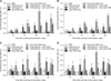
PCV-2-specific IgG titers in the serum were determined (Fig. 1A). From 21 to 35 days after priming immunization, significantly higher levels of IgG were detected in the group immunized with rAd/Cap/518 combined with CpG ODN compared to the animals immunized with rAd/Cap/518 alone (p < 0.05). No significant difference in IgG titers was observed between the group immunized with rAd/Cap/518 along with non-CpG ODN and the mice immunized with rAd/Cap/518 alone (p > 0.05).
IgA titers in saliva, lung, and intestinal lavage fluid
PCV-2-specific IgA titers in the saliva, lung, and intestinal lavage fluid were measured. From 21 to 35 days after priming immunization, significantly higher levels of IgA were detected in saliva from the group immunized with rAd/Cap/518 combined with CpG ODN compared to mice immunized with rAd/Cap/518 alone (p < 0.05). No significant differences in antibody responses were found between the group immunized with rAd/Cap/518 along with non-CpG ODN and the animals immunized with rAd/Cap/518 alone (p > 0.05, Fig. 1B).
Between 14 and 28 days after priming immunization, significantly higher IgA levels were detected in lung lavage fluid from the group immunized with rAd/Cap/518 combined with CpG ODN relative to animals immunized with rAd/Cap/518 alone (p < 0.05). Significant differences were no longer detected on day 35 (p > 0.05). Except for 21 days following priming immunization, no significant differences in antibody responses were found between the mice immunized with rAd/Cap/518 as well as non-CpG ODN and the group immunized with rAd/Cap/518 alone (p > 0.05, Fig. 1C). From 14 to 28 days post-priming immunization, significantly higher levels of IgA were detected in the intestinal lavage fluid from the animals immunized with rAd/Cap/518 and CpG ODN compared to the group immunized with rAd/Cap/518 alone (p < 0.05). Significant differences were no longer detected on day 35 (p > 0.05). From 14 to 21 days after priming immunization, no significant differences in antibody response were observed between the group immunized with rAd/Cap/518 combined with non-CpG ODN and mice that received rAd/Cap/518 alone (p > 0.05, Fig. 1D).
Analysis of T cell subsets
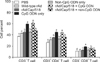
The frequencies of various T cell subsets within spleen lymphocyte populations were analyzed at 14 days after boosting immunization (Fig. 2). The frequency of CD3+ T cell subsets in the group immunized with rAd/Cap/518 and CpG ODN was significantly higher than that of the group immunized with rAd/Cap/518 alone (p < 0.05). In contrast, no significant difference was detected between the group that received rAd/Cap/518 with non-CpG ODN and the group immunized with rAd/Cap/518 alone (p > 0.05). Increased frequencies of CD3+CD4+ T cells and CD3+CD8+ T cells were similar to that of CD3+ T cells. No significant difference in the frequency of CD3+ T cells or CD3+CD4+ T cells was observed between the group immunized with CpG ODN alone and the mice that received rAd/Cap/518 with CpG ODN (p > 0.05). Furthermore, both groups showed significant differences in the frequencies of CD3+ T cells and CD3+CD4+ T cells compared the other groups that were not treated with CpG ODN (p < 0.05). Surprisingly, a higher proportion of CD3+CD8+ T cells was detected in the group treated with CpG ODN alone than the mice treated with rAd/Cap/518 and CpG ODN (p < 0.05). These observations suggested CpG ODN can stimulate the propagation of CD3+, CD3+CD4+, and CD3+CD8+ T cells in a non-specific manner, and may serve as a suitable adjuvant that efficiently promotes cellular immunity.
Intracellular cytokine analysis
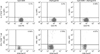
The percent of IL-2-secreting CD4+ T cells (IL-2+/CD4+ T) and IFN-γ-secreting CD8+ T cells (IFN-γ+/CD8+ T) was measured (Fig. 3). The percent of IL-2+/CD4+ T cells in the group immunized with rAd/Cap/518 and CpG ODN was 0.55%. In contrast, this percent was only 0.2% in the animals immunized with rAd/Cap/518 alone and 0.1% in the CpG ODN-treated group. Similarly, the percent of IFN-γ+/CD8+ T cells was 4.07% in the group immunized with rAd/Cap/518 and CpG ODN whereas this percent was only 2.79% and 0.54% in the groups treated only with rAd/Cap/518 or CpG ODN, respectively.
Evaluation of protection against PCV-2 challenge
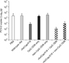
Two weeks following viral challenge, PCV-2 loads were measured by qPCR (Fig. 4). The viral load in the mice intranasally immunized with rAd/Cap/518 and CpG ODN rAd-Cap was 7-fold lower than that in animals immunized with rAd/Cap/518 alone although the difference was not statistically significant (p > 0.05). However, the viral loads in these two groups were significantly lower than those found in any of the other groups (p < 0.05).
Discussion
Approximately 70% of pathogens infect humans or animals via the mucosal route [37]. ucosal immunity therefore represents the frontline of host immune defense. Most commercially available vaccines are administered through the parenteral route, which can induce the production of high serum IgG levels. This may provide protection against clinical symptoms but cannot completely prevent local infection at mucosal entry sites or subsequent virus replication and shedding. For example, classical swine fever virus can chronically propagate in the tonsils [30] while pseudorabies virus can establish latent infection in the trigeminal ganglion after entry via the nasal mucosa [39] even in immunized animals possessing high levels of serum antibodies. Similarly, influenza virus [31] and Newcastle disease virus [35] can cause local infection in the respiratory or genital tract, leading to mild respiratory symptoms and continuous virus shedding in poultry that have been vaccinated intramuscularly. It is therefore imperative to develop effective mucosal vaccines that can induce protective immunity against mucosal invasion. However, the host usually strives to maintain mucosal homeostasis by responding to mucosal antigens through a tolerogenic mechanism as opposed to primary immune reactivity [9]. In this sense, induction of mucosal immunity still poses a considerable challenge.
To increase mucosal immunity and develop an effective PCV-2 vaccine, we have sought to improve the expression vector, delivery method, and immune adjuvant. In a previous study, we showed that mucosal immunization with recombinant adenoviral vectors expressing the major epitopes of the PCV-2 Cap protein (rAd/Cap/518) induce a PCV-2-specific humoral immune response and a Th1-type immune response at the mucosal site as well as a systemic level in mice [28]. To further improve the efficacy, the use of CpG ODN as an adjuvant was explored.
In the present study, the levels of serum IgG and mucosal IgA in the group intranasally immunized with rAd/Cap/518 together with 10 µg CpG ODN were significantly higher than those of the group immunized with rAd/Cap/518 alone. Frequencies of CD3+, CD3+CD4+, and CD3+CD8+ T cells in the group immunized with rAd/Cap/518 and CpG ODN were significantly higher than those for the group treated with rAd/cap/518 alone. However, CpG ODN alone significantly increased the frequencies of CD3+, CD3+D4+, and CD3+CD8+ T cells. This observation indicated that CpG ODN is an effective adjuvant capable of augmenting humoral and cellular immunity induced by rAd/Cap/518. Nevertheless, the impact of CpG ODN on the production of PCV2-specific neutralizing antibodies that were shown by others to prevent PMWS is still unclear [20].
T helper cells can be classified into two groups based on their function, responses to different cytokines, and ability to secrete various cytokines. IFN-γ and IL-2 contribute to the differentiation of Th1 cell types whereas IL-4 and IL-10 induce the differentiation of Th2 cells. Th1-type cellular immunity is considered to be more important for antiviral immune defense. Production of cytokines such as IL-2 and IFN-γ by splenic lymphocytes was enhanced by immunization with rAd/Cap/518 and CpG ODN, suggesting that mucosal delivery of CpG ODN induces high levels of Th1-type cytokines. In addition, the viral load after challenge in mice immunized with rAd/Cap/518 and CpG ODN was 7-fold lower than that in animals treated with rAd/Cap/518 alone. This difference wasn't statistically significant (p > 0.05). However, the viral load after challenge in mice immunized with rAd/Cap/518 and CpG ODN was significantly lower relative to that in the PBS placebo group. Many studies have shown that commercially available and experimental vaccines against PCV-2 can significantly reduce viremia, systemic viral loads, and shedding but cannot prevent or clear viral infection [28,29]. Much still has to be done before a more efficacious vaccine against PCV-2 can be developed.
The immunostimulatory activity of CpG ODN is determined by the structure of the CpG motif and other related factors. Active CpG ODN must contain unmethylated CpG dinucleotides flanked by two purines and two pyrimidines [22]. Other factors, including CpG dose, delivery systems such as the utilization of conjugate nanoparticles, liposomes, or biodegradable microparticles, and delivery route can also affect immunostimulatory activities [13,23]. Considering the characteristics of CpG and specificity of different mucosal administration routes, these variables should be further investigated to optimize the immunization regime and improve the adjuvant effect as well as vaccine potency.
In summary, we demonstrated that intranasal immunization with CpG ODN and rAd/Cap/518 markedly enhanced humoral immunity at both a systemic level and the mucosal sites compared to treatment with rAd/Cap/518 alone. In addition, cell-mediated immune responses were significantly increased and resulted in reduced viral loads. Thus, CpG ODN was found to be an effective immune adjuvant that can be intranasally delivered with rAd/Cap/518 to enhance host immunity.
 XML Download