

 PDF
PDF ePub
ePub Citation
 Print
Print


African horsesickness (AHS) is a frequently fatal infectious disease of equines [6,15] caused by the AHS virus (AHSV), an orbivirus belonging to the family Reoviridae [2]. Nine different serotypes of the virus have been identified [7,12]. AHSV is composed of seven structural proteins that include VP7, the highly conserved major core protein, and an outer capsid VP2 protein that plays a major role in virus neutralization and antigenic variability [1,11]. Several enzyme-linked immunosorbent assays (ELISAs) based on murine monoclonal antibodies (mAbs) have been developed for detecting AHSV and AHSV-specific antibodies [10,13,16]. Although murine mAbs [9] represent a major step towards standardized immunochemical reagents, they lack the physical link between the antibody and its genetic information inherent to phage displayed antibody [14], a feature that allows manipulation and even reconstruction of a single-chain variable fragment (scFv) gene. Assays for AHSV-specific antibodies are of little practical value since animals often die prior to the development of measurable antibody titers [10]. In the present study, we describe two phage-displayed scFvs that are suitable for the serotype- and serogroup-specific detection of AHSV in double antibody sandwich (DAS)-ELISAs. Lca12, a serotype-specific scFv with a detection limit of 2 ng purified AHSV-3 per well, was selected with directly immobilized, sucrose gradient-purified AHSV-3 [8] as previously described in detail [17]. The serogroup-specific scFv G7 was isolated with trapped AHSV-8. Both scFvs were acquired from the Nkuku library, a large semi-synthetic phage display library based on chicken antibody genes [17].
Panning with AHSV-8 was performed with two modifications of the methods previously described for isolating scFv Lca12 [17]. To select for antibodies suitable for detecting trapped AHSV in a DAS-ELISA, AHSV-8 was trapped by polyclonal IgG purified from the serum of a rabbit immunized three times with 50 µg of purified AHSV-3 (produced in house). Cross-reactive epitopes of conserved structural proteins such as VP7, the major core protein of AHSV [3], allow the use of IgG against any one of the serotypes for this purpose. Polysorp Immunotubes (Nalge Nunc International, USA) were first coated for 2 h at 37℃ with 10 µg/mL of the purified anti-AHSV-3 rabbit IgG [4] in phosphate buffered saline (PBS) and then blocked for 1 h at 37℃ with 2% (w/v) fat-free milk powder (Elite, South Africa) in PBS (MP/PBS). The tubes were then filled with an AHSV-8-infected baby hamster kidney (BHK; ATCC, USA) cell culture and incubated overnight at 4℃. The AHSV-8-infected cells were harvested in the culture medium after extensive cell damage was observed. The second modification was made to the usual pre-incubation of 5 × 10¹² library phage particles in MP/PBS supplemented with 0.1% (v/v) Tween 20 (MP/PBS/TW; Saarchem, South Africa) prior to panning [17]. For the present study, 200 µL pre-immune rabbit serum and 1/5 of the BHK cells from a 175 cm2 cell culture flask (Greiner, Germany) were added to reduce the possibility of selecting scFvs specific for rabbit IgG and/or BHK antigens. Monoclonal phage-displayed scFvs were produced and isolated as previously described in detail [17].
Microtiter plates for the DAS-ELISAs (Polysorb; Nalge Nunc International, USA) were coated overnight at 4℃ with 50 µL/well purified anti-AHSV-3 rabbit IgG [4] in PBS at a concentration of 10 µg/mL. All subsequent ELISA steps, up to the addition of the substrate solution, were incubated for 45 min at 37℃ for the serotype-specific assay and 1 h at 37℃ for the group-specific assay. Blocking was performed at 37℃ with 300 µL/well MP/PBS. After washing with PBS containing 0.1% (v/v) Tween 20 (PBS/TW), the antigen (50 µL/well) was added. Antigens were supplied by the World Organisation for Animal Health Reference Centre for AHS at the Onderstepoort Veterinary Institute (South Africa) with the approval of its Animal Ethics Committee. These antigens consisted of lyophilized Vero cell (ATCC, USA) suspensions infected with reference strains of AHSV, a lyophilized Vero cell culture infected with the Bryanston reference strain of equine encephalosis virus (EEV), and 12 Vero cell cultures declared AHSV-6-positive by the reference centre based on virus neutralization tests. These 12 cell cultures were inoculated with spleen and lung tissue from field horses to allow multiplication of the virus in samples that possibly contained too little virus for detection. A Vero cell homogenate, a Bryanston EEV reference strain, and MP/PBS were used as negative controls for the DAS-ELISAs. Lyophilized cell cultures that contained the reference strains were reconstituted to their original volumes in double distilled, de-ionized water.
After another wash with PBS/TW, the plate was coated with 50 µL/well phage displayed scFvs in a solution that consisted of equal volume of scFvs in centrifuged supernatant fluid and 4% MP/PBS/TW (0.2% Tween 20). The plate was washed again before 50 µL/well murine anti-M13 mAb B62-FE2 (PROGEN Biotechnik, Germany) diluted to a final concentration of 0.1 µg/mL in MP/PBS/TW was added. After washing with PBS/TW to remove unbound mAb, 50 µL/well horseradish peroxidase-labeled rabbit anti-mouse immunoglobulin (Dako, Denmark) diluted 1/1,000 in MP/PBS/TW was added to the wells. A final wash with PBS/TW was followed by the addition 50 µL/well of a substrate solution consisting of 10 mg o-phenylene diamine (Sigma, USA) and 5 µL of 30% hydrogen peroxide (Saarchem, South Africa) in 10 mL 0.1 M citrate buffer (pH 4.5). After 40 min at room temperature, the reaction was stopped with 50 µL/well 2 N H2SO4 (Associated Chemical Enterprises, South Africa) and absorbance was read at 492 nm using a BDSL Immunoskan microtitre plate reader (Biological Diagnostic Supplies Limited, UK).
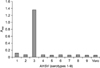
Phage-displayed scFv Lca12 with a detection limit of 2 ng trapped AHSV-3 per well [17] was selected with directly immobilized, purified AHSV-3 and facilitated serotype-specific detection of the homologous serotype with a DAS-ELISA (Fig. 1). This scFv produced an absorbance of more than 1.3 at 492 nm for AHSV-3 but an absorbance lower than 0.2 for the other eight serotypes as well as the control Vero cell homogenate. Phage-displayed scFv G7, selected with trapped AHSV-8, behaved as an antibody for serogroup-specific detection in the DAS-ELISA. This scFv generate absorbance values above 1.5 at 492 nm (Fig. 2) for all nine trapped AHSV serotypes and values below 0.1 for the Bryanston EEV strain, Vero cell homogenate, and MP/PBS negative controls. G7 was able to detect 10 ng of trapped AHSV-3 per well (result not shown) and therefore matched the detection limit (10 ng/well) of two murine mAbs described by Laviada et al. [10]. The latter two murine mAbs were not tested against AHSV-5 and AHSV-6 [10] while scFv G7 was found to be specific for all nine AHSV serotypes (Fig. 2). Using the same DAS-ELISA format, 12 Vero cell cultures that contained AHSV-6 from naturally occurring infections were all positive relative to the negative controls. The average absorbance value was at least 1.6 at 492 nm when scFv G7 was used as the detecting antibody (Fig. 3). The lowest absorbance value used to calculate the average was 1.2. A noteworthy feature of this scFv was the stability of the epitope to which it binds. Very similar signals were obtained with all 12 AHSV-6-infected Vero cell cultures despite the fact that some were stored at 4℃ for more than 8 months.
This study confirmed that the Nkuku library is a suitable source of immunoreagents for detecting AHSV with DAS-ELISAs. The scFv-based inhibition ELISAs developed by Fehrsen et al. [5] are appropriate for the serotype- and serogroup-specific detection of antibodies that bind to bluetongue virus. The VP7-specific murine mAb 1F1 [16] can also detect antibodies specific for all nine AHSV serotypes with an ELISA. In contrast to the latter two assays, we produced serotype- and serogroup-specific scFvs in the present study that can detect the causative agent of AHS. During AHS outbreaks in which animal deaths precede the production of measurable antibody titers, these two scFvs facilitate the rapid identification AHSV infection while the isolation of additional serotype-specific scFvs may considerably simplify virus serotyping.
 XML Download