

 PDF
PDF ePub
ePub Citation
 Print
Print


Introduction
Electroretinography (ERG) is a well-established diagnostic method for evaluating retinal function. This technique is non-invasive and requires no cooperation from the anesthetized patient [5,19]. It measures retinal sensitivity, photoreceptors in the outer retina, and associated pathways in the middle layers of the retina [25]. A typical ERG includes monitoring scotopic responses of dark-adapted eyes in dim (rod response) and bright light (combined rod-cone response), and photopic responses of light-adapted eyes with bright (single-flash cone response) and flickering (30 Hz flicker response) light stimuli.
Candidates for canine ERG include dogs of various ages with different diseases. The testing procedure may also be conducted for different purposes. This examination is essential for evaluating retinal function when it is difficult to perform funduscopy because of ocular media opacity. Components of the ERG change before presentation of ophthalmoscopical abnormalities in the retina [6,24]. Therefore, ERG recording is performed prior to cataract extraction [19] to experimentally evaluate retinal toxicity [10] and diagnose retinal disorders such as generalized progressive retinal atrophy (an inherited form of photoreceptor degeneration) and sudden acquired retinal degeneration associated with sudden visual loss due to acute photoreceptor death in dogs [18,19].
Establishment of normal ERG values is important for evaluating a subject's response. However, normal values of the different ERG components are difficult to define. There are many reports demonstrating that various factors affect ERG components including differences in protocols [19], stimulus intensity [15,16], and breed of the subject [8]. In human medicine, the International Society for Clinical Electrophysiology of Vision (ISCEV) [15,16] and the National Retinitis Pigmentosa Foundation Committee have attempted to standardize ERG protocols [9]. In veterinary medicine, the European College of Veterinary Ophthalmologists (ECVO) Committee has recommended a common setting [18]. The methodologies have been updated to standardize ERG responses and make them universally comparable. In our previous study, we also suggested the use of 3.0 cd·s/m2 flash-stimulation as a standard flash and 0.01 cd·s/m2 flash-stimulation as a rod flash for performing ERG with an LED-electrode in canines [14]. With these intensities, standard ERG values have been identified for some breeds of dog including beagles [13] and Shih-Tzus [8]. The ISCEV and ECVO Committee have proposed that normal ERG values should be expressed as normal ranges [15,18]. There are a few studies about the effect of aging on ERG findings in canines [5,11], but these investigations only reported the findings for dogs from birth to 2 years old. There are no reports about changes in ERG results for senile dogs.
In the present study, ERG was performed with an LED-electrode in beagle dogs, a widely used experimental animal. The goal of our investigation was to determine the normal ranges for different age groups under our flash conditions. We also investigated differences between results for the left and right eyes.
Materials and Methods
Animals
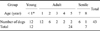
Eighty-six eyes in 43 laboratory-beagle dogs in Rakuno Gakuen Univeristy (Japan) [35 males, eight females; 2.8 ± 2.4 (mean ± SD) years old with an age range of 8 months to 8 years; body weight, 8.0~13.4 kg] were included in this study. The subjects were divided into three groups according to age. Table 1 shows the age distribution of the groups: 12 dogs less than 1 year old in the young group (8~10 months old); 24 dogs in the adult group (1~5 years old), and seven dogs over 6 years old in the senile group (7~8 years old). The animals were housed individually in the department of Small Animal Clinical Sciences, Rakuno Gakuen University, Japan, and fed commercial dry food (ED-1; Sanwa Kagaku Kenkyusho, Japan) and water. Physical, serological, and ophthalmic examination findings were normal for all dogs. Ophthalmic examinations, including pupillary light reflex, menace response, tonometry, slit-lamp biomicroscopy, and ophthalmoscopy were performed on the day prior to the study. Our investigation was conducted according to the Guidelines of the Experimental Animal Research Committee of Rakuno Gakuen University (Japan).
ERG equipment
A portable commercial system (LE-3000; Tomey, Japan) was used for ERG recording. This system includes a stimulus instrument, amplifier, recorder, and printer. The frequency band was 0.3~300 Hz. We used an ERG contact lens electrode with built-in diode light sources (LED-electrode, LED-electrode H2000; Kyoto Contact Lens, Japan) as an active electrode with a flash stimulator. The responses obtained for each recording were printed. The reference and ground electrodes were needle-type (disposable needle electrode; Tomey, Japan) and plate-type (ear electrode for LE-3000; Tomey, Japan), respectively.
ERG procedure
Prior to ERG recording, the subjects' pupils were fully dilated to over 12 mm with a solution of 0.5% tropicamide and 0.5% phenylephrine hydrochloride (Mydrin-P; Santen, Japan). After inducing mydriasis, the animals were adapted to the dark for more than 30 min in a dark room. ERG was performed under red light in a dark room.
ERG was conducted with the subjects in a prone position while sedated with a combination of 0.01 mg/kg medetomidine (Domitor; Zenoaq, Japan), 0.15 mg/kg midazolam (Dormicam; Astellas, Japan), and butorphanol 0.025 mg/kg (Stadol; Bristol-Myers Squibb, USA) injected intravenously. The LED-electrode was positioned on the bilateral cornea after topical anesthesia was induced with 0.4% oxybuprocaine hydrochloride (0.4% Benoxil ophthalmic solution; Santen, Japan) while protected with 1.5% hydroxyethylcellulose gel (Scopisol 15; Senju, Japan) as described our previous report [13]. A needle-type electrode was positioned subcutaneously in center of the frontal bone as a reference electrode. The electrodes were positioned in the configuration of an equilateral triangle formed by the LED electrode on each eye and the reference electrode. A plate-electrode was attached inside an auricle with conducting paste (EC2 Grass electrode cream; Grass, USA) as a ground electrode.
The ERG started by monitoring scotopic responses (rod and combined rod-cone responses) and photopic responses (single-flash cone and 30-Hz flicker responses) after light adaption with 25 cd/m2 of background light for 10 min. ERG data were obtained under the stimulus conditions as described in our previous study [13] with an LED-electrode (Table 2). After ERG recording, sedation of the subject was reversed with an intravenous injection of 0.05 mg/kg atipamezol hydrochloride (Antisedan; Zenoaq,
Japan).
ERG evaluation
Wave amplitude and implicit time were determined for each response. Amplitude of the a-wave was measured from the baseline to the peak of the first negative deflection, and that of the b-wave was measured from the peak of the a-wave to the largest positive-trough of the combined rod-cone response. Implicit times of both waves were measured from the onset of the flash stimulus to the peaks of the a- and b-waves. To evaluate the rod and single-flash cone responses, only b-wave components were measured. Amplitudes of the 30-Hz flicker were measured from the baseline to the positive peak and the implicit times were calculated from the light onset to the positive peak.
Statistical analysis
Normal values of implicit time and amplitude of each ERG procedure, including the b/a ratio of the combined rod-cone response, were statistically defined as the median value and range between the 2.5th and 97.5th percentiles (lower and upper limits). These were calculated as the mean value ± 1.96 SD. Amplitudes and implicit times for each age group were evaluated using a one-way factorial ANOVA with Tukey's test as a post-hoc test [21]. To compare differences between the left and right eyes, a paired t-test was performed. P-values less than 0.05 were considered statistically significant. All statistical analyses were conducted using GraphPad Prism 5 for Windows (GraphPad Software, USA).
Results
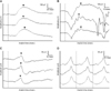
For all subjects, sedation was adequate for ERG examination. ERG results were recorded with certainty although slight eyeball rotation was observed, especially soon after sedative administration as described in our previous report. No anesthesiologic irregularities occurred and sedation was reversed safely in all subjects after ERG examination. Fig. 1 shows representative ERG waveforms obtained from one dog in each of the three age groups.
Normal ERG ranges
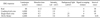
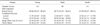
The ranges of implicit time and amplitude of the scotopic b-wave for the young, adult, and senile groups were 56.35~80.81 (median: 69.06) msec and 78.01~272.83 (median: 172.06) µV, 52.23~86.81 (median: 68.75) msec and 79.06~243.38 (median: 158.81) µV, and 56.25~80.21 (median: 67.69) msec and 84.06~248.50 (median: 150.56) µV, respectively. No statistically significant differences in the scotopic b-wave were found although decreases in amplitude were observed with age (Tables 3 and 4). Ranges of the implicit time and amplitude for the a-wave of the combined rod-cone response were 10.42~13.56 (median: 11.78) msec and 108.01~184.65 (median: 143.06) µV for the young group, 8.75~15.65 (median: 11.85) msec and 81.33~200.53 (median: 141.19) for the µV for the adult group, and 11.17~13.79 (median: 12.56) msec and 71.78~203.88 (median: 127.81) µV for the senile group. Ranges of implicit time and amplitude for the b-wave of the combined rod-cone response were 25.62~37.96 (median: 32.53) msec and 222.93~411.53 (median: 317.23) µV for the young group, 23.66~38.84 (median: 29.94) msec and 175.17~393.79 (median: 282.23) µV for the adult group, and 21.12~41.82 (median: 29.25) msec and 147.43~364.83 (median: 253.94) µV for the senile group. Differences in the combined rod-cone response between the adult and young groups (p < 0.05) as well as the young and senile groups (p < 0.005) were statistically significant.
The b/a ratios for the young, adult, and senile groups were 1.76~2.60 (median: 2.15), 1.68~2.40 (median: 2.06) and 1.40~2.38 (median: 1.90), respectively. Differences in the b/a ratio between the adult and young groups (p < 0.05) along with the young and senile groups (p < 0.0005) were statistically significant (Tables 3 and 4). Ranges of the implicit time and amplitude for the b-wave of the single-flash cone response were 23.02~27.18 (median: 22.38) msec and 37.73~73.25 (median: 53.09) µV for the young group, 23.95~27.25 (median: 25.50) msec and 32.78~80.84 (median: 55.06) µV for the adult group, and 22.15~27.45 (median: 24.78) msec and 32.36~71.32 (median: 51.19) µV for the senile group. No statistically significant differences in the amplitudes of the single-flash cone response between the groups were observed (Tables 3 and 4). Ranges of the implicit time and amplitude of the 30-Hz flickerfor the young, adult, and senile groups were 21.63~23.01 (median: 22.38) msec and 45.02~102.80 (median: 72.72) µV, 21.67~23.27 (median: 22.50) msec and 37.98~108.14 (median: 71.81) µV, and 21.31~23.59 (median: 22.16) msec and 49.44~113.10 (median: 83.94) µV, respectively. No statistical differences in the amplitudes of the 30-Hz flicker were observed. However, the median value for the senile groups was higher than those of the other groups (Tables 2 and 3).
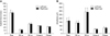
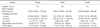
Differences between the left and right eyes
Mean ERG values for left and right eyes are shown in Fig. 2. No significant differences in the measurements were found between the left and right eyes.
Discussion
Normal ranges for ERG data obtained with an LED-electrode in beagle dogs were identified in the present study. These are considered reliable findings because a larger number of subjects (86 eyes from 43 beagle dogs) were evaluated compared to previous reports proposing normal ERG values for canines using "mean values" [8]. Different values for the b-wave amplitude and b/a ratio were observed according to age for the combined rod-cone response. Thus, normal ERG values should be expressed as the median and range. ERG ranges should be described for age groups in all species. Even rodents, which live for 1~3 years, have shown age-related differences for ERG variables. Beagle dogs are widely used as a companion and experimental animals. We propose that normal ERG ranges should be determined according to age in each clinic and laboratory using its own equipment because institutions usually have different systems or protocols for ERG.
Stimulus intensity of canine ERG varies among different reports although the ECVO Committee has proposed standard stimulus intensities [18]. The stimulus intensity for canine ERG with an LED-electrode was determined in our previous report [13]. A wide range of stimulus conditions has been used with different ERG systems. Stimulus intensities for combined rod-cone and rod responses have been determined with beagle dogs aged 1~3 years old according to ISCEV stimulus setting: a standard flash intensity (3.0 cd·s/m2) showing a b/a ratio of approximately "2" under scotopic conditions and -2.5 log of the standard intensity (0.01 cd·s/m2) for rod response [15]. Stimulus conditions for canine ERG are occasionally based on the ISCEV stimulus setting for humans [15]. However, intensity of the background light (25 cd/m2) is weaker than the intensity most recently recommended by the ISCEV [16]. Thus, normal ranges in the present study were identified under the same conditions, aside from the background light intensity, that were used for establishing the recommendations proposed by the ISCEV. We did not perform a detail analysis of the cone a-wave in the present study. A-waves in the cone response can directly demonstrate photoreceptor characteristics and may help analyze some rare disorders. Recording and analysis of the cone a-wave should be taken into consideration to understand the electrophysiology of patients with unknown photoreceptor disorders and other diseases.
In dogs, ERG responses gradually increase from 2~3 weeks old and within 4 months reach a level equivalent to that of a 1-year old dog [5]. However, there are no reports describing the changes in ERG findings with age. In previous reports on human ERG, shortening of the implicit time and increased amplitudes of the scotopic and photopic responses were observed from birth to 6 months old after which the values stabilized [1,7]. Amplitude of the human b-wave for the combined rod-cone and rod responses starts to decline when the subject is about 20 years old and around 50 years old [1,7], respectively, when nuclear sclerosis is obvious [20,23]. In the present study, the subjects were divided into three groups in order to detect the changes in ERG variables associated with age. One year was the cut-off age used to separate young from adult, and 6 years was set the cut-off age used to distinguish adult from senile subjects because nuclear sclerosis becomes evident in dogs at 6 years or older [22]. The detailed causes of age-related changes in ERG findings have not been fully elucidated thus far. The increasing number of retinal cells and outer segments of photoreceptors with retina growth occurring from birth is thought to be associated with ERG changes in humans [5]. It is assumed that the ERG changes in older individuals are caused by ocular media alterations (ocular media opacity or pupil size) with decreased effective intensity of the stimulus [1], decrease photoreceptor density [1,26], and bipolar or Müller cell death [2,3]. A reduction in photopigment sensitivity might be an influential factor in dogs [12].
In the present study, statistically significant decrease in b-wave amplitude and b/a ratio in the combined rod-cone response were observed in the adult and senile groups compared to the young group. No significant differences were observed in the rod response amplitude or implicit times. These findings were different from those reported in humans, b-wave amplitudes of photopic and scotopic ERG showed an obvious decrease with age [1]. The decreases in b-wave amplitudes in older dogs may be associated with bipolar and/or Müller cell death in the retina similar to humans [2,3]. Our previous report showed normal ERG values for Shih-Tzus with the same flash intensity as the one used in the present study, though the number of eyes were just 16 [8]. Findings from the study were that the amplitudes of ERGs in Shi-Tzu might be lower than that in the beagle dog. Thus, it is assumed that the ranges are different among breeds and it is desirable to determine normal ERG values for each dog breed.
Evaluation of the b/a ratio has been used conventionally to assess the retinal state [12]. In the present study, the b/a ratio using a flash intensity of 3.0 cd·s/m2 with an LED-electrode was approximately "2". It is known that the b/a ratio is decreased in cases of lens-induced uveitis associated with reduced b-wave amplitude [14]. Thus, ERG evaluation of the b/a ratio is strongly recommended, especially prior to cataract surgery. It is also known that a weaker flash stimulus increases or stabilized the b/a ratio with a reduction in a- and b-wave amplitudes [4,13]. ERG is therefore considered to be unaffected by nuclear sclerosis. At the very least, no obvious decline in b/a ratio is observed even when the b-wave amplitude is slightly decreased.
In the current study, no significant differences in values for the left and right eyes in the same subject were observed. Recent research has uncovered wide normal ranges for ERG variables, especially amplitude. Many factors such as electrode placement [17] and pupil size [16] may affect ERG amplitudes. It is difficult to judge slight abnormalities in the eye detected by ERG in different subjects during a single examination. However, it should be useful to compare the responses from both eyes in the same subject in order to detect an abnormality.
In summary, normal ranges for ERG variables measured with an LED-electrode in healthy beagle dogs of different ages were determined in the present study. Full-field ERG is an objective examination and widely used to assess the retinal function of dogs in which it is difficult to perform a subjective examination in veterinary clinics and experimental laboratories. ERG values might vary among different laboratories due to minor variations in recording equipment [4], electrodes [4,17], protocols [18,27], and animal breed [18]. The normal range for each breed should also be established in order for ERG to become a more valuable examination tool. It remains unclear when age-related changes in the retina begin to occur. Studies of ERG changes in an individual subject through their whole life could reveal chronological alterations in dog retina electrophysiology.
 XML Download