

 PDF
PDF ePub
ePub Citation
 Print
Print


Avian influenza viruses (AIVs), which consist of 16 hemagglutinin (HA) and nine neuraminidase (NA) antigenic subtypes, are maintained in wild birds and poultry throughout the world [10]. In 1994, a mildly pathogenic H5N2 AIV was isolated from a Mexican chicken (A/chicken/Mexico/26654-1374/94) and a highly pathogenic strain (A/chicken/Queretaro/14588-19/95) emerged the following year [3]. A large-scale vaccination campaign was implemented in 1995, and no further cases of highly pathogenic H5N2 AIV have been detected since 1996. However, low-pathogenicity AIV (LPAIV) strains belonging to the subtype H5N2 continue to circulate in some Mexican farms, which raises concerns about the possible emergence of highly pathogenic strains from nonpathogenic precursors.
AIV infection in ducks is typically asymptomatic. Viral replication primarily occurs in epithelial cells of the intestinal tract but can also take place in cells of the respiratory tract [11]. Viral excretion is consistent with an acute infection. In one study, mallard ducks (Anas platyrhyncos) were infected via oral and intratracheal routes with 107 50% egg infectious doses (EID50) of H3N6, H1N2, H4N1, or H5N2 AIV that had been previously isolated from mallard ducks. Viral excretion was detected by analyzing tracheal and cloacal swabs by titration. In all cases, viral excretion lasted 6~7 days, and most of the viral excretion was cloacal in all but one case (H5N2) [11]. More recently, mallard ducks were infected via an intra-esophageal route with 108.7 EID50 of an H7N7 AIV strain that had been previously isolated from wild congeners [4]. Viral excretion was detected by RRT-PCR for an average of 12 days and intermittent shedding was observed for another 4 days [4].
AIV strains that infect chickens have been described as non-pathogenic, mildly pathogenic, or highly pathogenic based on the virus' virulence in young adult chickens. In one study, leghorn chickens were infected via the conjunctival sac with 104.5-105 EID50 of four H7N2 LPAIV strains that had been isolated from congeners [5]. Viral excretion was monitored by analyzing tracheal swabs using RRT-PCR and typically lasted 7 days with intermittent viral shedding occurring for another 7 days [5].
In 2004, the LPAIV strain A/parrot/CA/6032/04 was identified in a psittacine bird in the United States [8]. Phylogenetic analysis of the HA gene grouped this virus with H5N2 viruses of a Mexican lineage. Leghorn chickens were infected intranasally with 106 EID50 of A/parrot/CA/6032/04. Viral excretion found in tracheal swabs lasted 7 days with only intermittent cloacal excretion on day 2 post-infection. Moreover, this viral strain was efficiently transmitted to control cagemates via direct contact. Pillai et al. suggested that the parrot AIV isolate may be a poultry-adapted virus that can easily replicate and circulate among domestic birds [8].
To gain a better understanding of the biological properties of H5N2 LPAIV (A/chicken/Mexico/2007) that is lethal in embryonated chicken eggs (CENID-Microbiología-INIFAP, unpublished data), we measured the rate and duration of excretion of this viral strain from Pekin ducks that were experimentally inoculated. Leghorn chickens were used as controls. Eight 3-week-old Pekin ducks (Cuernavaca, Morelos) and four 3-week-old chickens (Tehuacan, Puebla) were inoculated with 107 EID50 of LPAIV A/chicken/Mexico/2007 provided by The Mexico-United States Commission for the Prevention of Foot and Mouth Disease and Other Exotic Animal Diseases (CPA) that had been propagated in the allantoic cavity of specific-pathogen-free embryonated chicken eggs (SPF-ECE). Four birds of each species were also treated with 1 mL of allantoic fluid from non-inoculated SPF-ECE. The inoculum was apportioned among the oropharynx (0.6 mL), nares (0.1 mL each), and eyes (0.05 mL each). Animal care was provided as approved by the Institutional Committee of Animal Experimentation of the Facultad de Medicina Veterinaria y Zootecnia, Universidad Nacional Autónoma de México.
Three days after inoculation, no clinical signs of infection were observed in the Pekin ducks except for mild bilateral or unilateral conjunctivitis that resolved the following day. In contrast, the AIV-inoculated leghorn chickens had a mild respiratory syndrome that was characterized by sneezing, hyaline rhinorrhea, and mild conjunctivitis from days 2 to 7 post-inoculation. In addition, the inoculated chickens developed bristled feathers and depression from days 2 to 7 post-inoculation. No fatalities were observed in this study. None of the non-infected control birds of both species showed any clinical signs of infection throughout the experiment.
The amount of virus shed from the Pekin ducks and leghorn chickens infected with LPAIV A/chicken/Mexico/2007 was quantified based on the correlation between the amount of viral RNA detected by RRT-PCR and EID50 values [6]. For this procedure, oropharyngeal and cloacal swabs were collected at 12 hrs, 1, 2, 3, 7, 14 and 21 days post-inoculation (DPI) and individually placed in 3 mL of Dulbecco's minimal essential medium (Invitrogen, USA) with penicillin (10,000 IU/mL), streptomycin (10,000 µg/mL) and amphotericin B (20 µg/mL) as previously described [1]. The RRT-PCR procedure for matrix gene was used as previously described [9] with modification for antisense primer for this study. Pairs of pooled oropharyngeal and cloacal swabs were used for RRT-PCR with matrix gene primers (forward 5'-AGATGAGTCTTCTAACCGAGGTCG-3' and antisense 5'-AGGATTGGTCTTGTCTTTAGCCAT-3) and reverse hydrolysis probe (5'-TCAGGCCCCCTCAAAGCCGA-3'). To generate a standard curve, we prepared ten-fold dilutions of allantoic fluid that contained known titers of A/chicken/Mexico/2007 (from 103 to 107 EID50/mL). The average cycle threshold (Ct) values for each viral dilution were 35.92, 31.74, 28.62, 24.46, and 21.87, respectively. The sequence of the amplicon was obtained by chain termination method using the BigDye Terminator v3.1 Cycle Sequencing Kit (Applied Biosystems, USA) and it was identical to that of GenBank CY005832-1.
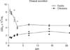
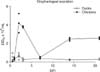
Pekin ducks that were inoculated with LPAIV A/chicken/Mexico/2007 excreted high but declining amounts of virus via the cloaca. During the first 3 DPI, viral excretion declined from 8.2 × 10⁶ to 4.3 × 10⁶ EID50/mL in the resuspended cloacal swabs (Fig. 1). Subsequently, viral excretion was maintained at a consistent level (approximately 4.0 × 10⁶ EID50/mL) in the four groups of infected ducks (eight individual animals). Compared to cloacal excretion, the infected ducks excreted much lower amounts of virus via the oropharynx, and a slightly higher level of excretion was observed during the first 2 DPI than afterwards (Fig. 2). It is worth noting that a low level of oropharyngeal excretion (approximately 0.3 × 10⁶ EID50 per mL) persisted from day 3 to 21 in the four groups of infected ducks (eight individual birds).
Compared to the Pekin ducks, chickens that were inoculated with LPAIV A/chicken/Mexico/2007 excreted lower amounts of virus via the cloaca. Nevertheless, the level of excretion from the two groups of infected chickens (four individual birds) increased from 1.4 × 10⁶ EID50 per mL on day 1 to 3 × 10⁶ EID50 per mL by day 15 and was maintained at a similar level until the end of the experiment on day 21 (Fig. 1). In contrast, viral excretion via the oropharynx was apparently biphasic in chickens (Fig. 2) with excretion increasing from day 0.5 (0.06 × 10⁶ EID50 per mL) to day 2-3 (4.0 × 10⁶ EID50 per mL) followed by a decline and subsequent rebound from day 7 (0.5 × 10⁶ EID50 per mL) to day 14 (2.5 × 10⁶ EID50 per mL). Remarkably, viral excretion via the oropharynx persisted at similarly high levels from day 14 until the end of the experiment (day 21) in the two groups of infected chickens (four individual birds). All non-infected chickens and ducks were negative for viral excretion.
Comparison of virus excretion from Pekin ducks and leghorn chickens inoculated with LPAIV A/chicken/Mexico/2007 demonstrated that the ducks primarily excreted the virus via the cloaca whereas the chickens generally excreted the virus via the oropharynx. This finding is consistent with results from previous studies showing that AIV infection in ducks is predominantly enteric while infection in chickens is mostly respiratory [11]. Additionally, cloacal excretion decreased in Pekin ducks while it increased in chickens. This finding may reflect the adaptation of AIV strains that enables the virus to use ducks as reservoirs. The duration of viral excretion in both species was at least 21 days. Remarkably, higher levels were observed in chickens than ducks during the second phase of oropharyngeal excretion. Viral excretion from both species inoculated with LPAIV strains is indicative of an acute infection that lasts up to 2 weeks [4,5,7,11]. Therefore, prolonged excretion from ducks and chickens infected with A/chicken/Mexico/2007 observed in the present study is atypical.
In the current investigation, we used RRT-PCR to measure the rate and duration of viral excretion based on its strong correlation with EID50 values [9]. RRT-PCR is able to detect noninfectious viral particles that are estimated to constitute as much as 90% of some virus preparations [2]. Further studies are necessary to directly address the infectivity and transmissibility of A/chicken/Mexico/2007 after the acute phase of infection and compare the strain used for this study with typical LPAIV strains.
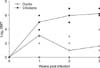
AIV hemagglutination inhibition (HI) antibody titers were determined in the birds' sera using four UHA of LPAIV H5N2 and expressed in geometric mean titer (GMT) Log2 as previously described [1] using A/chicken/Mexico/2007 antigen provided by CPA. Low HI antibody titers were detected in six out of eight infected ducks 1 week after inoculation (GMT 8.7; Fig. 3). By the following week, HI titers had declined in the inoculated ducks (3/8 positive, GMT 2) and remained at similarly low levels during week 3 (7/8 positive, GMT 3.1). In contrast, all of the inoculated chickens developed high HI titers after 1 week post-inoculation (GMT 32) and showed increasing antibody levels during the second (GMT 64) and third (GMT 76) weeks. All of the sera from the non-infected control birds were negative for HI throughout the experiment. The results from this experiment demonstrated that the Pekin ducks inoculated with A/chicken/Mexico/2007 showed a low and decreasing HI antibody response whereas the chickens had a high and increasing antibody response. These findings are consistent with those from previous studies in which low HI titers in ducks and high HI titers in chickens infected with H5 LPAIV strains were observed [7].
Prolonged excretion of LPAIV A/chicken/Mexico/2007 in Pekin ducks and leghorn chickens observed in the current investigation is atypical. The genetic basis for prolonged excretion of A/chicken/Mexico/2007 from the Pekin ducks and chickens is not known, and it will be necessary to perform additional studies of all of the viral genes that might influence this biological feature. Further studies are also required to directly address how prolonged excretion affects the infectivity and transmissibility of A/chicken/Mexico/2007 after the acute phase of infection.
 XML Download