

 PDF
PDF ePub
ePub Citation
 Print
Print


Introduction
Recently published literature has provided molecular evidence indicating that the dog as a highly suitable biomedical model for cancer research [22]. Canine lymphoma is biologically and phenotypically similar to a high-grade variant of non-Hodgkin's lymphoma (NHL) in humans called diffuse large B-cell lymphoma (DLBCL) [2]. Canine lymphoma is the most common hematopoietic tumor (83%) in dogs. The annual incidence of canine lymphoma is ~84/100,000 dogs among animals 10 to 11 years old and accounts for up to ~24% of all cases of malignant neoplasia [11]. Multicentric lymphoma affecting the peripheral lymph nodes is the most common form in dogs although the disease can arise at virtually any location. Similar to human NHL, canine lymphoma can result from the malignant transformation of either B or T cells. Approximately 75~80% of lymphomas are B cell in origin [11]. In humans, the incidence of B-cell lymphoid neoplasm is about 26/100,000 per year with approximately 67,000 cases/year diagnosed in the United States [14].
Canine lymphoma is treated with the same drugs as human NHL using L-CHOP protocol, which has been considered as the most commonly administered reagents including vincristine, cyclophosphamide, L-asparaginase, doxorubicin, and prednisone. In general, complete remission is achieved in ~80% of treated dogs with median survival times of 290 days; only 10% of dogs survive 2 years or longer [12]. The vast majority of canines succumb to recrudescent disease that is resistant to all known chemotherapy drugs.
The considerable range of survival times is due to the fact that lymphoma subtypes have different prognoses [16] and may require different treatments. For example, animals with canine T-cell lymphoma exhibit shorter survival times than ones with B-cell lymphoma [7]. Further knowledge beyond whether a lymphoma is of B or T cell origin is urgently needed to optimize specific treatment plans, understand the molecular underpinnings of chemotherapy resistance, and develop more targeted, less toxic treatment strategies.
Recent stem cell biology studies have suggested that some cancers contain stem-like cells (cancer stem cells or CSCs) that exist as a small subpopulation within tumor tissues [18]. A side population (SP) assay consisting of Hoechst 33342 staining and subsequent flow cytometic analysis has recently been used to identify both malignant and normal stem cells [15]. This SP assay is based on the observation that stem cells express high levels of specific ATP-binding cassette (ABC) transporters that actively remove drugs from cells and protect against cytotoxic agents. Hoechst 33342 dye accumulates within the cytoplasm of most differentiated cells. However, this does not occur in stem cells because the dye is actively removed by an ABC transporter called breast cancer resistance protein (BCRP) [15]. Cells that contain a low level of Hoechst 33342 fluorescence are referred to as an SP that can be separated by flow cytometry-assisted cell sorting (FACS). SP fractions from normal and malignant tissues are enriched with stem cells [15].
The current study was performed to explore the potential value of the SP assay for identifying and characterizing putative CSCs in canine lymphoid malignancies. Results from this investigation indicated that the SP assay could identify a subpopulation in canine lymphoma cells. This subpopulation was not abrogated by treatment with chemical inhibitors of ABC transporters. Further studies on methodologies for CSC identification and characterization are warranted.
Materials and Methods
Canine neoplastic lymphoid cells
Two well-characterized canine lymphoid B-cell lines (GL-1 and 17-71 cells) and a canine T-cell lymphoma cell line (CL-1) provided by Dr. Steven Suter (North Carolina State University, USA) were used in this study. The cell lines were maintained at 37℃ in a humidified atmosphere in RPMI (Roswell Park Memorial Institute) medium supplemented with 10% FBS (Fetal Bovine Serum), 10 mM HEPES, 1 mM sodium pyruvate, and 100 U/100 mg/mL penicillin/streptomycin (Sigma Aldrich, USA). GL-1 cells represent an immature B-cell leukemia while 17-71 cells represent a B-cell malignancy.
Neoplastic lymphoid cells were obtained from the lymph nodes of dogs diagnosed with lymphoma at North Carolina State University and Seoul National University (Korea). The diagnosis of canine B-cell lymphoma was made based on the clinical signs, cytology of fine needle aspirates from the enlarged peripheral lymph nodes, and flow cytometry (for immunophenotyping). For this study, neoplastic B-lymphoblasts were isolated via fine needle aspiration from the enlarged peripheral lymph nodes of five dogs during initial staging (before chemotherapy was initiated). The neoplastic cells were cultured overnight in RPMI supplemented with 10% FBS, 10 mM HEPES, 1 mM sodium pyruvate, and 100 U/100 mg/mL penicillin/streptomycin (Sigma Aldrich), and then stained with Hoechst 33342 dye.
SP assay
All of the lymphoid cells were grown in RPMI medium supplemented with 100 U/100 mg/mL penicillin/streptomycin (Sigma Aldrich) and 10% FBS. The Hoechst 33342 staining procedure was based on the method described by Goodell et al. [1] with slight modification. Briefly, the cells (106 cells/mL) were re-suspended in the pre-warmed media containing 2% FBS and then incubated for 90 min at 37℃ with Hoechst 33342 stain (5 µg/mL final concentration; Sigma, USA) with intermittent mixing. After incubation, the cells were centrifuged at 480 × g for 5 min and washed in a cold 2% FBS/PBS solution. Hoechst 33342 staining was observed using a FACSan flow cytometer (Becton-Dickinson, USA) at 357 nm to detect Hoechst Blue with a 424/44 broad pass (BP) filter and Hoechst Red with a 675/20 BP filter. At least 50,000 events within the live gate were examined to define an SP region.
Validation of the SP fraction
SP regions were generally confirmed by treatment with chemical inhibitors of an ABC transporter, ABCG2 (ATP-binding cassette, sub-family G, member 2, also known as BCRP). Before the flow cytometric analysis, the cells were treated with 25, 50, or 100 µM verapamil hydrochloride (Sigma, USA) or 10 µM fumitremorgin C (EMD Biosciences, USA) at 37℃ for 0 ~ 20 min prior to Hoechst 33342 staining in order to monitor the prevention of Hoechst 33342 dye efflux.
Western blot assay
Cellular lysates were prepared by re-suspending the canine neoplastic lymphoid cells in Mammalian Protein Extraction Reagent (Thermo Fisher Scientific, USA) containing protease (Roche Diagnostic, USA) and phosphatase inhibitors (Thermo Fisher Scientific) at 4℃. The lysates were then sonicated for 5 min by using a Vibra-Cell sonicator (Fisher, Canada), centrifuged for 10 min at 23,000 × g at 4℃, and concentrated using a Micron cellulose filter (EMD Millipore, USA). Protein concentration was measured using a detergent-compatible protein assay reagent (Bio-Rad Laboratories, USA). Gel electrophoresis as well as membrane transfer, blocking, and incubation with antibody were performed as previously described by Kai et al. [5]. Primary antibodies used for this study were specific for ABCG2 [1 : 500 dilution, rat monoclonal antibody (BXP-53) against BCRP/ABCG2; Abcam, USA], phosphorylated (p)-glycoprotein [1 :75 dilution, mouse monoclonal antibody (C219); EMD, USA], Nanog (1:600 dilution, rabbit polyclonal antibody; Abcam), and Bmi (1: 1,000 dilution, mouse monoclonal antibody; Abcam). Chemiluminescence was generated using Enhanced Chemiluminescent (ECL) Western Blotting Substrate (Thermo Fisher Scientific). The blots were stripped with Restore Western blot stripping buffer (Thermo Fisher Scientific) and re-probed with antibody against GAPDH (Glyceraldehyde 3-phosphate dehydrogenase) as an internal loading control.
Results
SP assay for detecting neoplastic lymphoid cells
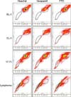
Canine lymphoma cells recovered from three cell lines and the lymph nodes of five clinical cases of canine lymphoma were subjected to an SP assay. The clinical cases were diagnosed as canine B-cell lymphoma based on clinical signs, cytology of fine needle aspirates from the enlarged peripheral lymph nodes, and flow cytometry for immunophenotyping. Following Hoechst 33342 staining, the subsequent flow cytometric analysis revealed a clearly delineated subpopulation of cells (Fig. 1). Staining intensities and patterns of the subpopulation cells observed with flow cytometric analysis were consistent with those of SP cells previously described in published study [5]. The subpopulation accounted for 0.13% of GL-1 cells, 4.07% of 17-71 cells, and 0.73% of CL-1 cells. Cells prepared from the clinical cases of canine lymphoma contained much higher percentages of the subpopulation cells ranging from 68 to 78%.
The SP fraction should be reduced or abrogated by treatment with verapamil hydrochloride, an inhibitor of ABCG2 [5]. Unexpectedly, treatment with verapamil hydrochloride did not affect the subpopulation cell percentage for the canine lymphoma cells we examined (Fig. 1). Moreover, fumitremorgin C, another specific inhibitor of ABCG2 [17], did not affect the abundance of subpopulation cells among the canine lymphoma cells.
Expression levels of ABC transporters and stemness genes
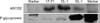
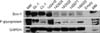
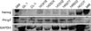
The canine lymphoma cells were subjected to Western blotting to measure the expression levels of ABC transporters and stemness-related genes. High expression levels of ABCG2 were detected in 17-71 and CL-1 cells while only a low level was observed in GL-1 cells (Fig. 2). The levels of p-glycoprotein were significantly higher in the GL-1 than the 17-71 or CL-1 cells (Fig. 2). ABCG2 were highly expressed in the clinical canine lymphoma cell samples, but the expression levels of p-glycoproteins were lower in most of these samples. Bmi-1 was highly expressed in the canine lymphoma cells while most clinical samples exhibited lower levels of this factor (Fig. 3). Nanog expression was rarely detected in the canine lymphoma cells we examined while NCCIT (Human embryonic carcinoma cell line) cells expressed a high level of Nanog (Fig. 4).
Discussion
CSC research is still in its infancy largely due to inherent drawbacks of current CSC identification methods. Flow cytometry using cell surface markers have been successfully applied to mice and human samples to characterize stem cell populations [9]. Different cell surface markers for isolating and targeting stem cells, including CD133, CD24, CD44, CD34, and integrins, have been described [9,10,18,21]. The work being done to identify biomarker surrogates for CSCs is rapidly progressing. Despite the extensive studies on various tissues and species, stem cell markers are scarce, the assays are difficult to standardize, and the actual hierarchy of the assays remains to be defined. In order to circumvent the limitations of existing assays using surrogate cell surface markers, extensive efforts have been made to develop alternative methods that could be universally applied to various tissue types. The most promising of these alternative methods is the SP assay consisting of Hoechst 33342 staining and subsequent flow cytometry [13]. Recently, the ALDEFLUOR assay exploiting cellular aldehyde dehydrogenase 1 (ALDH1) activity was developed and has been evaluated as a tool for CSC identification [10]. The present study was performed to evaluate the potential value of the SP assay for identifying and characterizing putative CSCs in canine lymphoma cell populations.
The SP assay is based on the overexpression of an ABC transporter, ABCG2, in stem cells [1]. This molecule removes fluorescent dyes such as Hoechst 33342 or Rhodamin 123 from stem cells, a property not found in differentiated cells. Cells with the capacity to remove dyes have been referred to SP cells as they fall to the "side" of the bulk of positively labeled cells in flow cytometric plots [1]. Previous studies have demonstrated that the SP fractions from normal and cancer tissues are enriched with cells possessing stem cell properties [3,15,20]. On the other hand, more recent investigations have suggested that the SP assay can enrich CSCs only partially and not exclusively [5,21]. The present study showed that the SP assay identified a wide range of SP fractions among canine lymphoma cells. An alternative SP assay using another dye, Rhodamin 123, could also help identify the SP of canine lymphoma cells.
It was not unexpected that the SP fraction of canine lymphoma cells was not altered by treatment with verapamil hydrochloride or fumitremorgin C. Membrane-spanning ABC transporters, ABCG2 and P-gp, are highly expressed in normal and cancerous stem cells [6]. SP contents in cancer cell populations should be significantly reduced by treatment with verapamil hydrochloride, a specific inhibitor of ABCG2, suggesting that the expression of ABCG2 is a major molecular determinant of the SP cell phenotypes [20]. However, the SP fraction of the canine lymphoma cells was not reduced or abrogated by treatment with verapamil hydrochloride. In humans, verapamil hydrochloride is a specific inhibitor of ABCG2. Verapamil hydrochloride exerts inhibitory effects on p-glycoprotein function by competing with substrate drugs for binding p-glycoprotein. Fumitremorgin C is known as a potent and selective BCRP inhibitor [17]. Treatment with this compound did not affect the SP fraction of canine lymphoma cells. This may be due to species-specific differences in the inhibitory effects on ABC transporters [19]. Alternatively, the SP fraction of canine lymphoma cells may have unknown means to resist to the inhibitory effects of the compounds.
Dogs provide an excellent model of naturally occurring human DLBCL for a variety of reasons [2]. Canines provide additional advantages over traditional murine xenograft models in that they possess a spectrum of genetic diversity that is not present in most laboratory mouse strains. This allows application of genomic strategies similar to those used in humans such as subclassification of tumors according to gene expression patterns [4]. Furthermore, naturally occurring DNA polymorphisms can help identify variants that influence lymphoma development or progression, thereby providing genetic clues for understanding disease biology [8]. An additional advantage is that canine lymphomas are an excellent model of chemoresistant disease. Despite treatment with chemotherapy regimens similar to those used for humans, dogs invariably die from their disease and so represent the segment of humans in most need of new treatment strategies. Finally, clinical data from dogs can be acquired within a much shorter time frame than from studies of human DLBCL because dogs naturally experience more rapid disease progression and relapse.
In summary, the SP assay evaluating canine lymphoma cells failed to detect an appropriate subpopulation of cells that were enriched with CSCs. Further studies of CSCs in canine lymphoma cell populations by other methodologies using cell surface markers or molecular markers should be attempted in order to help understand the disease pathogenesis. Characterization of putative canine lymphoid CSCs could provide further insight into the molecular underpinnings of both lymphoid carcinogenesis and chemotherapy resistance. This could lay the groundwork for the development of a novel CSC-specific therapeutic regimen.
 XML Download