

 PDF
PDF ePub
ePub Citation
 Print
Print


Introduction
Staphylococci resistant against methicillin and other antibiotics have frequently been reported in pets [5,27]. These microorganisms are opportunistic pathogens that may colonise the skin and mucosae of humans and other animals. Staphylococcus is currently divided into coagulase-positive and coagulase-negative species. The pathogenicity of coagulase-negative Staphylococci (CNS) has long been underestimated because they were associated with more chronic or subacute infections when compared to coagulase-positive Staphylococci (CPS) [20]. However, the etiological role of CNS in prosthesis and foreign body infections is increasingly being recognised in human medicine [12,13,23]. In pets, the pathogenic potential of these microorganisms has not yet been clearly recognized, although there have been some reports of infections related to methicillin-resistant CNS in cats and dogs [11,24].
Few studies have addressed the composition of staphylococcal populations on the mucosae of healthy cats and dogs [8,9]. Previous investigations of the staphylococcal species diversity in these animals have focused on clinical isolates [34], mainly CPS [17], or described the distribution of well defined antibiotic resistance within a limited number of staphylococcal species [1,38]. However, these studies were carried out before 2005, when Staphylococcus (S.) pseudintermedius had not yet been described. In fact, this species had probably been reported in all previous studies as S. intermedius, leading to confusion regarding its actual occurrence in pets [14,21,22,29,30]. S. pseudintermedius has recently been suggested as the most relevant and prevalent CPS dog coloniser, and there have been increasing reports on its pathogenicity and methicillin resistance [15].
To date, CNS strains in pets has been neglected; however, the recent development of new molecular techniques has allowed accurate identification of CNS [3,31], which will eventually lead to a better understanding of these bacterial species. Additional knowledge regarding CNS carriage in animals will be of benefit, because these bacteria might represent a pool of antibiotic resistance for CPS species. Indeed, horizontal gene transfer of staphylococcal chromosome cassette mec (SCCmec) between CPS and CNS species has been documented [19].
In the last decade, several authors have suggested that pets may be reservoirs of antibiotic resistant bacteria [18,25,26]. This assumption was mainly based on studies reporting antibiotic resistance in clinical CPS isolates from dogs and humans in close contact [28,37]. However, a clear picture of the distribution, diversity and multi-drug resistance of both CPS and CNS species in pets is lacking, as is the role of cats and dogs as reservoirs of antibiotic resistance.
The purpose of the present study was to gain insight into the distribution of commensal staphylococcal species of healthy cats and dogs and determine the occurrence of multi-drug resistance in both CNS and CPS. We also explored risk factors associated with the carriage of these microorganisms by pets.
Materials and Methods
Study design and settings
Samples were collected between March 2008 and December 2009 from four different Swiss cantons (Berne, Ticino, Vaud and Zurich). Only healthy pets with no overt acute disease at the time of sample collection were enrolled in the study. The pets either lived in or visited nursing homes for pet-therapy or lived in households. The selection strategy differed between community and nursing homes. Pets in the community were included in the study based on convenience sampling in households (n = 196) in four Swiss cantons representing the northern, southern, central and western part of Switzerland. Additional pets (n = 239) were recruited from cats and dogs visiting a total of 12 veterinary practices in the same regions for routine vaccinations. Nursing homes were selected by two-stage random cluster sampling from an exhaustive list of nursing homes located in the four Swiss cantons as reported in [16]. In randomly selected nursing homes, all pets matching the inclusion criteria and present at the time of sample collection (n = 98) were enrolled in the study. Informed written consent was obtained from all pet owners prior to the start of the study, and the investigation received the approval for animal experimentation from the Cantonal and Swiss Federal Veterinary Offices (authorisation reference No. 01/2008-02/2008).
Sample collection
Nasal and ear swab samples were collected using cotton swabs (Amies agar gel 108C and 110C; Copan, Italy) that had been soaked in a physiological 9% NaCl solution. For collection, a swab was introduced 1~2 cm in the nostril, while a second swab was introduced as deeply as possible in the ear channel of each animal. The collected samples were then stored in transport medium at room temperature and analyzed for the presence of Staphylococci within 24~48 h of collection. A questionnaire collecting information regarding the demographic and health status of the pets was filled in by the owners (available on request).
Sample analyses
Both swabs were streaked onto Mannitol Salt Agar (Chapman 2 - MSA 2; bioMérieux, France), after which they were incubated for 48 h at 37℃, enriched in MRSA broth supplemented with 6 µg of oxacilllin (48 h at 37℃) and cultured on Gelose ChromID S. aureus (SAID; bioMérieux) for 48 h at 37℃. All morphologically different colonies were isolated and catalase positive, Gram positive coccal bacteria were frozen in skim milk at -℃ until further analyses.
Isolates were grown on blood agar for 24 h and then identified by matrix-assisted laser desorption ionisation-time of flight mass spectrometry (MALDI-TOF MS) using an Axima Confidence spectrometer (Shimadzu-Biotech, Japan) in positive linear mode (m/z = 2,000 to 20,000) [10]. The identity of isolates that could not be identified by MALDI-TOF MS (24%) was confirmed by sequencing of the amplified partial rpoB gene [31].
Phenotypic antibiotic resistance to 24 different drugs was assessed by the Kirby-Bauer method on Mueller-Hinton blood agar (MHS2; bioMérieux). The following antibiotics were tested: penicillin (10 units), ampicillin (10 µg), oxacillin (1 µg), cefazolin (30 µg), gentamicin (10 µg), tetracycline (30 µg), erythromycin (15 µg), clindamycin (2 µg), vancomycin (30 µg), trimethoprim-sulfamethoxazole (1.25 + 23.75 µg), ciprofloxacin (5 µg), amoxicillin and clavulanic acid (20 + 10 µg), ceftazidim (30 µg), imipenem (10 µg), tobramycin (10 µg), fusidic acid (10 µg), rifampicin (30 µg), chloramphenicol (30 µg), cefoxitin (30 µg), kanamycin (30 µg), doxycyclin (30 µg), mupirocin (5 µg), linezolid (30 µg) and quinopristin-dalfopristin (15 µg). An inducible clindamycin resistance test ("D-zone" test) was also carried out for all isolates. Results were interpreted according to the Clinical and Laboratory Standards Institute (CLSI) guidelines [4,32], and intermediate results were classified as resistant. Multi-drug resistance (MDR) was defined as resistance to at least three drugs belonging to three different antibiotic classes [16]. Additionally, the presence of the mecA gene, which confers methicillin resistance, was investigated by polymerase chain reaction (PCR) on all isolates that showed phenotypic resistance to oxacillin [6,7]. We considered isolates from the same animal as being different strains if they belonged to different staphylococcal species or their phenotypic antibiotic resistance profiles differed.
Statistical analyses
Sample size calculation was based on the assumption that 5% of pets carried at least one MDR staphylococcal strain and that the intra-class correlation coefficient (rho) in these settings was 0.15. We used the cluster sample equation developed by Bennett et al. [2] for all sample size calculations. Assuming that each nursing home with pets owned or was visited by three animals on average, sample collection in 42 different nursing homes would have provided 126 pets. Accordingly, the expected precision for the prevalence estimate of MDR in pets would have a standard error of 2.2%, and a 95% confidence interval (CI) = 0.68~9.3%.
The characteristics of the cats and dogs were compared to check for consistent differences in the demographics and health status of the different populations sampled. A Chi-square test (Fisher's exact test when expected observations < 5) and 95% CI were used for this comparison. We reported the prevalence of Staphylococci and MDR Staphylococci and the distribution of antibiotic resistance among different staphylococcal species together with the median number of resistances to different antibiotic classes. Univariable logistic regression models with MDR staphylococcal carriage status of the pet as the outcome variable of interest were applied to explore risk factors. Unadjusted odds ratios (OR with 95% CI) were calculated as a measure of association. Statistical significance of each explanatory variable was determined by a likelihood-ratio test (LRT). We included in a multivariable model all variables with LRT p values ≤ 0.2 from the univariable analysis. All statistical analyses were performed using STATA 9.0 (Stata Corporation, USA).
Results
Demographics and staphylococcal carriage in pets
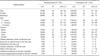
We collected samples from 533 healthy pets (277 cats and 256 dogs), 98 of which lived in or visited nursing homes at least once a week and 435 that lived in the community. The demographics of the two populations studied are reported in Table 1. Parameters such as sex, age, sterilisation, otitis in the last year, and antibiotic treatment showed different distributions between the nursing home and community settings, but the 95% CI estimates of these parameters overlapped (Table 1). We did not carry out stratified analyses of the samples because the overall frequencies of MDR in nursing homes (15/98) and in the community (76/435) did not differ significantly (χ2 = 0.27, p = 0.6).
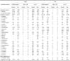
Staphylococci were detected in 60% (320/533) of pets, and 17% (92/533) of all animals carried at least one MDR strain. There were no significant differences in MDR carriage between pet species [14.8% (95% CI: 11.0~19.5) in cats and 20.0% (95% CI: 15.5~25.4) in dogs; χ2 = 2.1, p = 0.14] (Table 2). In cats, most CNS were MDR (39/41), whereas the proportion of MDR in CPS was small (1/41). Conversely, MDR CPS (20/51) and MDR CNS (28/51) carriage was almost equal in dogs (Table 2). Additionally, we observed species-specific differences (χ2 = 63.69, p < 0.001) in the proportion of S. pseudintermedius carriage, with 27% (70/256) of dogs and 3% of cats (8/277) harbouring this species. No difference in S. aureus carriage was observed between the two pet species (13/256 dogs and 14/277 cats, respectively).
Staphylococcal isolates
We isolated 284 staphylococcal strains (176 from the nostrils and 108 from the ears) from dogs and 300 (153 from the nostrils and 147 from the ears) from cats (Table 3). We were able to identify 94.5% (552/584) of all isolates at the species level. Two S. schleiferi isolates from two cats were identified only at the species level. CNS species accounted for 60% (172/284) of all isolates in dogs and 86% (258/300) in cats (Table 3).
In cats, the total number of CPS strains was lower (22/300) than in dogs (98/284). Among the CPS strains, S. pseudintermedius was more frequently isolated from dogs [(85/98), 87%] than from cats [(8/22), 36%], whereas S. aureus was more frequent in cats [(14/22), 63%] than dogs [(13/98), 13%]. No other CPS were isolated.
The diversity of CNS was high, with 22 different species in dogs and 24 in cats (Table 3). S. felis was only isolated from cats, in particular from their nostrils, and it represented 31% of all CNS isolates (41/132). Other CNS recovered in relevant proportions from both pets were S. epidermidis, S. warneri, S. hominis, S. xylosus and S. equorum (Table 3)
Antibiotic resistance
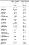
The mecA gene was present in 6% (11/172) of dog and 3% (7/258) of cat isolates. We did not recover any MDR S. aureus (Table 3). MDR, with a few strains showing resistance to eight different antibiotic classes, was detected in bacteria at proportions of 21% (36/172) in dogs and 16% (42/258) in cats. MDR was observed in S. pseudintermedius isolated from both pet species with resistance to up to six different antibiotic classes, but no methicillin resistance was seen (Table 3).
About 50% of all isolates in dogs and 30% in cats showed phenotypic resistance to penicillin and ampicillin (Table 4). Fusidic acid and erythromycin resistance were detected in 31% and 25% of dog and 28% and 19% of cat isolates, respectively. Additionally, 15% of all strains isolated from dogs were resistant to tetracycline and 11% to kanamycin. Clindamycin resistance was reported from 16% of dog and 15% of cat isolates (Table 4).
Exploratory analysis of risk factors
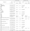
Univariable exploratory analysis revealed that a stay in a veterinary clinic in the last year was associated with increased risk of colonisation by MDR Staphylococci (OR = 2.4, 95% CI: 1.1~5.2, p value LRT = 0.04; Table 5). We included species, canton, stay in veterinary clinic in the last year, and antibiotic treatment in the last 3 months in the multivariable analysis. No missing data for these variables was observed for all 465 records. When accounting for other variables, we observed an influence of the cantons (geographic origin) on the carriage of MDR Staphylococci (p value LRT = 0.02). Additionally, cats had a lower risk of being carriers of MDR Staphylococci, whereas a stay in a veterinary clinic in the last year and antibiotic treatment in the last 3 months were associated with a higher risk, although these differences were not statistically significant (Table 5).
Discussion
This study provides detailed information on staphylococcal carriage in healthy cats and dogs and on drug resistance of these bacteria to different antibiotic classes for the first time since the description of S. pseudintermedius. We showed that S. pseudintermedius was recovered from the mucosae of healthy dogs more frequently than from those of healthy cats. Previous hospitalisation (at least one night in a veterinary clinic) was a risk factor for the carriage of MDR Staphylococci in pets using the univariable approach. The multivariable model showed that geographical distribution of the animals in the four cantons had an influence on the carriage of MDR staphylococci, which might reflect different pet health care and prescription practices of veterinarians in different regions of Switzerland.
Identification of the Staphylococci was carried out by MALDI-TOF MS, which provides reliable and rapid identification of the taxa in the S. intermedius group (S. delphini, S. intermedius and S. pseudintermedius) [10]. Previous investigations of the staphylococcal population of the mucosae of cats and dogs were based on phenotypic characterisation of the isolates, which may have led to misidentification of some closely related staphylococcal species [22,39].
We isolated MDR staphylococcal strains from healthy cats and dogs; however, MDR was not always associated with the presence of the mecA gene. In this study, resistance of strains to different antibiotic classes ranged from very low proportions (e.g., 1~2% resistance to ciprofloxacin in cats and dogs) to high values (11%) for kanamycin resistance in dogs. Methicillin resistance is of particular interest, because it confers resistance to all beta-lactams and is also often linked to resistance to other antibiotic classes; however, other resistances are also relevant in clinical settings, and infections resulting from MDR opportunistic pathogens are a critical problem to clinicians because they limit the choice of active antibiotic treatments [36].
It should be noted that our study has some limitations. Specifically, the exploratory analysis of risk factors was carried out by combining all staphylococcal species and information on pet-therapy animals as well as household pets, even though the risk associated with the carriage of MDR Staphylococci belonging to several species might differ between groups. This approach was necessary because the numbers for given combinations of investigated risk factors and animals carrying different MDR staphylococcal species were small. Pet management factors in the three months preceding the study were reported by the owners; therefore, a recall bias might be present. However, we do not consider this potential bias to be important because one can reasonably expect pet owners to recall whether or not a pet had visited a veterinary clinic during the preceding three months. We did not collect data on the number of different antibiotic treatments and the length of treatments, and the analysis of these data might have revealed other risk factors. In addition, we defined MDR as resistance of a strain to at least three antibiotics of different classes. Official guidelines (e.g., CLSI and EUCAST) lack a clear and standard criteria to define a staphylococcal strain as MDR, which reduces the possibility of carrying out meaningful comparisons with published data [16]. Despite the limitations of an exploratory univariable approach, our results confirm findings from published studies regarding factors associated with the carriage of MDR Staphylococci in pets, and in particular, the importance of previous hospitalisation, which was already reported as a risk factor for acquisition of both MRSA and MRSP in pets [33,35].
Our study has shown that carriage of multi-drug resistant Staphylococci in healthy cats and dogs is common; thus, clinical therapy guidelines would benefit from an approach that is not only focused on methicillin resistance, neglecting the presence of other resistances. The monitoring of antibiotics use in veterinary clinics could provide an overview of possible future trends of antibiotic resistance in pets. In veterinary medicine, further studies investigating the dissemination of antibiotic resistance determinants would benefit from considering the possible role of reservoirs of CNS in their spread.

 XML Download