

 PDF
PDF ePub
ePub Citation
 Print
Print


Introduction
Repair of large bone defects resulting from trauma, tumors, osteitis, delayed unions, non unions, ostoectomies, arthrodesis, and multifragmentary fractures is a current challenge for surgeons and investigators [3,8,40]. Therefore, application of various bone graft substitutes including autografts, allografts, xenografts, polymers, ceramics and some metals have been employed to promote bone reunion [14,19]. Allogenic, demineralized bone matrix (DBM) has been used for several decades in human surgery for treatment of nonunions, osteomyelitis, and large defects resulting from removal of the benign tumors [20]. The process of demineralization with hydrochloric acid destroys, but also decreases antigenic stimulation and may enhance the release of bone morphogenic proteins (BMPs) [30]. BMPs stimulate transformation of local undifferentiated mesenchymal into osteoblasts (osteoinduction), and the collagenous framework of the DBM particles allows migration of the newly regenerated tissue into the site (osteoconduction). Extensive research continues to identify BMPs that might be osteoinductive, and these are being readied for clinical application [5,7,22,28]. Beyond their role in osteoinduction, certain BMPs and DBM particles have shown the potential to aid in repair of osteochondral defects [23,31]. Advantages of DBM over other substitutes include inherent osteoinductive capacity (unlike tricalcium phosphate and hydroxyapatite) and availability in large amounts.
Omentum is a very important element to supply vascularization to implants [32]. The presence of abundant blood vessels in omentum is an ideal source of nutrients, oxygen, and angiogenic and growth factors that create a proper microenvironment for tissue induction [15,26]. Proper vascular flow increases the oxygen concentrations, resulting in the production of osteoprogenitor cells from perivascular mesenchymal cells [10,27].
The present study was conducted to compare the effects of xenogenic bovine fetal DBM, commercial DBM, omentum, omentum-DBM, cortical autograft and xenogenic cartilage powder on healing of an experimental bone defect in a dog model to determine the best material for bone healing. In the present study, cortical autograft and commercial DBM were used as positive standards to evaluate the effects of our home-made DBM and cartilage powder on bone healing procedures.
Materials and Methods
Animals
Seven male adult mongrel dogs that were 2 to 3 years old, 26.2 ± 2.5 kg and free of evident infectious or parasitic illnesses were used in this study. The experimental protocols were approved by the Animal Care and Experiment Committee of the University and were in accordance with the ethics standards of the Principles of Laboratory Animal Care.
Preparation of calf fetal demineralized bone matrix
DBM was prepared from the midshafts of the long bones of a 4-month-old Holstein calf fetus that were collected from a local slaughterhouse. The bones were collected aseptically, and the soft tissues were removed before storage at -70℃. The bones were later cut into 1 cm pieces with a Stryker saw under saline (0.9% NaCl) solution lavage, after which they were stored at -70℃ until further use. The pieces were later thawed in 200-proof ethanol and air dried, after which they were milled (Universal Mill A-20; Tekmer, USA) and placed through a sieve to collect 2- to 4-mm pieces. Next, the pieces were decalcified in 0.6 mol/L HCL at 4℃ for 8 days under constant agitation. Demineralization was subsequently evaluated by radiography and calcium analysis [39]. Loss of density radiographically was used to subjectively evaluate demineralization. In addition, random samples of DBM were dried at 95℃, weighed, and then ashed at 600℃ for 24 h. These samples were subsequently dissolved in 0.6 mol/L nitric acid and analyzed by atomic absorption spectrophotometry to determine the percent calcium per gram dry weight (% Ca: DW) [12,29]. Demineralization was considered adequate when samples were no longer visible radiographically and when the calcium content was less than 1% [38]. Following demineralization, all bone pieces were rinsed in sterile water and placed in phosphate buffer overnight. The bone pieces were then rinsed and the pH was adjusted to 7.3. Finally, the samples were placed in ethanol, the ethanol was allowed to evaporate overnight, and the pieces were packaged aseptically and stored at 4℃.
Preparation of bovine fetal cartilage powder
Epiphyseal cartilage of the long bones of a 4-month-old Holstein calf fetus were collected, washed three times in 95% ethanol for 15 min, rinsed in ether for 15 min, and finally air dried overnight. The cleaned and dried growth plate was then milled (Universal Mill A-20; Tekmer) to obtain 400~700 µm granules, air dried and stored in sterile plastic containers at 4℃ until being used for implantation. The entire process was performed under sterile conditions (except for the milling) and a sample was cultured to demonstrate that the specimens contained no bacterial or fungal contamination.
Cortical autograft granules
During tibial drilling to create defects, the protruding granules from beside the drill were collected for further use as autograft cortical granules.
Omentum free graft
For omental free graft preparation, the abdominal cavity was approached through a 3-cm ventral midline incision midway between the umbilicus and pelvic inlet, after which the free end of the greater omentum was located and exteriorized from the abdominal cavity. A 5 × 5-mm piece of the omentum was then isolated by two catgut ligatures and cut free from the remaining omentum.
Surgical technique
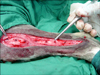
The dogs were sedated with acepromazine (0.05 mg/kg intrasubcuticularly) and anesthesia was induced with ketamine (10 mg/kg intravenously) and diazepam (0.25 mg/kg intravenously). The animals were then intubated and 1% to 2% halothane was used to maintain anesthesia with spontaneous breathing. During the procedure, saline (10 mL/kg per hour) was substituted through an intravenous catheter, and all operative procedures were conducted under general anesthesia. In all dogs, the left hind limb from the stifle to the metatarsal region was prepared for aseptic surgery. The tibia was exposed via a medial approach and a circular bone defect of 4 mm in diameter was made (Fig. 1). Ostectomy was then performed with an electrical motor and seven 4-mm carbon burr under continuous irrigation with physiologic serum. Finally, the defects were filled with autograft, commercial DBM (Osteotech, USA), calf fetal DBM, omentum, omentum-calf fetal DBM and cartilage powder. The implanted site was changed between materials in each dog in a Latin square design.
Post operative evaluations
Radiological evaluation
Lateral view radiographs were taken on the 1st day and then weeks 2, 4, 6 and 8 post injury using a step-wedge to calibrate the radiodensity. The radio-opacity of the implanted area was then scored using the range of 0 (minimally opaque) to 4 (most opaque) by an investigator blinded to treatment mode.
Histopathological evaluation
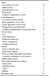
Eight weeks after operation the dogs were euthanized for histopathological evaluation, which was carried out on all harvested specimens. Briefly, the left hind limb was harvested and dissected free of soft tissues. Sagital sections containing the defect were then cut with a slow speed saw, after which each slice was fixed in 10% neutral buffered formalin. The formalin-fixed bone samples were then decalcified in 15% buffered formic acid solution and processed for routine histological examination. Next, two 5 µm thick sections were cut from the centers of each specimen and stained with Hematoxylin and Eosin. Finally, the sections were blindly evaluated and scored by two pathologists according to Heiple's scoring system [18] (Table 1).
Results
There was no intraoperative and postoperative death during the study. None of the dogs sustained a fracture of the tibia.
Radiographic findings
14th postoperative day
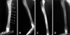
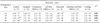
On the 14th postoperative day, statistically significant differences (p < 0.05) were observed between the control group with autograft (p = 0.03), commercial DBM (p = 0.03), calf fetal DBM (p = 0.02) and cartilage (p = 0.01) groups, and the control group was significantly inferior to the other groups. Additionally, the omentum group was significantly inferior to the autograft (p = 0.02), calf fetal DBM (p = 0.05) and cartilage (p = 0.03) groups. In addition, the omentum-calf fetal DBM was significantly inferior to the autograft (p = 0.02), calf fetal DBM (p = 0.03) and cartilage (p = 0.01) (Fig. 2, Table 2) groups.
42nd postoperative day
On the 42nd postoperative day, statistically significant differences (p < 0.05) were observed, with the control group being significantly inferior to the autograft (p = 0.04), commercial DBM (p = 0.3), calf fetal DBM (p = 0.02), omentum (p = 0.05), omentum-calf fetal DBM (p = 0.05) and cartilage group (p = 0.04). At this stage, the calf fetal DBM group was significantly superior to the omentum (p = 0.01) and omentum-calf fetal DBM (p = 0.03) groups (Fig. 2, Table 2).
Histopathological findings
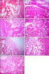
There was no significant difference between the histopathological sections of the lesions of the animals of different groups in terms of bone healing criteria. None of the grafted materials elicited a significant inflammatory reaction. As shown in Fig. 3A-G, at 8 weeks post-surgery, histological examination demonstrated the presence of the regenerated bone with a typical structure of the trabecular bone in the defect site of all groups. Although the statistical analysis did not show any significant differences between groups, the autograft, commercial-DBM and calf fetal DBM showed superior intense trabecular bone formation relative to the control, omentum and omentum-calf fetal DBM groups (Fig. 3, Table 3).
Discussion
Autogenous bone still remains the "golden standard" of bone graft material in all facets of orthopedic surgery and is commonly used as a standard to which allografts and graft substitutes are compared [1,2,11,13,25,27]. In the present study, the autograft was found to be the best implant upon radiological evaluation. Additionally, histopathological evaluation showed that it had intensive proper thickened trabecular bone and did not elicit any inflammatory reaction.
The bone inductive activity of the demineralized bone matrix (DBM) has been well established [33,34,37]. Additionally, the addition of the autologous bone marrow and/or autograft to DBM has been shown to provide an immediate source of osteogenic precursor cells at the implant site that is believed to provide an additional biochemical contribution to osteogenesis [6,34,37]. DBM also appears to support new bone formation through osteoconductive mechanisms [24]. The primary osteoinductive components of DBM are a series of low-molecular-weight glycoproteins that include bone morphogenetic proteins (BMPs). Decalcification of cortical bone exposes osteoinductive growth factors buried within the mineralized matrix, thereby enhancing the bone formation process [35]. Specifically, these proteins promote the chondroblastic differentiation of the mesenchymal cells, which is followed by new bone synthesis and endochondral osteogenesis [35,36]. In this study, the results of commercial-DBM and calf fetal DBM did not differ significantly from the autograft at 8 weeks post-injury. Overall, these results suggest that grafted xenogenic commercial-DBM and calf fetal DBM have osteoinductive activity, which may occur through the release of BMPs, similar to that of the autogenous cortical bone grafts. However, previous studies have shown that the cortical autograft has more osteoconductive properties and less osteoinductive activity than DBM material [4,21]. DBM also appears to support new bone formation through osteoconductive mechanisms [24]. No significant differences were found upon histopathological evaluation between the animals of different groups, and none of the graft material elicited a significant inflammatory reaction. It has been reported that the demineralization process destroys the antigenic materials in bone, making DBM less immunogenic than the mineralized allograft [17], and cortical autogenous bone graft does not induce an immunological reaction by the host [4], which was confirmed by the results of the present study.
Omentum is considered a major source of supplying new vascularization into the implants [32]. Proper vascularization by omentum provides a valuable source of nutrients, oxygen, and angiogenic and growth factors, and creates a proper microenvironment for this graft implantation, further tissue maturation and bone formation [15,26]. A sufficient vascular flow effectively increases the oxygen concentration, and induces the production of osteoprogenitor cells from the perivascular mesenchymal cells [10,26]. During angiogenesis, the vascular endothelial growth factor (VEGF) increases capillary permeability, supplies hormones and growth factors [9] and maintains high levels of oxygen concentration, and all of these mechanisms [15] may have played a role in the ossification process of the grafted area of the animals investigated in the present study. Omentum and omentum-calf fetal DBM groups were found to be superior to the control group; however, the lesions of these two groups were inferior to those of the autograft, commercial-DBM and calf fetal DBM groups. These findings suggest that the osteoinductive and osteoconductive properties of the autograft, commercial-DBM and calf fetal DBM were more powerful than those of the omentum and omentum-calf fetal DBM. In the present study, it was expected that the omentum-calf fetal DBM would lead to better bone formation criterion, but its effects were not superior to those of the omentum group. Rather, the calf fetal DBM was covered with omentum, which obscured the proper action of the calf fetal DBM.
It has been stated that the autogenous diced cartilage could act as a framework for deposition of bone in defects [16]. In the present study, the authors applied xenogenic processed cartilage as a graft material for bone healing and found that it led to satisfactory bone healing in comparison to the control, omentum and omentum-calf fetal DBM groups. These findings suggest that this technique has great potential for induction of bone formation in bone defects.
Histopathological evaluation did not show any significant differences after 8 weeks in the present study even though statistical differences between groups were expected. There were likely earlier significant differences between the histological features of the lesions of different groups over the preceding postoperative intervals; however, histopathological evaluation was not conducted at earlier postoperative intervals because of limitations imposed by the ethics committee. Therefore, histopathological studies during the inflammatory, proliferative and remodeling phases of fracture healing as well as investigations of the type of the inflammatory cell constituents, osteoblasts proliferation and maturation, angiogenesis, collagen synthesis, presence or absence of cartilaginous materials, quantity and quality of mineralization and many other criteria are recommended.
In the present study, the sites of material implantation were changed in a chi square manner in the defected area, and each material was implanted in the same site at least once, but no differences related to the site of implantation were observed. Therefore, statistical analysis was not performed to identify differences related to the site. It is possible that the lack of significant differences in histopathological findings among materials may have been related to hole size (4 mm) in our study, and that large-sized holes (e.g., 10 mm in diameter) may show significant differences in histopathological findings. Based on the radiological findings of the present study, the autograft, commercial-DBM, calf fetal DBM and calf fetal cartilage demonstrated superior osteogenic potential in healing of the tibial bone defect in a dog model. The omentum and omentum-DBM groups were superior to the control group, but were inferior to the autograft, commercial-DBM, calf fetal DBM and calf fetal cartilage groups. The histological findings of the present study did not reveal any superior bone healing capability among groups at this stage.

 XML Download