

 PDF
PDF ePub
ePub Citation
 Print
Print


Introduction
Low-dose-rate (LDR) irradiation is both beneficial and harmful to human life. For example, LDR irradiation therapy for cancer is known to stimulate antioxidant capacity, repair of DNA damage, apoptosis, and induction of immune responses [4], and LDR irradiation has been shown to mitigate lymphomagenesis and prolong the life span of AKR/J mice [17,18]. Conversely, low-dose (LD) irradiation is reportedly involved in carcinogenesis, and LD irradiation (lower than 2.0 Gy) has been shown to promote tumor growth and metastasis by enhancing angiogenesis [19]. Therefore, the present study was conducted to determine the effects of LD and LDR irradiation on gene expression and cell metabolism using the AKR/J lymphoma mouse model.
AKR/J mice carrying inserted murine leukemia viral oncogenes are known to develop thymic lymphoma during later periods of life and eventually die [10,14]. Additionally, it is well known that the viral oncogene insert in the thymus of AKR/J mice acts as an initiator of DNA damage and tumor formation. If damaged DNA in AKR/J mice is not repaired properly, tumors will progress, resulting in death. DNA damage in the thymocytes of AKR/J mice can be further enhanced by exposure to high-dose (HD) and high-dose-rate (HDR) radiation. Conversely, the number of thymocytes showing efficient DNA repair was found to be higher in mice exposed to LD and LDR radiation [7], suggesting that LDR irradiation could have triggered the expression of DNA repair genes.
DNA repair-related genes have been shown to play an important role in the growth, life span, and immune system of an organism. Indeed, it has been shown that the absence of functional Lig4, a DNA repair enzyme (R278H/R278H mutation), can lead to growth retardation, decreased life span, severe cellular sensitivity, and blocks in T and B cell development in response to HD irradiation [15]. HD-irradiation-induced double-strand breaks have been demonstrated to promote PI3K-independent AKT phosphorylation mediated through MRE11-ATM-RNF168 signaling [6]. The formation of the MRN (Mre11, Rad50, and Nbs1) complex, which participates in DNA repair pathways, is known to be activated by LD irradiation [16]. However, the mechanisms and pathways underlying cancer regulatory DNA repair signaling triggered by LDR irradiation remain unclear. Therefore, this study was conducted to investigate the regulation of DNA repair-associated genes by LDR irradiation using a naturally occurring tumor model in AKR/J mice. Herein, we identify immune response, nucleosome organization, and the peroxisome proliferator-activated receptors (PPAR) signaling pathway in the thymus of LDR-irradiated mice using the enrichment analysis of Gene Ontology (GO) terms and Kyoto Encyclopedia of Genes and Genomes (KEGG) pathways. Our data also demonstrate that thymuses of LDR-irradiated mice specifically upregulate the expression of Lig4 and RRM2, which are genes involved in DNA repair process, as well as H2AX and ATM, which are proteins known to recruit DNA repair factors.
Materials and Methods
Animals
Seven-week-old female AKR/J mice (24 ± 1.37 g) were purchased from Shizuoka Laboratory Center (Japan) and maintained under specific-pathogen-free conditions. Male mice were not used because fighting is known to occur frequently in male mouse groups, which could affect the characteristic features of AKR/J mice. Animals were maintained at a temperature of 23 ± 2℃ and a relative humidity of 50 ± 10% with a 12-h lighting schedule (200~300 Lux; 08:00 to 20:00). After a 1-week adaptation period, 15 mice were placed in a polycarbonate cage and provided with γ-sterilized pellets and autoclaved water ad libitum. Five mice each were assigned to the sham-irradiated group, HDR-irradiated group and LDR-irradiated group. All procedures were reviewed by the institutional Animal Care and Use Committee at the Radiation Health Research Institute (RHRI) of the Korea Hydro and Nuclear Power Company, and mice were treated in accordance with governmental guidelines and the guidelines of the RHRI for the care of animals.
HDR and LDR irradiation
A γ-ray generator (IBL 147C; CIS Bio-International, France) was used to generate HDR irradiation (137Cs, 0.8 Gy/min). Eight-week-old female mice were irradiated with a total dose of 4.5 Gy, which was selected based on a study by Yoshida [23]. Both HDR and LDR irradiation were started at 8 weeks of age. For HDR irradiation, mice were irradiated at a total dose of 4.5 Gy at 0.8 Gy/min and then returned to their cages until analysis. For LDR irradiation, mice were reared in a long-term LDR irradiation facility equipped with a 137Cs source and exposed at 0.7 mGy/h until the cumulative dose reached 2.1 Gy, which took approximately 130 days. The HDR cohort received their dose at 8 weeks of age, but was analyzed at 130 days after the start of radiation (26.5 weeks of age) to enable direct comparison of the cumulative effect of HDR- vs. LDR-irradiated mice. The sham-irradiated mice for both groups were reared in the SPF facility until they reached 26.5 weeks of age. Sham-irradiated mice were maintained under the same conditions as the experimental groups, except that radiation was not administered.
Sampling
At 130 days, we collected the thymuses from sham-, HDR (0.8 Gy/min, single dose of 4.5 Gy)-, and LDR (0.7 mGy/h, cumulative dose of 2.1 Gy)-irradiated mice and preserved them in liquid nitrogen.
Thymic lymphoma
To confirm the diagnosis of thymic lymphoma, a portion of the thymus was fixed in 10% buffered formalin, sectioned, and stained with hematoxylin and eosin. Thymic lymphoma was diagnosed histopathologically by Dr. D.Y. Kim (Laboratory of Pathology, School of Veterinary Medicines, Seoul National University, Korea). We also measured the relative thymus weight 130 days after HDR and LDR irradiation.
Microarray analysis
The RNA of five thymuses collected at 130 days after sham treatment, HDR and LDR irradiation was collected and analyzed by whole genome microarray using an Agilent oligo microarray (44K, 60-mer oligonucleotide). Microarray slides were then scanned with a GenePix 4000B scanner (Axon Instruments, USA), after which the scanned images were analyzed using the GenePix v6.0 software to determine the signal intensities of the spots. Raw data were normalized by the median normalization method, while the expression values of HDR- and LDR-irradiated mice were normalized to those of sham-irradiated mice. The differentially expressed genes were then classified by Benjamini-Hochberg's false discovery ratio method. We also selected DNA repair-, iron transport-, ribonucleotide reductase family-, and apoptosis-related genes from the differentially expressed genes on the basis of references. GO terms and KEGG pathways for the differentially expressed genes were analyzed using the GeneSpring GX 11.5.1 (Agilent Technologies, USA) and DAVID Bioinformatics tools [9].
Quantitative reverse transcription polymerase chain reaction (RT-PCR)
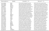
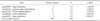
We conducted quantitative PCR (qPCR) analysis to validate microarray data for thymuses of sham-, HDR- and LDR-irradiated mice, respectively (n = 4). To accomplish this, 1 µg of RNA was reverse transcribed using the Iscript cDNA synthesis kit (Bio-Rad Laboratories, USA). PCR was then performed in duplicate with SYBR green (Qiagen, Germany) and a 7500 real-time PCR machine (Applied Biosystems, USA). The relative abundance of specific messenger RNA was calculated by normalization to 18S ribosomal RNA using the 2-ΔΔCt method [1]. The primers are listed in Table 1.
Results
LDR irradiation may influence a mild lymphomagenesis in AKR/J mice
We previously showed that the survival rate of LDR-irradiated AKR/J mice was greater than that of HDR-irradiated AKR/J mice [17]. To investigate whether LDR irradiation could influence lymphomagenesis in AKR/J mice, we collected the thymuses of sham-, HDR-, and LDR-irradiated mice at 130 days after the start of irradiation. The rationale for selecting thymic tissue samples at day 130 was that AKR/J mice began dying at approximately 130 days after irradiation; thus, any transcriptional changes induced by irradiation could be detected more easily than at later time points, when mice were overwhelmed with heavy tumor burden. The cause of death was not apparent to us; however, this provided a reasonable time point to analyze differential expression of DNA repair-related genes in the thymuses of sham-, HDR-, and LDR-irradiated AKR/J mice and determine the effects of irradiation during lymphomagenesis. At the time of sacrifice, we normalized the thymus weight to the whole body weight. The relative thymus weight of LDR-irradiated mice (0.017 ± 0.017) was found to be lower than that of the sham-(0.026 ± 0.041) and HDR-irradiated mice (0.023 ± 0.019; Fig. 1A). We also observed the development of thymic lymphoma in all mice. In accordance with macroscopic observation, histopathological examination revealed that neoplastic cells in the thymuses of sham- and HDR-irradiated mice were pleomorphic with marked anisocytosis and anisokaryosis, whereas both the cells and their nuclei were relatively small and uniform in LDR-irradiated mice (Fig. 1B). These data are in accordance with our assumption that LDR irradiation influenced naturally occurring mild lymphomagenesis in AKR/J mice.
Expression profiles of thymic DNA repair genes in HDR- and LDR-irradiated AKR/J mice
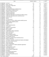
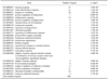
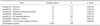
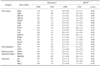
We performed microarray analysis for gene expression in the thymuses of sham-, HDR- and LDR-irradiated AKR/J mice collected at 130 days after irradiation. Enrichment analysis of the GO terms and KEGG pathways with respect to differentially expressed genes was then performed using GeneSpring GX 11.5.1 and DAVID Bioinformatics Resources v6.7. The most significant GO terms and KEGG pathways in HDR-irradiated AKR/J mice represented transcription, transcriptional regulation, cell cycle, and cancer pathways, whereas immune response, nucleosome organization, and the PPAR signaling pathway were represented in LDR-irradiated AKR/J mice (Table 2, 3, 4, 5). We next investigated the expression of DNA repair, iron transporter, ribonucleotide reductase family, and apoptosis. Thus, we performed qPCR analysis for validating their gene expression of sham-, HDR- and LDR-irradiated mice (Table 6). The expression value of HDR- and LDR-irradiated groups was normalized to that of the sham-irradiated group. In addition, microarray analyses showed that LDR irradiation led to increased gene expression of transferrin receptor 1 (Tfrc), ribonucleotide reductase M1 (RRM1), ribonucleotide reductase M2 (RRM2), and ribonucleotide reductase M2B (RRM2B). Moreover, the expression of genes related to base excision repair (Ogg1, Gtf2h2, Ung, Neil3, ERCC8, and ERCC6), cell-cycle regulation (Terf2), double-strand break repair and homologous recombination (Rad51L1, Mbd4, and Lig4), mismatch repair (Msh2 and Msh3), and nucleotide excision repair (Lig1) were upregulated. Among these genes, we validated the changes in expression of DNA repair- and ribonucleotide reductase family-related genes by qPCR. As shown in Table 6, gene expression of Lig4 and RRM2 was upregulated in the thymus of LDR-irradiated AKR/J mice. Furthermore, transcriptional levels of the genes encoding H2AX and ATM, proteins known to recruit DNA repair factors, were shown to be upregulated in the thymus of LDR-irradiated AKR/J mice. However, expression of apoptosis-related genes such as p53, Bax, and Parp1 were comparable between HDR- and LDR-irradiated mice. Therefore, LDR irradiation might have triggered the upregulation of DNA repair-related genes in the thymus of AKR/J mice, which could result in decreased thymic lymphoma mass and increased survival.
Discussion
In the present study, we investigated the effects of LDR irradiation on the expression of DNA repair-related genes in AKR/J mice bearing thymic lymphoma using microarray and qPCR. Our previous studies showed that the survival curve of LDR-irradiated mice was significantly different from that of mice that received HDR irradiation [18]. Moreover, the relative thymus weight of LDR-irradiated mice was smaller than that of sham- and HDR-irradiated mice at 130 days after the start of irradiation. These findings suggest that progression of thymic lymphoma may have been delayesd in LDR-irradiated mice relative to sham- and HDR-irradiated mice, which is consistent with the histological analysis of thymic samples. These data are also consistent with recent findings demonstrating that LDR irradiation (10 cGy/year) can prolong the lifespan and suppress the incidence of lymphoma in SJL/J female mice [12]. However, Tanaka reported that very low LDR irradiation (20 mGy) had no influence on life span [21]. Our recent data showed that a cumulative total of 1.7 Gy LDR irradiation had little effect on the relative thymus weight and thymic apoptosis of ICR mice (unpublished data). Thus, we assume that tumor-model mice, not normal mice, are more sensitive to LDR irradiation. In addition, our previous study showed that mice irradiated with LDR applied at a cumulative dose of 4.5 Gy had a higher survival rate and a lower incidence of thymic lymphoma than sham-mice and mice irradiated with HDR in a single dose of 4.5 Gy [18]. Taken together, these findings suggest that LDR irradiation influences mild lymphomagenesis in AKR/J mice.
We conducted enrichment analysis of GO terms and KEGG pathways with respect to the differentially expressed genes in HDR- and LDR-irradiated AKR/J mice. In LDR-irradiated mice, GO terms and KEGG pathways represented immune response, nucleosome organization, and the PPAR signaling pathway. Interestingly, the cancer pathway was specifically represented in HDR-irradiated mice. These findings suggest that LDR irradiation influences immune response, nucleosome organization, and the PPAR signaling pathway whereas, HDR irradiation accelerates tumorigenesis in AKR/J mice. Herein, we describe a selective effect of LDR irradiation on DNA repair-related genes; specifically, Lig4, RRM2, H2AX, and ATM. DNA ligase 4 repairs DNA double-strand breaks [22] and Lig4-deficient mouse embryonic fibroblasts promote genomic instability and frequent nonreciprocal translocation [5]. Therefore, upregulation of Lig4 may have participated in the repair process of damaged DNA in the thymus of LDR-irradiated AKR/J mice. RRM2 is a downstream target of the ATM-p53 pathway that mediates radiation-induced DNA repair [8]. The inhibition of ribonucleotide reductase activity by 3-aminopyridine-2-carboxaldehyde thiosemicarbazone has been shown to impede DNA damage repair mechanisms and increase the radiation sensitivity of human cervical cancers to LDR irradiation [11]. In addition to RRM2, transcriptional levels of H2AX and ATM, which are known to be recruited DNA repair factors, were also upregulated at 130 days after LDR irradiation. ATM protein is recruited to the site of DNA damage, where it recognizes double strand breaks [13, 20]. Recruited ATM also phosphorylates H2AX [2], which then suppresses the transformation of developing thymocytes, presumably by facilitating the assembly of DNA repair complexes such as Nbs1, 53 bp1, and Brca1 on damaged DNA [3]. Collectively, our data suggest that the activation of DNA repair processes is likely to control tumorigenesis.
In summary, the results of this study highlight the importance of DNA repair processes by LDR irradiation during tumor development and progression in AKR/J mice. The precise function of DNA repair genes subsequent to LDR irradiation still requires validation through examination of post-transcriptional and post-translational events. Nevertheless, our results provide significant insight into the molecular basis underlying transcriptional events by LDR irradiation, and it is expected that they will provide genetic markers for diagnosing cancers and enhancing the efficacy of cancer radiotherapy.

 XML Download