

 PDF
PDF ePub
ePub Citation
 Print
Print


Introduction
During surgery, air enters the pleural space through diaphragmatic defects; pneumothorax can subsequently develop [9,10]. This condition can be a fatal if it occurs in both hemithoraxes due to mediastinal rupture [4,6,12]. Complete elimination of pneumothorax is the main goal following diaphragmatic repair [3,16]. Thus, thoracic drainage techniques have been developed to eliminate pneumothorax following diaphragmatic rupture closure [7,9-11].
Thoracic drainage techniques (trocar-assisted or hemostatic forceps-assisted thoracostomy tube insertion) have been used to evacuate air from the pleural cavity [2]. Drainage can be achieved using a plastic catheter or tube with a small diameter. For transdiaphragmatic thoracostomy tube insertion, a catheter or tube is inserted through the diaphragmatic defect before one or two sutures are placed in the diaphragm [10].
The mediastinum is formed by serous membranes called the pleurae that are attached to the dorsal portion of the diaphragm and extend to the esophagus. The caudal mediastinal pleura make a strong deflection to the left, return to the midline, and attach to the sternum. The line of diaphragmatic attachment to the caudal mediastinum follows the left convex portion of the diaphragm [6]. There are considerable variations in the shape, size, and site of traumatic diaphragmatic hernias [1].
A thoracic evacuation technique involving the intentional creation of a large puncture in the caudal mediastinum can be used for complete pneumothorax elimination from both hemithoraxes if contralateral pneumothorax is suspected. Therefore, the current study was conducted to introduce a novel technique that combined transdiaphragmatic thoracostomy tube placement with intentional perforation of the caudal mediastinum as a way to eliminate pneumothorax from both hemithoraxes. Additionally, four thoracic evacuation techniques to eliminate pneumothorax following diaphragmatic defect closure were compared.
Materials and Methods
Cadavers
Forty cadavers (n = 40; weight range, 3.0 to 9.0 kg) of dogs euthanatized due to different reasons were donated by the humane society and used for this study. Respiratory system problems and thoracic or abdominal injuries were not evident in any of the dogs prior to euthanasia.
Procedures
All cadavers were randomly assigned to one of four groups. A polyvinyl chloride thoracostomy tube with a 4.7-mm outer diameter (PVC Thoracic Catheter; Sewoon Medical, Korea) was used for all insertion procedures. The same surgical team (Department of Veterinary Surgery, College of Veterinary Medicine, Konkuk University) inserted thoracostomy tubes into the pleural space of the cadavers. One of four thoracic evacuation techniques were performed on each group (10 cadavers in each group): (1) thoracic drainage through a left diaphragmatic defect and a small puncture intentionally made in the caudal mediastinum (DD-SP), (2) thoracic drainage through the left diaphragmatic defect and large puncture intentionally made in the caudal mediastinum (DD-LP), (3) thoracic drainage through both the left diaphragmatic defect and intact contralateral diaphragm with a small puncture intentionally made in the caudal mediastinum (DI-SP), and (4) thoracic drainage through both the left diaphragmatic defect and intact contralateral diaphragm with a large puncture intentionally made in the caudal mediastinum (DI-LP).
DD-SP and DD-LP techniques
An 11 no scalpel (Surgical blade; Ailee, Korea) was used to create a 5-cm vertical defect in the muscular portion of the left side of the diaphragm. The defect was created 1 cm away from the left abdominal wall to avoid iatrogenic rupture of the caudal mediastinum. A small (approximately 1-cm long) or large (approximately 4-cm long) puncture was created in the caudal mediastinum using a large curved forceps (Kelly hemostats; Medicom, Korea). The forceps was passed through the caudal mediastinum and entered the contralateral hemithorax. A large puncture was enlarged by opening the forceps pincers. The diaphragmatic defect was continuously sutured with 3-0 polyglyconate (Maxon; Syneture, USA). The thoracostomy tube was inserted through the diaphragmatic defect before the last two sutures in the diaphragm were tightened. After thoracostomy tube placement, these sutures were tightened and tied.
DI-SP and DI-LP techniques
Diaphragmatic and caudal mediastinal holes were made as described above. Additionally, a 2-cm diameter purse-string suture with 3-0 polyglyconate (Maxon; Syneture, USA) was placed in the muscular portion of the intact contralateral diaphragm. The diaphragm was punctured in the center of the purse-string suture with an 11 no scalpel. A thoracostomy tube was then inserted through the incision into the hemithorax. After tube placement, the purse-string suture was tightened and tied. The diaphragmatic defect was closed as described above.
Pneumothorax elimination
After unilateral or bilateral transdiaphragmatic placement, the thoracostomy tubes were attached to a commercial three chambers thoracic drainage system (Thora-Seal III; Covidien Animal Health and Dental Division, USA). The capillary tube of the water seal chamber was submerged 2 cm below the surface of the water level in the bottle. The capillary tube of the suction chamber was submerged 10 cm below the surface of the water level in the bottle and attached to a vacuum line via a regulator (Vacutron; Allied Healthcare Products, USA). The suction chamber was maintained with 10 cm H2O to provide consistent thoracic suction and the regulator was turned on. Consistent thoracic suction was maintained for 2 minutes as previously described [19].
Measurement of intrapleural pressure (IPP)
Before transdiaphragmatic thoracostomy tubes placement, a catheter (TURKEL Safety Thoracocentesis System; Covidien Animal Health and Dental Division, USA) was inserted into each hemithorax in the ventral third portion of the bilateral thoracic walls through a tiny skin stab incision made over the sixth intercostal space. The ends of a transducer (pressure monitoring kit; Biosensors International, USA) were connected to a monitor (patient monitor; Votem, Korea) and the catheter with a 3-way stopcock to measure and record IPP. The transducer was positioned at the level of the table on which the cadaver was placed, and zeroed with the 3-way stopcock opened to air and closed to the cadaver. Next, the 3-way stopcock was opened to the cadaver, and the pressure was recorded after stabilization (1 min after manipulation). Bilateral IPP was measured in each hemithorax at three different times: before making the diaphragmatic defect, after closing of the diaphragmatic defect, and after air evacuation via the thoracic drainage system.
Statistical analysis
All data are expressed as the mean ± standard deviation (SD). A one-way ANOVA was used to compare mean cadaver weights of the DD-SP, DD-LP, DI-SP, and DI-LP groups. IPP differences were compared using a two-way ANOVA. For the dependent variables of IPP difference, puncture size (small or large) and suction type (unilateral or bilateral) were evaluated for an interaction effect by use of a two-way ANOVA. Data analysis was performed with SAS software (version 9.1; SAS Institute, USA). P values ≤ 0.05 were considered significant for all comparisons.
Results
Cadaver weight
Average cadaver weight (mean ± SD) was 4.8 ± 1.5 kg, 4.7 ± 0.9 kg, 4.4 ± 0.7 kg, and 4.7 ± 1.6 kg for the DD-SP, DD-LP, DI-SP, and DI-LP groups, respectively. There was no significant difference in cadaver weight among the groups (p = 0.916).
IPP
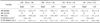
Mean IPP values for each group are presented in Table 1. Puncture size and suction type affected the differences between IPP in the left and right hemithoraxes after air evacuation via the unilateral or bilateral tubes using the thoracic drainage system (p = 0.0012). A greater difference between IPP values for the right and left hemithoraxes after air evacuation was detected for the DD-SP group compared to the DI-LP (p < 0.0001), DD-LP (p < 0.0001), or DI-SP (p < 0.0001) groups. However, no significant difference was observed between the DI-LP animals compared to the DD-LP group (p = 0.8229), the DI-LP group compared to the DI-SP group (p = 0.5031), or DD-LP dogs compared to the DI-SP animals (p = 0.3731).
Puncture size and suction type had no interaction effect on differences in IPP values before making the left diaphragmatic defect and after air evacuation from the left hemithorax (p = 0.4279). In contrast, puncture size and suction type had an interaction effect on differences in IPP values before making the defect and after air evacuation from the right hemithorax (p = 0.0296). Differences in IPP before making the defect and after air evacuation from the right hemithorax were significantly greater in the DD-SP group than the DI-LP (p < 0.0001), DD-LP (p = 0.0011), or DI-SP (p < 0.0001) groups. However, no significant differences were detected between the DI-LP group compared to DD-LP animals (p = 0.0835), the DI-LP dogs compared to the DI-SP group (p = 0.7239), or the DD-LP animals compared to the DI-SP group (p = 0.1630).
Discussion
Currently available options for removing intrathoracic air after diaphragmatic hernia repair include transdiaphragmatic thoracostomy tube placement through the diaphragmatic defect or intact contralateral portion of the diaphragm, transthoracic chest drainage through the lateral thoracic wall, hyperinflation of the lungs, and a combination of these methods [2,4,7,8,10]. Thoracic drainage techniques do not ensure communication between right and left hemithoraxes via a puncture made in the caudal mediastinal pleura. Furthermore, small mediastinal punctures might not facilitate proper elimination of bilateral pneumothorax after diaphragmatic defect closure. Our results suggest that a large puncture intentionally made in the caudal mediastinum was more effective than a small puncture for eliminating pneumothorax from the pleural cavity after repairing a diaphragmatic defect. Successful evacuation achieved with the DI-SP and DI-LP techniques indicate that thoracic evacuation through both the diaphragmatic defect and intact contralateral diaphragm would facilitate proper elimination of bilateral pneumothrax after diaphragmatic defect closure.
Previous studies reported that traumatic diaphragmatic hernias on the left side are twice as common as ones on the right side, and 15% of traumatic diaphragmatic hernia cases involve bilateral or multiple sites [10,13]. Depending on the diaphragmatic defect location, the caudal mediastinal pleura may tear concurrently with diaphragm rupturing. Tears in the left leaf of the caudal mediastinal pleura may frequently occur since left-sided hernias are more common than right-sided ones. If the caudal mediastinum tears concurrently when the diaphragm rupture is too small to enable communication between the left and right hemithoraxes, thoracic evacuation through a large puncture created in the caudal mediastinum or thoracic evacuation through both the diaphragmatic defect and intact contralateral diaphragm as described in this report is recommended.
Insufficient postoperative elimination of pneumothorax after suturing the diaphragmatic rupture can be life-threatening [13,15,17,18]. Garson reported that postoperative pneumothorax contralateral to the affected hemithorax that persisted due to insufficient elimination of intrathoracic air after diaphragmatic herniorrhaphy was the cause of five out of 27 fatalities [8]. In the present study, pneumothorax remained in the right hemithorax after thoracic evacuation in cadavers that underwent transdiaphragmatic thoracostomy tube placement through the diaphragmatic defect with a small puncture made in the caudal mediastinum. In contrast, no pneumothorax was evident in either the right or left hemothorax of cadavers that underwent thoracic evacuation with a large puncture created in the caudal mediastinum. These results suggest that insufficient elimination of intrathoracic air, a potentially fatal complication associated with the thoracic drainage technique, can be attributable to the presence of a small caudal mediastinal puncture.
Air is easily introduced into the right hemithorax through small punctures in the caudal mediastinum due to pressure-gradient force between atmospheric pressure and negative pressure in the thoracic cavity after removing displaced abdominal organs thought the left-sided diaphragmatic defect. However, air introduction between the right and left hemithoraxes through a small puncture in the caudal mediastinum might not be possible without pressure-gradient force after suturing the diaphragmatic rupture. In addition, air pressure decreases as the velocity of air moving through the small defect in the caudal mediastinum increases according to Bernoulli's principle [5]. Soft tissues around the small defect in the caudal mediastinum including those of the pleura, fat, and lung may also block air movement from the right hemithorax to the left hemithorax as attempts are made to eliminate intrathoracic air through the thoracostomy tube placed in the left hemithorax.
A large puncture created in the caudal mediastinum provides adequate communication between the hemithoraxes, and air can be subsequently removed by thoracic tube aspiration. This type of puncture prevents air from being trapped in the hemithorax opposite to the diaphragmatic defect, making it easy to eliminate pneumothorax from both hemithoraxes. In the present study, the difference between IPP in the right and left hemithoraxes after evacuating air with DD-SP was likely due to air that was trapped in the right hemithorax when the small puncture was obstructed by surrounding tissues. The lack of differences between IPP in the right and left hemithoraxes after evacuating air with DD-LP suggested that a complete opening between the hemithoraxes was consistently maintained. In addition, comparison of the four thoracic drainage techniques indicated that intrathoracic air evacuation via a unilateral tube in conjunction with a large puncture created in the caudal mediastinum was as effective as thoracic evacuation through bilateral tubes.
Abnormally high or low IPP can interfere with proper lung expansion [14]. In the present study, IPP in the right hemithorax after DD-SP was higher than that before the procedures. IPP in the right hemithorax of the DI-SP and DI-LP groups and left hemithorax of the DD-SP, DI-SP, and DI-LP groups were lower. Additionally, DD-LP showed similar IPP to that before procedures. With abnormal IPP, attempts by the dogs to inhale or exhale might be unsuccessful, and death from asphyxia could result.
When creating a large puncture in the caudal mediastinum for the thoracic evacuation technique, one must ensure that the finger penetrates the left and right leaves of caudal mediastinal pleura without damaging the esophagus, vagus nerve, or caudal vena cava. Penetration of the left and right leaves of the caudal mediastinal pleura can be confirmed by touching the opposite lung surface. The aforementioned structures are enclosed in the dorsal portion of the caudal mediastinal pleura [6]. Damage to these structures can be avoided by limiting the placement of the iatrogenic puncture to the ventral portion of the caudal mediastinal pleura [6].
Contrary to our hypothesis, a large puncture made in the caudal mediastinum and thoracic evacuation through both the diaphragmatic defect and intact contralateral intact diaphragm were more likely to result in complete pneumothorax elimination from the pleural cavity after repairing the diaphragmatic defect. Therefore, we concluded that a large mediastinal puncture or thoracic evacuation through both the diaphragmatic defect and intact contralateral diaphragm would facilitate proper elimination of bilateral pneumothorax after diaphragmatic defect closure in dogs with a small puncture in the caudal mediastinum. Unilateral evacuation via a transdiaphragmatic tube passing through the intact diaphragm contralateral to the closed defect should be effective as long as a large mediastinal puncture is created.
 XML Download