

 PDF
PDF ePub
ePub Citation
 Print
Print


Introduction
Canine hip dysplasia (CHD) is a common disease that normally affects dogs of medium to large breeds. The main features of this pathology are joint laxity, which leads to subluxation of the femoral head, and subsequent degenerative joint disease (DJD) [30].
The preventive surgery described in the current literature is Juvenile Pubic Symphysiodesis (JPS), and surgeries performed to prevent subsequent DJD include Triple Pelvic Osteotomy (TPO), Double Pelvic Osteotomy (DPO) and Intertrochanteric Osteotomy (ITO) [21,25,29,33]. The goal of these techniques is to improve the acetabular coverage of the femoral head through an axial rotation of the acetabulum (JPS, TPO and DPO) or a reduction of the femoral inclination angle (fIA) and consequent repositioning of the femoral head within the acetabulum, as described for the ITO. The technique selected is based on localization of the main abnormalities on the hip joint, which can be either acetabular or femoral [27].
The ITO is described as specific for femoral dysplasia in which the femoral inclination angle is greater than 146° when measured using the Hauptman A method [13,21]. More recent studies have affirmed that CHD is not usually associated with an increased femoral inclination angle [2,14,24]. Moreover, recent human medical literature shows that the reduction of mechanical stress and the improvement of joint congruency are the aims of intertrochanteric varus osteotomy for dysplastic osteoarthritis of the hip [35].
In this study, we investigated whether varisation of the femoral inclination angle could help reduce subluxation and improve the acetabular coverage of the femoral head, even if its angle is not increased. In our study, the reduction of the femoral angle necessary to move the femoral head closer to the center of the acetabulum and improve hip congruency was calculated for each individual patient. To accomplish this, a rapid radiographic measurement method was proposed to quantify the wedge of bone to be removed for femoral varisation in dogs.
The goal of this study was to assess the precision of the proposed method of measurement exclusively from a radiographic point of view for dogs that underwent ITO and presented hip dysplasia with joint incongruency and subluxation of the femoral head associated with normal or increased femoral inclination angle.
Materials and Methods
In the present study, radiographs of the hip joints of seven privately owned dogs with early stage hip dysplasia that had undergone intertrochanteric osteotomy (five unilateral and two bilateral procedures) were investigated.
Study inclusion criteria were radiographic signs of CHD with absence or mild to moderate evidence of DJD (score 0 to 4) [16], joint incongruence, coxofemoral subluxation, and normal or increased femur inclination angle. Dogs between 8 to 13 months of age, especially large breeds, were enrolled.
Radiographic and clinical evaluation of each dog was carried out preoperatively, immediately postoperatively, at 1~2 months after surgery (first recheck evaluation) and 4~6 months postoperatively (second recheck evaluation). Signalment, leg operated, discomfort and lameness (score 0 to 4) were recorded in standard files. The postoperative DJD score was only evaluated at the second recheck. Scores were as follows: 0 = none, 1 = mild, 2 = moderate, 3 = severe, 4 = extreme.
Radiographic valuation
Radiographs of the hip joints were taken in standard ventrodorsal projection. The hind limbs were positioned so the femurs were parallel and extended, and each patella was centered in the trochlear groove of its respective femur with a symmetrical pelvis. Radiographs were performed with dogs under heavy sedation, except for immediate postoperative radiographs, which were taken when dogs were anesthetized.
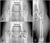
The computed radiography image (Fujifilm Global Corporation, USA) was used to diagnose coxofemoral subluxation and take measurements of the Norberg angle (NA), percent coverage (PC) of the femoral head by the acetabulum as described by Belkoff in 1989 and fIA by the symmetric axis-based method (symax) [3,18,23] (Fig. 1). The reference values were NA = 105°, PC = 50% and fIA = 127.6° [8,23,28]. Only dogs that had NA < 105°, PC < 50%, and DJD scores of 0 to 2 (out of 4) were selected for ITO. Values of fIA were not a limit to inclusion.
Measurement method
All radiographic measurements were performed using a computer program for image analysis (Digimizer; MedCalcSoftware, Belgium). The size of the bony wedge to be removed with ITO was then quantified on the radiographs as follows.
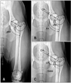
Two circles were drawn touching the silhouette of both the proximal and distal femoral epiphysis in three points, and a first line was traced between the centers of these circumferences. A third circle was drawn on the silhouette of the acetabulum, and a second line was traced between the center of the acetabulum and the center of the proximal femoral epiphysis. The angle formed by the two lines was calculated and designated as the planned femoral inclination angle (pfIA) (Fig. 2A).
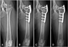
The difference between fIA and pfIA was measured and recognized as the correction angle (CA1) (Fig. 2B). The correction angle was subsequently shifted to the intertrochanteric side, immediately proximal to the lesser trochanter (CA2) (Fig. 2C). Overall, this resulted in a triangle being drawn. The length of the base traced on the medial cortex of the femur corresponded to the base of the bony wedge to be removed by the surgical procedure, which was measured using a surgical ruler. After surgery the new femoral inclination angle nominated effective femoral inclination angle (efIA) was measured by the symax method to confirm the success of the ITO (Fig. 3A). Finally, two circles on both the femoral head and acetabulum were traced to verify that joint congruency was achieved (Fig. 4).
Statistical analysis
All measurements were performed by the same operator at each point time to avoid interobserver variation errors [12]. All data were given as the mean ± standard deviation (SD) and median. Nonparametric Wilcoxon tests were applied to identify significant differences between the preand postoperative values of NA and PC, between fIA and efIA, between pfIA and efIA, and between DJD scores. Values of p < 0.05 were considered significant.
Results
The sample consisted of radiographs of five male and two female dogs to give a total of nine hip joints that were treated surgically and radiologically investigated. The following dogs were evaluated: 2 Maremmano-Abruzzese Sheppards, 2 Golden Retrievers, 2 Border Collies, and 1 Cane Corso. The mean body weight ± SD at surgery was 29.3 ± 8.6 kg (range, 18 to 44 kg; median 28) and the mean age ± SD was 10 ± 1.7 months (range, 8 to 13 months; median 11). The mean (± SD) ages at the first and second recheck evaluations were 11.3 ± 1.7 months and 15.7 ± 1.8, respectively. The mean value (± SD) of lameness prior to surgery was 1.9 ± 0.8 (range, 1 to 3; median 2), while it was 1.2 ± 0.4 (range, 1 to 2; median 1) at the first recheck and 0.2 ± 0.4 (range, 0 to 1; median 0) at the second recheck. There were no recorded intra or postoperative complications.
The mean ± SD values of the NA, PC and fIA, assessed by the methods previously described (Figs. 1 and 3), were as follows (Table 1).
Norberg angle
The mean ± SD value of the preoperative NA was 78.9° ± 7.5 (range, 67° to 92°; median 78°). After surgery, the mean ± SD of the same parameter increased to 92.2° ± 6.8 (range, 82° to 101°; median 90°). The NA values increased significantly (p < 0.05) after surgery; however, the values recorded at the first (91.2° ± 8.5) and second (92.2° ± 6.6) recheck did not differ significantly.
Percent coverage
The PC values were 36.9% ± 5.2% (range, 24% to 41%; median 38%) and 50.6% ± 8.3% (range, 40% to 65%; median 49) before and after surgery, respectively, and this increase was significant (p < 0.05). PC values of 49.1% ± 6.9% and 48.9% ± 7.7% estimated at the first and second recheck, respectively, did not differ significantly.
Angle of inclination
The preoperative value of fIA was 127.6° ± 2.6 (range, 125° to 132°; median 127°), while the pfIA was 115.9° ± 2.5. The mean ± SD of the femoral angle -efIA- obtained after surgery, was 111.3° ± 6.4. No significant difference (p > 0.05) was observed between pfIA and efIA, whereas the efIA was significantly decreased when compared to fIA (p < 0.05) after surgery. The mean degrees of efIA were 108° ± 7 and 108° ± 10 at the first and second recheck, respectively. No significant difference was recorded between the immediate postoperative and recheck evaluations.
Discussion
Some studies have stated that restoring the correct percentage of acetabular coverage of the femoral head results in forces acting on any given area of the bone and cartilage of the hip joint being well redistributed. This redistribution produces a reduction of stress throughout the cartilage surfaces in a hip that is mechanically compromised and a consequent slowing of the progression of the DJD, which is usually associated with the CHD conditions [1,9,19,20]. Indeed, this is the rationale for JPS, TPO and ITO procedures; however, outcomes of the evaluations regarding their efficacy are discordant [4,5,9-11,15,16,22,26].
Current research is focused on improvement of the JPS, TPO and DPO techniques, but no recent studies have been published regarding the ITO to the best of our knowledge. Indeed, there are few records regarding clinical and radiographic evaluations of intertrochanteric osteotomy, and only two studies published in 1990 and 1997 affirming that no benefit to DJD progression is obtained after application of this technique [6,7,11,21,34]. In the present study, preoperative X-ray examinations revealed an absence of moderate signs of DJD (such as inclusion criteria). Moreover, X-ray re-check conducted at six months after surgery showed no progression to only a slight increase of DJD, with only one case showing a severe progression of osteoarhtrosis (score 3).
The beneficial role of varus osteotomy for treatment of hip osteoarthritis has been recognized in human medicine since the 1920s, and it is reported that osteotomy delays the need for hip replacement in young patients with osteoarthritis by more ten years [35].
We investigated whether varisation of the femoral inclination angle in presence of coxofemoral joint laxity could lead to radiographic improvement of congruency and stability of the hip and a subsequent reduction of stress on the cartilage surfaces.
To support our theory, NA and PC were analyzed prior to and after surgery, and the results of our analysis demonstrated that a significant improvement (p < 0.05) of each value was obtained. Accordingly, there was a remarkable decrease of subluxation and joint incongruency in all patients (Fig. 3).
The mean value of NA recorded immediately postoperative was 92.2° (82° to 101°). This value differs greatly from the optimum value of 105°, and was close to the cutoff point of 92.6° (88.2° to 94.8°) reported by Tomlinson to discriminate between normal and dysplastic hips in Golden Retrievers [32].
The mean PC values reached 50%, which is considered to be a good percentage of coverage for normal hips [28].
The outcome of this analysis confirmed that a reduction of the femoral inclination angle, even in patients in which it was within the normal range, led to a decrease in subluxation of the femoral head and to radiographic improvement of hip congruency and containment. The preoperative NA and PC values observed in the present study were lower than those reported by Tomlinson [31]. We assumed that the variations were due to differences in computer systems for image analysis and to the method used for PC measurement. Moreover differences in values recorded in the postoperative checks could also be due to the different types of surgery conducted (ITO vs. TPO).
The aim of ITO described in veterinary literature is to reduce the coxa valga through a varisation of the femoral inclination angle measured by the Hauptman method A (146° ± 5) to achieve an angle of 135° [13,21]. The amount of varisation is standardized to be 10° less than normal, and is measured intraoperatively using a surgical goniometer in such procedures [21].
We conducted femoral intertrochanteric varus osteotomy in dysplastic dogs that did not present coxa valga to achieve a position of the femoral head closer to the center of acetabulum and an adequate acetabular coverage with the same rationale of intertrochanteric varus or valgus osteotomy described in human medicine [17,35]. The mean fIA of our sample was 127.6° ± 2.6, which was less than the mean value reported in literature (133.5° ± 4.7) for non-dysplastic hips [2]. The projected inclination angle was measured with the symmetric axis-based method because we consider this to be more precise based on a study conducted by Rumph and Hathcock in which a value of 127.6° was reported [23].
Owing to these different inclusion criteria, it was necessary to find new marker points to produce the right positioning of the femoral head more deeply into the acetabulum. To accomplish this, the center of the acetabulum was drawn on the radiograph and the angle of varisation was calculated specifically for each individual patient, without any standardization. This option allowed adequate joint congruency to be obtained.
Moreover, we proposed a precise and simple procedure for measurement of the bony wedge to be removed for femur varisation. In fact, it was possible to proceed intra-operatively following measurement of the base of the wedge using just a surgical ruler, without a surgical goniometer.
Statistical analysis of the values of the pfIA measured before surgery and the efIA obtained after surgery revealed no significant difference. These findings confirmed the accuracy of the proposed method to plan the correct varisation degree to be applied.
It should be noted that this study has several limitations. We demonstrated the effects of ITO on the position of the femoral head within the acetabulum based on NA and PC measurements taken in a small sample population. A greater sample size and longer follow up are necessary before we can draw more definite conclusions.
In conclusion, assessment of DJD progression of both hips (treated and contralateral) should be considered in future studies. However, these preliminary results indicate that intertrochanteric varus osteotomy should be re-considered for the treatment of early stage hip dysplasia in dogs with radiological signs of joint incongruence.


 XML Download