

 PDF
PDF ePub
ePub Citation
 Print
Print


Introduction
Pigs have become useful animal models for studying organ xenotransplantation and human disease because of their physiological similarities to humans. Due to these important characteristics, in vitro cultures (IVCs) of porcine oocytes are very crucial for studying pre-implantation embryo development and the production of transgenic animals. To increase the success rates of oocytes maturation and blastocyst formation in vitro or improve cloning efficiency after embryo transfer, it is essential to produce high-quality matured oocytes. Therefore, in vitro maturation (IVM) systems involving porcine oocytes for in vitro embryo production must be improved. Many research studies have attempted to enhance IVM systems by improving techniques performed during all stages of pre-implantation development. Among the most important factors influencing the developmental potential of embryos produced in vitro are the culture conditions including external oxygen concentrations that affect both oocyte maturation and embryo development [18].
Reactive oxygen species (ROS) are endogenously produced by oocytes and embryos during in vivo development and IVC. Examples of ROS include oxygen ions and peroxides, particularly superoxide anions and hydroxyl radicals that are generated during the process of oxygen reduction. Environmental stress can dramatically increase ROS levels. This may result in significant damage to cell membranes and plays a role in apoptosis. Additionally, the over-production of intracellular ROS in mammalian embryos during IVC is generally thought to be detrimental to embryo development [15]. Because pre-implantation embryos are particularly sensitive to ROS damage [14], the deleterious effect of ROS result in developmental inhibition [9]. In previous studies of pigs, in vitro fertilization (IVF) embryos with increased levels of ROS were found to have low developmental competence and increased DNA fragmentation [21]. Many reports have focused on overcoming these detrimental effects of ROS on embryo development. For example, reducing oxygen tension [16,20] or treatment with antioxidants [7] during IVM or IVC improves embryo development. Therefore, it is important to protect oocytes against oxidative stress during IVM. One approach is to supplement the medium with antioxidant compounds during IVM.
Quercetin [2-(3,4-dihydroxyphenyl)-3,5,7-trihydroxy-4H-chromen-4-one] is a plant-derived flavonoid mainly found in fruits and vegetables. In mammals, flavonoids exert various biological and pharmacological effects [10]. Several studies have indicated that quercetin may have anti-inflammatory and antioxidant properties [8] due to its free radical scavenging and metal chelating activities [23]. In addition, this compound exerts a potent antioxidant effect. On the other hand, quercetin can also elicit pro-oxidant effects [3]. A relationship has been demonstrated between the free radical scavenging activity and anti-carcinogenic and anti-inflammatory properties of quercetin [17,32].
Studies have been performed to evaluate the physiological functions and biological activities of quercetin in humans and animals [1,13]. However, there is limited information available on the effect of quercetin on oocyte maturation and embryonic development in pigs. Quercetin has been found to exhibit both estrogenic and anti-estrogenic effects in pigs in vitro, suggesting that this compound has different potential impacts on reproductive function [28]. Given this information, the objective of the present study was to examine the possible effects of quercetin supplementation during IVM and IVC on pig oocyte maturation and developmental competence of parthenogenetically activated embryos. For this, we monitored the nuclear maturation, ROS levels of porcine oocytes, embryo cleavage, and blastocyst formation of parthenogenetic embryos.
Materials and Methods
Experimental design
In order to identify the effective quercetin concentration for improving oocyte maturation (experiment 1), IVM medium was supplemented with four concentrations (0, 1, 10, or 50 µg/mL) of quercetin during the entire 44-h maturation period. For experiment 2, we evaluated the effects of the same four concentrations of quercetin in IVM medium on the parthenogenetic development of embryos. In experiment 3, we assessed the effects these concentrations of quercetin in IVM medium on the ROS levels in oocytes to assess the effect of antioxidant of quercetin.
Oocyte collection and IVM
Pig ovaries were collected from a local slaughterhouse and transported to the laboratory in a 0.9% (w/v) NaCl solution at 30~35℃ within 3 h. Follicular contents from the antral follicles (3~6 mm in diameter) were aspirated using an 18 gauge needle attached to a 10 mL disposable syringe. The contents were pooled in a conical tube and allowed to settle for a few minutes at 39℃. The sediment was removed and diluted with Dulbecco's phosphate buffered saline (Invitrogen, USA) containing 1% penicillin/streptomycin (pen-strep; Invitrogen, USA). Cumulus-oocyte complexes (COCs) which has compact, multilayered cumulus cell and homogeneous cytoplasm were selected and washed about 3 times. They then were transferred to IVM medium containing TCM-199 supplemented with 10 ng/mL epidermal growth factor, 0.57 mM cystine, 0.91 mM sodium pyruvate, 5 µg/mL insulin, 1% (v/v) pen-strep, 1 µg/mL gonadotrophin, and 10% porcine follicular fluid. For the first 22 h only, the IVM medium contained 0.5 µg/mL follicle stimulating and 0.5 µg/mL luteinizing hormones. The COCs were cultured at 39℃ in 5% CO2 in air. After 44 h of maturation, the oocytes were denuded of cumulus cells by pipetting with 0.1% hyaluronidase in Tyrode's albumin lactate pyruvate (TALP) medium [19]. The denuded oocytes were then treated according to the experimental design.
Assessment of meiotic maturation of the mature oocytes
After culturing for 44 h, the denuded oocytes were stained with Hoechst 33342 in TALP medium. The stage of meiotic maturation was determined according to the presence or absence of the first polar body (metaphase II) viewed under UV light with an inverted microscope (Nikon, Japan) equipped to perform epifluorescence.
Parthenogenetic activation of mature oocytes and in vitro culturing
At 44 h of IVM, metaphase II oocytes were parthenogenetically activated. Briefly, the denuded oocytes were equilibrated for 1 min in 0.26 M D-mannitol-based activation solution supplemented with 0.1 mM MgCl2, 0.1 mM CaCl2, and 0.5 mM HEPES. The oocytes were then transferred to a chamber between two electrodes spaced 3.2 mm apart and overlaid with activation solution. The oocytes were activated by electric stimulation with a single direct current pulse of 2.0 kV/cm for 60 µsec using a BTX Electro-Cell Manipulator 2001 (BTX, USA). A group of approximately 20 to 30 parthenogenetically activated oocytes were cultured in 500 µL porcine zygote medium-5 (Funakoshi, Japan) for 7 days at 39℃ in a humidified atmosphere with 5% CO2, and 5% O2. The cleavage and blastocyst formation rates were checked at 48 and 168 h of IVC, respectively.
Assessment of embryo quality
Blastocyst quality was assessed by Hoechst staining of the inner cell mass and trophectoderm cells according to standard procedures. Briefly, the blastocysts were washed in HEPES-buffered TALP medium and then incubated with TALP medium containing 25 µg/mL bisbenzamide (Hoechst 33342) stain for 15 min at 39℃. The stained blastocysts were mounted onto glass slides under a cover slip and counted while examined with an inverted microscope (Nikon, Japan) equipped to perform epifluorescence.
Measurement of ROS levels
Oocytes were sampled 44 h after IVM to determine intracellular ROS levels. The level of ROS in each oocyte was measured according to the 2',7'-dichlorodihydrofluorescein diacetate (DCFH-DA) method described in our previous study [19]. Briefly, 20 oocytes from each treatment group were incubated in the dark for 30 min in TALP medium supplemented with 10 µM DCFH-DA. After incubation, the oocytes were washed in TALP medium, placed onto a glass slide, and covered with a cover slip. Fluorescent emissions (405~435 nm for excitation and 515 nm for emission) from the embryos were recorded as TIFF files using a cooled CCD camera attached to a fluorescence microscope (Axio Photo; Carl Zeiss, Germany). The recorded fluorescent images were analyzed using NIH image software 1.55 (National Institutes of Health, USA) and the number of pixels was determined after color inversion as previously described [19].
Statistical analysis
All statistical analyses were performed using Prism software (ver. 4.0; GraphPad, USA). A one-way ANOVA followed by Tukey's test was used to measure statistical differences among the groups. P-values < 0.05 were considered to be statistically significant. Data are expressed as the mean ± SE.
Results
Effect of quercetin on porcine oocyte nuclear maturation
About 782 oocytes were used for five replicate trials to evaluate the effects of quercetin on nuclear maturation during IVM. The polar body extrusion rate was not significantly different among the controls and groups treated with 1 or 10 µg/mL quercetin. In contrast, this rate was significantly lower (p < 0.05) for the oocytes given 50 µg/mL quercetin (50.7% ± 1.9) compared to the control (81.23% ± 1.0) and other groups (Table 1).
Effects of quercetin on the subsequent in vitro development of porcine oocytes
A total of 672 oocytes underwent IVM in media supplemented with four concentrations of quercetin. The oocytes were parthenogenetically activated and in vitro development was evaluated. Quercetin supplementation had no effect on the first cleavage frequency or cell number per blastocyst (Table 2). However, a significantly greater (p < 0.05) proportion of blastocysts developed into oocytes when the IVM medium was supplemented with 1 µg/mL quercetin. Addition of 1 µg/mL quercetin to the IVM medium improved the frequency of blastocyst development, but both the cleavage frequency and rate of blastocyst formation of oocytes treated with the highest concentration of quercetin (50 µg/mL) were significantly depressed compared to all other groups (Table 2).
Measurement of ROS concentrations in porcine oocytes
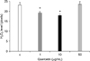
For experiment 3, about 230 oocytes were used in four replicate trials to evaluate the effects of quercetin on the levels of ROS during IVM. The relative hydrogen peroxide (H2O2) contents of oocytes matured in medium supplemented with 1 or 10 µg/mL quercetin ranged from 13.0 to 14.4 pixels, and were significantly lower than those in oocytes matured in the control medium (Fig. 1; 28.2 pixels, p < 0.05). However, the relative H2O2 contents were not significantly different between the control and oocytes treated with 50 µg/mL quercetin.
Discussion
In the present study, the effects of quercetin as an antioxidant during IVM and IVC on oocyte maturation and embryonic development after parthenogenetic activation were examined, and the intracellular levels of ROS were measured. Our results demonstrated that quercetin enhances the in vitro development of porcine oocytes. In quercetin-treated oocytes, ROS concentrations were significantly lower after maturation. Additionally, the rate of blastocyst formation was significantly higher in quercetin-treated oocytes that were activated and further cultured compared to the control group. On the other hand, excessive doses of quercetin were detrimental to oocytes and embryos without reducing the levels of ROS.
In recent years, interest in quercetin has gradually increased along with other flavonoids due to its health promoting activities that likely result from antioxidant effects in humans and animals. Nevertheless, the overall biological impact of quercetin remains controversial, mostly due to the limited information about its bioavailability, endogenous dynamics, and relative contribution of different types of conjugates. Some studies have showed that quercetin can mediate cancer cell apoptosis [22,25]. Furthermore, these investigations have indicated that quercetin can selectively induce apoptosis of cancer cells and not normal cells. Other research has shown that quercetin can protect against oxidative stress by decreasing ROS generation through its antioxidant activity in normal human cells [2,11]. Our study demonstrated that quercetin at the optimal concentration acts as an antioxidant and positively affects the maturation of porcine oocytes and in vitro embryonic development.
ROS are generated by embryonic metabolism or in the surrounding environment during IVC [19], and are detrimental to embryonic development [12]. Increased ROS levels are associated with the two-cell embryo block in mice [24]. It has been suggested that increased ROS concentrations may lead to apoptosis during embryo culturing [9]. Among different ROS, H2O2 at high concentrations induces apoptosis [26]. Therefore, this study was conducted to monitor the levels of H2O2 within oocytes to indirectly assess ROS toxicity. We also examined parthenogenetic embryonic development to evaluate oocyte competence rather than in vitro fertilization or somatic cell nuclear transfer. The reason for this was because parthenogenetic activation can be used to evaluate oocyte developmental competence in vitro without confounding factors from sperm and a variety of other reagents introduced during the in vitro procedure. Furthermore, parthenogenetic activation is relevant to cloning research because artificial oocyte activation is an essential component of nuclear transfer protocols [19].
The concentrations of quercetin used in this study (1, 10, and 50 µg/mL) were selected based on previous results from a study of porcine granulosa cells [28]. In this study, 50 µg/mL quercetin was found to inhibit progesterone production, modify estradiol-17β production, and interfere with angiogenesis in granulosa cells by inhibiting vascular endothelial growth factor production, implying that quercetin may have a negative influence on ovarian physiology. Despite the detrimental effect of quercetin on embryos, our study showed that treatment with adequate concentrations of quercetin (1 and 10 µg/mL) improved embryonic development, but it was not clear whether quercetin directly affects embryo development by decreasing ROS toxicity. We also found that the rates of oocyte maturation and blastocyst formation were substantially reduced with a high concentration of quercetin (50 µg/mL) although the expansion and total cell number of blastocysts were not adversely affected, demonstrating that quercetin has dose-specific effects on oocytes.
We suggest that the reduction of oocyte maturation and blastocyst formation rates with this concentration of quercetin may result from unresponsive signaling to oocytes and embryos or direct embryo toxicity owing to excessive levels of flavonoids. Some groups have reported toxic effects of other flavonoids on embryos from different species [4-6,27]. In contrast, several investigations have shown that supplementation of porcine IVM medium with antioxidants such as selenium, vitamin E, and ascorbic acid decreases ROS levels while enhancing the developmental competence of IVF embryos and parthenotes [21,29,30]. Another study recently demonstrated that treatment with anthocyanin, a type of flavonoid, in IVM media improves the developmental competence of cloned pig embryos, most likely by increasing glutathione synthesis and reducing ROS levels [31]. Compared to a previous study conducted with other antioxidants, the effects of quercetin on the maturation rate, blastocyst rate, and ROS generation rate in oocytes we observed were less potent.
In our investigation, no beneficial effect of quercetin treatment on the first polar body extrusion rate during IVM was found even though quercetin effectively reduced ROS levels. However, a beneficial effect of quercetin treatment at less than 10 µg/mL was observed during subsequent culturing to the blastocyst stage. Therefore, it may be that the antioxidant effect of quercetin on oocytes was maximized during subsequent IVC, and thus, as suggested by previous studies and the present investigation, ROS may play a pivotal role in regulating oocyte maturation and embryonic development.
In conclusion, treatment of porcine oocytes with quercetin had a significant effect on embryonic development. At low levels, quercetin reduced intracellular ROS levels but was detrimental at high concentrations. It is not clear whether this low level of quercetin is optimal in pigs. In addition, the rate of blastocyst formation was significantly increased by quercetin treatment whereas there was no increase in the number of blastocyst cells. Further studies are needed to determine the optimal concentration of quercetin and to ascertain the beneficial effects of this compound on pig embryo development.


 XML Download