

 PDF
PDF ePub
ePub Citation
 Print
Print


Introduction
Cranial cruciate ligament (CrCL) rupture is the most common cause of lameness in dogs [13,14,21]. In large breed dogs, many surgical treatments have been proposed; however, few reports exist in the literature regarding CrCL rupture in small breed dogs in which, conservative management was the preferred method of treatment. Along this same line, Vasseur [31] showed that conservative management has beneficial effects in 85.7% of patients.
Due to the long recovery period of conservative management techniques, however, surgery is now preferred for the treatment of CrCL rupture in small breed dogs [17]. The goal of surgery is to stabilize the stifle joint, preserve range of motion, and prevent osteoarthritis (OA) [22]. Intracapsular, extracapsular, and tibial osteotomy procedures have been described [2,5,6,8,12,16-18,26-28,33]. Indeed, many surgical methods have been proposed, but no specific procedure is considered optimal [1].
Gait evaluation of dogs is generally obtained using qualitative analysis methods through direct inspection examination and/or video recording [20]. Further, gait analysis can be carried out using a system featuring multimodal (kinetic, kinematic and electromyographic) evaluation and three-dimensional measurements [34]. Even though investigations obtained using force platforms are the most reliable, it is also possible to obtain important quantitative data supporting the clinical evaluation of patients by using specific questionnaires completed by the owners [11,30].
The aims of this paper were to describe a new surgical technique for the extracapsular stabilization of the CrCL rupture through transposition of a strip of biceps femoris and to objectively evaluate the results by using specific questionnaires focused on the stifle joint.
Materials and Methods
Clinical data were obtained from dogs that were presented in 2009. The inclusion criteria were based on the diagnosis of CrCL insufficiency in small breed dogs (weight ≤ 15 kg).
Each patient underwent the same protocol: orthopaedic evaluation, preoperative X-ray examination, surgery, and clinical follow-up carried out at 1, 3, and 12 months. Postoperative radiographs were performed at 3 (nine patients) and 12 months (six patients). Medical records including all clinical data were compiled for each dog. During the follow-up evaluation, a multiple-choice questionnaire was completed by the owners.
Surgical technique
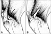
All patients were anesthetized and placed in lateral recumbency with the affected side uppermost. A craniolateral approach to the stifle joint was carried out; the incision extended from the distal third of the femur to the proximal third of the tibia. The aponeurotic portion of the biceps femoris muscle was exposed, and the margin between the biceps femoris and fascia lata was recognized. This was followed by identification of the cranial insertion of the biceps femoris muscle, after which two incisions were made through the flap that would be used subsequently (Fig. 1A).
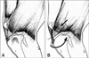
A triangular pedicle flap was prepared; one side was the cranial margin of the biceps muscle separated from the caudal margin of the vastus lateralis by a 3 cm incision, whereas the other side was formed by a 2 or 3 cm incision along the muscle fibers of the caudal portion of the distal biceps itself (Fig. 1B). The resulting flap was moved up, and by applying gradual traction in a cranial-medial-distal direction, it was transposed towards the tibial tuberosity and maintained extension of the limb. The flap was located next to the patellar ligament, where it was sutured with a 2-0 monofilament glycomer (Fig. 2A). In this manner, the biceps femoris muscle assumed a portion of its contractile fibers, and the slope was similar to that of the CrCL.
Intraoperatively, both tibial thrust and drawer tests were carried out to assess the degree of cranial shift of the tibia. If the drawer sign was still evident, tension of the biceps femoris flap was increased so as to completely counter the cranial displacement of the tibia towards the femur. The flap was sutured to the lateral margin of the patellar ligament as close as possible to the tuberosity. Neither arthrotomy nor arthrocentesis were carried out, and routine suturing was performed. All patients underwent antibiotic and analgesic therapy, and a soft bandage was placed over the wound. The owners were instructed to limit physical activity of the dogs to a minimum for approximately 15 days before allowing resumption of normal levels of activity.
Medical records and owner questionnaire
Medical records compiled by the clinician included various parameters: stifle pain, patellar-femoral crepitus, joint stability, range of motion, swelling, muscle mass, and lameness. These parameters were divided into three different subscales: visual examination, manual examination, and X-ray evaluation of OA.
Each owner completed a questionnaire [4] consisting of 24 questions divided into three subscales: pain, stiffness, and limb function. Five choices were given for each of the six subscales (three medical record and three questionnaire subscales) regarding the presence of clinical signs (4: always, 3: obvious/often, 2: moderate/sometimes, 1: mild/rarely, 0: absent/never).
Data processing
The data were normalized using a standardization test and were transformed into a score from 0 to 100.
The normalized score was calculated as shown below:
Normalized score = 100 - total score of each subscale × 100 / possible maximum score for the subscale
Our results were classified as: excellent (81~100), good (61~80), poor (41~60), or failed (0~40). The score obtained for each subscale was added to that of the same subscale for each clinical case, and the mean values, calculated at each postoperative examination, were plotted on a graph to assess healing.
Finally, to statistically analyze the significance of the differences between the mean ± SD of each pre and postoperative variable, Student's t-test was applied. Values of p < 0.01 were considered significant.
Results
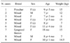
Nine monolateral BFTs were performed on nine small dogs (seven females and two males) of different breeds (one miniature Pinscher, one Epagneul Breton, and seven mixed breeds) with a mean weight of 11.48 kg (range: 6 to 15 kg) and a mean age of 7.77 years (range: 4 to 11 years) (Table 1). All BFTs were performed by the same experienced surgeon, and the procedure required, on average, 20 min.
There were no recorded intraoperative complications, and only two patients (cases 5 and 7) postoperatively had a subcutaneous seroma, which was subsequently drained.
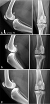
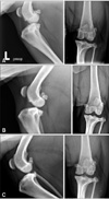
All nine interventions resulted in complete healing with good postoperative results at 3 months; cases 6, 7, 8, and 9 showed only mild lameness after prolonged movement within the first postoperative month. At all time points during the 1-year follow-up (1, 3, and 12 months), no recurrences of stifle instability were reported (Figs. 3 and 4).
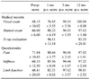
The results for medical records and owner questionnaire are shown in Table 2.
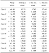
In assessing the overall outcome of the questionnaire as well as the clinical evaluation for each patient, six out of nine were judged as excellent at 1 month after surgery (from 83.67 to 90.01). This figure increased to nine dogs at 3 months (from 91.26 to 98.29) and was maintained at 12 months (from 95.26 to 100) (Table 3). In this specific evaluation, the X-ray-score was not included since its high value prior to surgery could modify the clinical score.
Both the mean value of each item as well as the overall scores of each dog recorded before surgery and at 1, 3, and 12 months postoperatively showed statistically significant improvements (p < 0.01).
Discussion
Extracapsular surgical methods performed for stabilization of the stifle joint aim to restore the functions of the ligament without its anatomical replacement. Further, these surgical procedures are all aimed at limiting mechanical cranial displacement of the tibia towards the cranial direction [30].
The main extracapsular stabilization procedures described in the literature are Flo technique, widely used in dogs of all sizes, fibular head transposition, and the anchoring system in dogs and cats [5,8,17,28].
The inclusion criteria of this paper included all dogs weighing ≤ 15 kg in which cranial cruciate ligament insufficiency was diagnosed. The mean age of the patients was 7 years and 8 months, confirming studies on incidence of the disease [7,35]. All dogs included in the study had no concomitant orthopaedic diseases, although CrCL rupture is often associated with medial patella luxation [3]. In this study, we only tested patients presenting with CrCL rupture in order to evaluate the outcome of BFT.
The main mechanism of BFT is to limit laxity of the stifle joint, during both the static and dynamic phases, by offering a new anchor on the tibial tuberosity. BFT had a dual effect; static stabilization was applied to simulate the direction of the CrCL, and active contraction of the biceps femoris prevented slipping of the cranial tibial plateau, resulting in dynamic stabilization. In addition, the muscle acted on the tibial tuberosity with a force directed caudally and externally, and the transposed muscle flap held the tibia and prevented cranial slippage and intrarotation.
Preoperative X-ray examination showed mild signs of OA in cases 2, 4, 6, 8, and 9. In addition, a 3 month X-ray re-check showed no progression of OA, whereas at 12 months, X-ray evaluation showed a slight increase in case 4.
Radiographic examination is an objective measure of OA secondary to joint instability [32]. X-ray examination performed at 1 year after surgery (case 2) showed small apical patellar osteophytes, which implies that OA progression was very slow. On the other hand, X-ray sessions on case 4 indicated no OA progression at 1 year after surgery as result of good joint stability.
As reported previously, there is no relationship between the functional capacity of dogs presenting with OA and radiographical evidence of disease [9]. Therefore, correct therapeutic planning should consider the numerous factors that cause disability [15]. In the present study, lack of lameness was indicative of joint stability achieved after surgery. However, the tibia was in the cranial position in some cases (cases 2 and 4) after surgery. This could be the consequence of decreased tension of the muscle-tendon flap on the patellar ligament following surgery, and could not be attributed to discomfort, lameness, or joint instability. Besides, active contraction of the biceps femoris could provide dynamic stabilization.
At 3 and 12 months, none of the dogs showed lameness, and all presented with good joint stability, absence of crepitus, and satisfactory recovery of muscular tone. At 12 months, cases 1, 8, and 9 were checked by means of a telephone questionnaire and interview, and the owners noted no complications.
The use of questionnaires derives from human medicine and aims to evaluate patients presenting with joint disease [1,4,10,19,24]. Questionnaires represent an effective and rapid detection method that is also easy to administer on a large-scale, although it requires the cooperation of the patient (human medicine) or owner (veterinary medicine) [25]. Regarding pets, it is essential that the questionnaire be completed by whoever is most familiar with the animal since he/she is the only one who can detect slight changes in appearance and behavior [30]. Constant monitoring of normal animal behaviors is considered to be simple, safe, and of prognostic relevance to the survey.
Based on our outcomes, we could confirm that BFT led to complete recovery of the stifle joint within 3 months. During the follow-up, an improved clinical condition was notable in the first month, followed by normalization within the next 2 months.
Medial meniscus injuries and CrCL rupture often occur in combination in large dogs [12,23,29]. However, in small dogs, the incidence of meniscal injury is not defined [17]. In a recent paper, Kunkel et al. [17] did not find any meniscal disorders in 16 small dogs and cats treated for CrCL rupture. Despite the small study population, Kunkel et al. [17] considers meniscal injuries to be less frequent in small dogs. In addition, full examination of the joint is often difficult and incomplete in small dogs.
In this report, arthrotomy was not performed, and we were limited by the small study population. Therefore, further studies should be carried out based on recently developed extracapsular techniques. However, these procedures are often more invasive, involving the drilling of holes in the condyles and tibial plateau in order to permit the passage of anchor wires [5,17]. Further, additional studies on a larger population should be carried out.
 XML Download