

 PDF
PDF ePub
ePub Citation
 Print
Print


Introduction
Conventional diagnostic imaging modalities such as radiography [23] and ultrasonography [10,14] used primarily in veterinary medicine cannot reliably identify lesions affecting the middle or inner ear. Today, advanced noninvasive diagnostic techniques such as computed tomography (CT) and magnetic resonance imaging (MRI) are used to evaluate the inner or middle ear [1,4,5,9,13,15,22]. In particular, virtual otoscopy (VO) enables non-invasive 3D endoluminal imaging of the complex inner ear structure. The computer-rendered images allow constant luminal visualization that permits navigation along the inner surfaces in a manner similar to conventional fiberoptic endoscopy. Virtual endoscopy has been studied extensively and was recently used in practice as a tool for radiological diagnosis, surgical planning, and teaching in human medicine [3,16,20,21].
Virtual endoscopy techniques for examining the ear are being developed because the images allow improved visual recognition of the fine temporal bone structures. VO of the middle ear is generally still not used in veterinary medicine. Previously, VO alone was investigated as a means for generating anatomic reference images of the canine middle ear in normal dogs [6]. However, no information on VO images has been reported in veterinary medicine. Therefore, the aim of this study was to evaluate the feasibility of VO through visualization of the tympanic cavity including ossicles in experimentally-induced otitis media and clinical canine patients with fluid accumulation in the tympanic cavity.
Materials and Methods
Animal preparation and induction of otitis media
Seven healthy male beagles (1 to 4 years old, 7.5 to 10.7 kg; Chemon, Korea) were included in our study. Additionally, we evaluated two canine patients, one Maltese (8 years old, 3.4 kg) and a mixed breed dog (6 years old, 5.8 kg), with otitis media based on clinical features and CT images that revealed hyperatteuated density indicative of fluid- or soft tissue-filled tympanic bulla. The investigation was carried out in accordance with the guidelines of the Animal Care Committee of the College of Veterinary Medicine, Chonbuk National University (Korea).
All of the animals were administrated dexamethasone [1.0 mg per animal via intramuscular injection (Dexason; Daesung, Korea)] 12 h before inoculation. While under general anesthesia with Zoletile (Virbac, France) five of seven healthy beagle dogs were inoculated using a 'mouse syringe' (SamwooKurex, Korea) with a culture of coagulase negative 5 × 10⁸ Staphylococcus aureus-571 (UOP) provided by National Veterinary Research and Quarantine Service (Korea) via the transtympanic membrane bilaterally (a total of ten ears) as previously described [11]. The two remaining healthy dogs were inoculated with the same amount (1.0 mL) of saline solution. After inoculation, clinical signs including indications of ear pain, scratching, head shaking, and cerumen production were observed daily for 3 weeks.
Diagnostic imaging
Radiographic examination (lateral and dorsoventral projection) with the conventional X-ray machine (Acoma, Japan) and CT scanning were performed prior to and 15 days after inoculation by multidetector CT scanner (Somatom sensation 16; Simense, Germany). All of the animals were fasted for 24 h prior to CT scanning and premedicated with atropine (Daewon, Korea) before induction of general anesthesia. Radiography and CT studies were performed with the two canine patients. General anesthesia was induced and maintained with propofol (Provive 1%; Claris Lifesciences, India) delivered intravenously.
For CT scanning, the dogs were placed in a sternal recumbent position using a position bag to support the head and cervical spine. Animals with a fluid-filled tympanic cavity were also placed in a dorsal recumbent position. The head was extended with the hard palate parallel to the CT table, and fixed in this position with medical tape (3M Micropore; 3M, USA). Transverse images were obtained with multi-detector-row CT scanners (Somatom Sensation 16 and Somatom Plus 4; Siemens, Germany). The CT scanning parameters were 120 kV, 60 mA, 0.5 sec/rotation, 90-mm field of view with a 1-mm scan slice thickness with no intervening gap, one helical pitch, and a 512/512 matrix size. A total of 40 to 50 axial CT images (window width = 4,000; window level = 400) were obtained per scan sequence. The images were then transferred to a computer equipped with commercially available software (Rapidia; Infinit, Korea) that can produce virtual endoscopic images. Two radiologists (KC Lee and HS Kwak, Chonbuk National University) interpreted the high-resolution CT (HRCT) images as well as the virtual endoscopic images. The threshold values varied from -450 to 1,300 depending on the part of the optimally visualized structures. Bony and/or soft tissue surfaces could be visualized according to the needs of the evaluator. Navigation from the external canal to the tympanic cavity using VO was possible by moving the arrows on the axial CT images in three different planes: transverse, sagittal, and dorsal. Navigation was also possible in the rostral or caudal, dorsal or ventral, and left or right directions and on all sides of middle ear. Identity of the structures visible on the VO images was verified by pointing to a spot in the VO view. The position of this spot was then simultaneously shown on the two-dimensional (2D)-CT images.
Image evaluation
The three-dimensional images were reviewed by two experienced radiologists (H.S. Kwak and K.C. Lee, Chonbuk National University). Image quality with respect to the nine anatomic structures examined was scored using the following grading system: 0, anatomical structure not detectable; 1, anatomical structure detected but not clearly defined; and 2, anatomical structure clearly defined.
Results
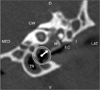
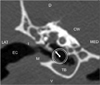
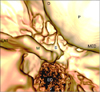
Eight out of 10 ears inoculated with the bacterial pathogen exhibited obvious clinical signs of infection while the two control dogs did not show any clinical signs of ear disease. CT scanning revealed fluid-filled tympanic cavities in three affected ears of three dogs inoculated with the infectious agent. No remarkable findings were observed on the radiographs in any of the animals. Qualitative measurements of the major inner region structures of all 10 ears are shown on Table 1. Conventional CT imaging revealed soft tissue density occupying the tympanic bulla, indicative of otitis media, in the experimentally infected animals and clinical canine patients (Figs. 1~3).
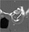
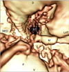
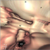
The head of the malleus, incus, stapes, and vestibular window were distinguishable but not clearly viewed in the experimental dogs. In the clinical patients, most of the structures were not distinguishable except for the malleus. The rostral process of the malleus was barely distinguishable. Internal structures such as the manubrium of the malleus, promontory, cochlea window, and septum bulla were identifiable on the VO images without interference by the fluid inside the bulla cavity (Figs. 4 and 5). Ossicles of the clinical patients, however, were not discernable from one another (Figs. 3 and 6).
Discussion
Several techniques can be used to facilitate detailed visualization of the middle ear. Indirectly, advanced imaging modalities such as CT and MRI provide useful information for diagnosing and evaluating middle ear problems [15,17] in veterinary medicine. Artifactual effects cause the fluid-filled bulla to appear thicker than that of the normal bulla on axial CT images. However, no correlation between the MR signal intensity of material within the middle ear and final diagnosis have been noted. CT and MR images must therefore be interpreted with caution [2].
Middle ear endoscopy provides an excellent direct view of the surgical micromorphology and pathology of the middle ear [7]. Video otoscopic treatment can also reduce the need for conventional surgery [19]. In humans, complications associated with otoendoscopy may include iatrogenic fracturing of the lenticular process of the incus, non-healing of atrophic thin tympanic membrane, and non-healing of a previous tympanoplasty graft [24].
VO is an alternative method to observe the inside of the middle ear. Although a topographical image produced by VO cannot depict color shades of the canal itself, which can be detected by fiberoptic endoscopy [12,26], VO has been widely investigated in human medicine as a supplement to diagnostic imaging and surgical planning [8,12,18,25]. In veterinary medicine, the utility of VO for examining the inside ear was recently assessed in normal dogs [6]. No study, however, has reported on the application of VO in dogs with an infectious ear disease such as otitis media.
In the present study, VO was used to visualize middle ear structures to assess the fluid-filled tympanic bulla observed in the experimental dogs and clinical patients with otitis media. As reported in a previous VO study in normal dogs, one advantage of VO is that this technique provides more topographical information of the inner ear than 2D-CT [6]. Furthermore, only simple post-processing of CT data is needed to produce a VO image.
The primary focus in this study was visualization of the tympanic cavity including ossicles using VO images. Similar to a previous investigation [6], some important technical adjustments, such as using different viewing angles along with the optimal use of thresholds and color shading that affect the final VO results, were required for our study. Most of the main bony structures in the middle ear could be identified. However, tiny muscles and nerves were not observed on the VO images as expected.
The visibility and clarity of the inside ear including almost all the manubrium of the malleus, promontory, cochlea window, and septum bulla in the view from the tympanic bulla toward the ossicular chain. in our study were similar to previous reported results in normal dogs [6]. For the most part, the structures seen in the normal ear cavity were also visualized in this present study, and the incus and stapes could be seen after threshold adjustment. Interestingly, the promontory, cochlea window, and septum bulla in the middle ear were identifiable regardless of fluid accumulation. As in the normal study with normal dogs, the manubrium as well as the head, neck, and process of the malleus could be easily distinguished, but not in the patient with otitis media. The bony structure of ossicles was not discernable since the ossicles were not clearly observed on the axial CT images for the patient. It is believed that post-processed 3D-CT images, including ones generated by VO, are greatly dependent on raw data from the axial CT image. In addition, the small structure of the ossicles and pathologic changes may contribute to the poor ossicles imaging in the clinical patients. In other words, once the Hounsfield number is perceptibly different between the structures, VO would be available to visualize the interest regions in any circumstances. Visualization of the inside structure like ossicles through threshold adjustment in VO was not interfered by soft tissue and fluid density within the tympanic bulla. Meanwhile, most of the fluid should be removed to assure a clear scope path that facilitates unobstructed visualization of the canal with fiberoptic otoscopy.
This study had a couple limitations. First, a small number of experimental dogs with a few affected ears were involved in our investigation. Additionally, these animals did not have any remarkable CT features of otitis media (such as sclerosis along with thickening and possible lysis of the tympanic bulla) except for fluid-filled tympanic cavities. Finally, only two canine patients were included in this study that were examined with a different CT machine which inherently provided slightly different spatial resolution.
Although VO did not provide enough data to make a definitive diagnosis of middle ear problems, this technique could be used to evaluate cases with fluid-filled tympanic cavities frequently observed with ear disease such as otitis media. Our findings also demonstrated that VO can be consistently used to obtain information about anatomical structures in the middle ear. Further studies should be conducted with various breeds of dogs, including small-sized animals, with a variety of ear diseases such as natural occurring otitis media, interna, polyps, and aggressive infiltrative disease.

 XML Download