

 PDF
PDF ePub
ePub Citation
 Print
Print


Peste des petits ruminants (PPR) is a contagious disease of ruminants, particularly sheep and goats, caused by the PPR virus (PPRV). PPR morbidity and mortality vary depending on the environment. Symptoms of PPR include fever, pneumonia, ocular and nasal discharge, anorexia, necrotizing erosive stomatitis, enteritis, diarrhea, and death [13].
PPR has been reported in Nigeria and Uganda, and has remained endemic in these countries [6,14] where it affects the economy of households that raise small ruminants as a source of livelihood [4]. The World Organisation for Animal Health recommends collecting swabs of conjunctival discharges, nasal secretions, buccal and rectal mucosae, and unclotted blood for reverse transcription (RT)-PCR detection, and sera for serological diagnosis. Rapid and specific diagnosis by PCR has become possible with the techniques developed by Forsyth and Barrett [7] using F gene-specific primers and Couacy-Hymann et al. [3] using primers specific for the N gene.
The objective of our research was to detect PPRV in ocular and nasal swabs, buffy coats, and tissues of sheep and goats from Karamoja region of Uganda and north-central Nigeria suspected of having PPR. This was accomplished with F and N gene-specific primers using RT-PCR. We also evaluated the suitability of clinical samples and determined the most appropriate sample type for PPRV detection.
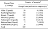
A total of 314 samples (274 oculo-nasal swabs and 40 buffy coat samples) were collected from five districts of the Karamoja region (Uganda) in 2009 as part of a cross-sectional study of a PPR outbreak occurring in 2007. In addition, we included 33 tissue samples (spleen, liver, lungs, intestine, and lymph node) from sheep and goats from the north-central states (Kaduna, n = 7 and Plateau, n = 26) of Nigeria presenting symptoms suggestive of PPRV infection. These tissue samples were collected in 2007 and 2009, and stored at the Central Diagnostics of the National Veterinary Research Institute (Nigeria). A total of 347 samples (Table 1) from Nigeria and Uganda were analysed. Lyophilized freeze-dried live PPR vaccine (Nig 75/1) obtained from the Jordan Bio-Industries Centre (Jordan) was used as the reference virus for the analysis. All samples were kept at -70℃ at the National Animal Disease Diagnostic and Epidemiology Centre of the Ministry of Agriculture Animal Industry and Fisheries (Uganda) prior to sample preparation and the RT-PCR assay.
Blood collected in tubes containing anticoagulant (BD Vacutainer; BD Biosciences, USA) were centrifuged at 2,000 × g for 5 min at 4℃~8℃. The plasma was decanted, and the buffy coat was collected and stored at -70℃ for antigen detection. Ocular and nasal swabs in 0.5 mL RNAlater (Qiagen, Germany) were vortexed and centrifuged at 8,000 × g for 5 min at 4℃.
The supernatant was decanted into sterilized tubes and stored at -70℃ until RNA extraction. One gram of tissue from each Nigerian sample was weighed and homogenized using a tissue homogenizer (Omni TH; Omni international, USA) or a sterile glass mortar and pestle (Glass Tenbroeck Tissue Grinders; Omni international, USA). After homogenization, 9 mL of PBS was added to make a 10% tissue suspension and centrifuged at 8,000 × g for 5 min at 4℃. The supernatant was decanted into a sterile tube and stored at 4℃ until RNA extraction.
Viral RNA was recovered from the ocular and nasal swab, buffy coat, and tissue samples using a QIAamp Viral RNA Mini extraction kit (Qiagen, Germany) according to the manufacturer's instructions. Five µL of extracted RNA were reverse transcribed into cDNA as previously described by Kerur et al. [9]. Ten µL of the PCR product from each reaction were mixed with 1 µL of 6× DNA loading dye (Thermo Scientific, USA) and separated along with a 50-bp DNA molecular weight marker (GeneRuler; Thermo Scientific, USA) by electrophoresis at 80 V for 45 min in 1× TBE buffer (10× Tris-Boric acid-EDTA; BioRad, USA).
Out of all 347 samples collected and analyzed, 95 (27.4%) and 68 (19.6%) produced positive PCR results with the F1/F2 and NP3/NP4 primers, respectively. Among the 314 Ugandan samples, 38 swabs and 40 buffy coat samples (24.8%) were positive for the F gene while 13 swabs and 40 buffy coat samples (16.9%) were positive for the N gene. The 33 Nigerian samples which were all from tissues yielded 17 (51.5%) and 15 (45.5%) positive PCR results using the F or N gene-specific primers, respectively. Among the Nigerian and Ugandan samples, 52 (15.0%) were positive for both the F and N genes; 40 of these were buffy coat samples.
Among the seven different locations where samples were collected, 50, 60, 103, 45, 56, seven, and 26 samples originated from Abim, Kaabong, Kotido, Moroto, Nakapiripirit, Kaduna, and Plateau, respectively. A total of nine (18.0%), 11 (18.3%), 13 (12.6%), 22 (48.9%), 23 (41.1%), seven (100.0%), and 22 (84.6%) samples from the Abim, Kaabong, Kotido, Moroto, and Nakapiripirit districts, and Kaduna and Plateau states, respectively, were positive for the PPRV antigen.
Results of PCR assay for PPRV detection in swab (ocular and nasal), buffy coat, and tissue (spleen, liver, lungs, lymph node, and intestine) samples using F and N gene-specific primers suggested that the choice of sample type and primers is vital for optimizing the efficiency of diagnosing PPR in sheep and goats. Buffy coat samples were found to be the best for molecular diagnosis, yielding a positivity rate of 100% by RT-PCR. Arguably, NP3/NP4 primers were expected to generate higher positivity rates because of the higher level of nucleoprotein gene transcripts in infected tissues compared to F gene expression [8]. The sensitivity of PCR for analysing buffy coat samples may be due to the fact that PPRV is propagated in the lymphoid and epitheliod tissues, leading to severe leucopoenia and immunosuppression that supports secondary and opportunistic infections [2,11]. Nevertheless, the positivity rate for oculo-nasal swabs and tissues indicated the presence of PPRV in these specimens.
In the current study, PPRV was detected in 95 (27.4%) and 68 (19.6%) of all samples tested using F and N gene-specific primers, respectively (Table 2). PPR is endemic in Nigeria and Uganda, and the appropriate molecular techniques and type of sample are crucial for rapid diagnosis [7]. Arguably, differences in positivity rates may be due to sampling method, sample size, and detection method (type of primer used). The results of our study agree with those of Kerur et al. [9] who reported a PPRV detection rate of 40% for clinical samples from India using RT-PCR with F1/F2 primers. Nanda et al. [12], Couacy-Hymann et al. [3], and Kerur et al. [9] have also used F1/F2 and NP3/NP4 primers for molecular diagnostic and epidemiologic studies.
All 40 buffy coat samples collected in Uganda were positive for both the F and N genes. This study revealed that buffy coat samples had higher positivity rates irrespective of the primers used. In contrast, F gene-specific primers were more sensitive for detecting PPRV in swabs compared to the N gene-specific primers. Nucleoprotein gene transcripts are more abundant in infected tissues compared transcripts of the fusion gene required for viral attachment and transmission on epithelial tissues. The ability of primers to detect the virus could be affected by the stage of infection and time of sample collection [1].
An important finding of this investigation is that the PPRV can be detected in buffy coats as well as ocular and nasal swabs. All of these types of samples are easy to obtain. This is important especially in countries where the disease is endemic and mortality rates are relatively low. Under these conditions, post-mortem samples are not readily available for diagnostic purposes [7].
All the primers used in this study were able to detect PPRV in three different types of samples. However, buffy coat samples had higher positivity rates, suggesting that at least two different primer sets should be used for PCR-based diagnosis since some samples were positive for the F gene and not the N gene while others were positive for both. Previously, N gene-specific primers have been reported to be more sensitive than F gene-based primers due to the higher level of PPRV N gene transcription compared to the F gene [8]. However, these data are different from the results of our study due to different sample sizes.
At the district and state level, antigen profiling showed that small ruminants in Moroto and Nakapiripirit were more at risk for PPR than animals from Abim, Kaabong, and Kotido. High levels of antigen were also detected in animals from Nigeria. This is in agreement with a study carried out in the savannah belt of Nigeria by Ezeokoli et al. [6] showing that up to 88% of sheep and goats were infected but the disease rate was found to be very low (0.7%). Mouaz et al. [10] also reported a low infection rate of less than 1% among animals from the Giza governorate in Egypt. This could be attributed to the type of samples used and diagnostic tests performed. The Nigerian results in this present study also agree with those of Yener et al. [15] who found viral antigen in 40% of tissue samples from the Bitlis and Van regions using immunohistochemical detection.
Control and prevention of PPR in the Karamoja region of Uganda and north-central states of Nigeria is very difficult, largely because of the nomadic animal husbandry practices which facilitate the spread of the virus [5].
In summary, the results from our study demonstrated that buffy coat samples are best for increasing the sensitivity of detecting PPRV using molecular diagnostic techniques and for the active surveillance of endemic PPRV infection. We also found that the use of two primer sets will increase the number of positive samples compared to a single primer set.
 XML Download