

 PDF
PDF ePub
ePub Citation
 Print
Print


Peste des petits ruminants virus (PPRV), a member of the genus Morbillivirus, is the causative agent of a highly contagious viral disease of small ruminants. PPRV is grouped genetically into four lineages based on a partial fusion (F) protein gene analysis. The disease is enzootic in West Africa, the Middle East, Arabian Peninsula, and parts of Asia [5]. Outbreaks of peste des petits ruminants (PPR) in wild animals [15] or in zoological collections [10] could be of considerable significance for virus perpetuation. Recently, the potential of PPR occurrence in different animals like camel, cattle, buffaloes has been debated. PPR seroprevalence in cattle, buffaloes [3], camels, Bharals (Pseudois nayaur), and other wild animals or ones in zoological collections [10] have been used to study the natural transmission of PPRV among these animals under field conditions [1]. Subclinical or inapparent cases of PPR in animals may reveal novel characteristics of the epidemiology and transmission of PPRV. However, the presence of infectious virus in these cases has not yet been reported except in a few hosts like gazelles [2] and camels [11]. In the present study, the PPRV genome was detected in tissues from an Asiatic lion that died of trypanosomiasis [confirmed by the Centre for Animal Disease Research and Diagnosis (CADRAD), Indian Veterinary Research Institute (IVRI), India]. The isolated virus was characterized by comparing sequences of the nucleocapsid (N), fusion (F), matrix (M), and hemagglutinin (H) genes with ones of PPRVs that were previously published [5].
Pooled tissue samples from the spleen, liver, kidney, lung, and heart of the deceased Asiatic lion from Gujarat, India was submitted to the CADRAD for diagnosis. A portion of the samples was also sent to the IVRI (India) for virological evaluation. According to the post-mortem examination, there were no gross and histopathological changes indicating a specific diagnosis. During the necropsy, a large amount of sero-sanguineous fluid was found within the body cavities. No apparent internal or external gross lesions of diagnostic significance were noticed in any organs. Samples were initially screened for PPRV antigen by a sandwich ELISA kit [13]. Marginal positivity was observed as indicated by an optical density of 0.172 vs. the negative control (0.120) and background (0.105) values. Total RNA extracted with an RNeasy Mini Kit (Qiagen, Germany) was subjected to a one-step RT-PCR assay [4] in the presence of PPRV and canine distemper virus (CDV)-specific primers as previously described [4,7-9]. The sample was positive for PPRV and negative for CDV. Subsequent amplification, cloning, and sequencing of the partial N (351/368 bp), M (191 bp), and F (372 bp) gene sequences confirmed the presence of PPRV (GenBank accession No. JN632530 and JN632531).
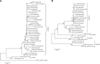
Sequence analysis of the partial F gene (372 bp) using the NCBI-BLAST (USA) server and MegAlign programs (DNAStar, USA) showed 100% identity with PPRV recovered from experimentally infected cattle (GenBank accession No. EF641264). Overall identities of 98% and 100% were observed with all Indian PPRV isolates, including that from Gujarat, irrespective of their origin. Among the isolates from Gujarat, the Patan/Gujarat/2005/sheep isolate had a high sequence identity (99%) to this isolate on comparison with F gene sequences. Similarly, analysis of the partial N gene sequence revealed a 98~99% identity with all Indian PPRV isolates and 99% identity with the Gujarat isolates. A phylogenetic tree based on a partial 372 bp sequences of the F gene (Fig. 1A) and 425 bp sequence of the N gene (Fig. 1B) was created using MEGA 4 [14] to demonstrate the relationships with other PPRV strains/isolates. This analysis showed clustering of all the Indian isolates together into a branch (lineage IV) separate from the Nigerian (lineage I) and ICV89 (lineage II) isolates as previously reported [5].
PCR assay was conducted to verify that the tissues were indeed from an Asiatic lion than from sheep or goats origins. The DNA recovered from the tissue samples of the Asiatic lion was subjected to Ple 46 locus based species-specific satellite marker PCR using Ple 46 Fwd and Ple 46 Rev primers as previously described [12]. The PCR products were separated on a 3% agarose gel (data not shown). Specific amplicons between 111~119 bp in size were observed, indicating that the samples were from an Asiatic lion/big cat species. The 10% triturated homogenate filtered supernatant was used to infect a 48-h-old confluent Vero cell (ATCC) monolayer in 25 cm2 flask as per standard protocols. The cells were monitored for the appearance of morbillivirus-specific cytopathic effects (CPEs). After a week of incubation, the infected cells were further passaged blindly and periodically examined for visible CPEs. Virus was successfully isolated (designated PPRVInd.Guj.Lion/2007) from the cells at passage 4 and identified by virus-specific RT-PCR.
For further molecular characterization, PCR amplification, sequencing, and sequence and phylogenetic analyses of the open reading frames (ORF) of structural genes (N, M, F, and H) from the virus isolated at passage 7 were carried out as previously described [5] to understand the genetic relationship with other PPRVs (GenBank accession No. JN632532~JN632535). Comparative sequence and phylogenetic analyses of the four gene (N, M, F and H) sequences from the lion isolate showed a close resemblance to Indian isolates and clustered into Asian lineage IV as previously reported [5]. Predicted amino acid sequence analyses of the N, M, F, and H genes also showed no significant difference in sequence alignment compared to other Indian Asian lineage IV isolates. In general, six of the seven residues in the H gene are presumed to be important for measles virus hemagglutinin-signalling lymphocyte activation molecule (SLAM) receptor interactions [6]. Along with the isolate from our study, these residues are conserved within the Nigerian and other PPRV isolates (Y529, D530, R533, F552, Y553, and P554) as reported earlier [6].
Percent identities of 97.7~99.8%; 98.8~100%; 99.4~99.9% and 99.5~99.8% were observed among the N, M, F, and H genes of Indian PPRVs respectively when analyzing the nucleotide sequences. Similarly, percent identities of 97.7~99.8%; 98.2~99.7%; 99.1~99.8%; and 99.0~99.7% were observed among the N, M, F, and H genes of Indian PPRVs respectively when analyzing the predicted amino acid sequences.
PPRV is a pathogen that infects small ruminants including ones in the wild, but PPRV seroprevalence has also been reported in other species [1]. Earlier studies on PPR indicated that it is enzootic in India [3,5]. Favorable climatic conditions may promote virus survival, spread of the virus, and distribution of seasonal outbreaks. Furthermore, the role of wildlife in the epizootiology of PPR has not been fully elucidated. In India, systematic attempts to isolate and characterize the virus from wild animals were seldom performed.
The occurrence of PPR in a subclinical form in cattle and buffaloes assumes epizootological significance [3]. In a similar manner, the detection of PPRV in the tissue samples from as Asiatic lion may be of significance. Detection of PPRV antigen/nucleic acids in tissues from the Asiatic lion was indicative of subclinical/inapparent infection. Such cases of infection could be due to close contact with other infected animals or contaminated fomites. The animal might have been seroconverted which has been reported for other infected animals [3] and could reveal new insight into PPRV epidemiology and transmission. In general, morbilliviruses have the propensity to adapt to new host species, which can be explained by the deterministic role of a conserved receptor (SLAM) and could be of paramount importance. Earlier studies on PPR showed that wild ruminants may play an important epidemiological role as a source of infection for domestic animals [10,15] and the reverse situation may also be possible. However, further random screening or methodical sero-screening, virus detection, and genome sequencing analysis of inapparently infected wild animals will help elucidate the disease prevalence among wild animals including ruminants.
To the best of our knowledge, this is the first report of detection and partial genetic characterization of PPRV isolated from Asiatic lion tissues. Our findings may provide insight into the emergence of PPR in a new species. Greater emphasis should be placed on continuous serological and clinical surveillance of PPR in wild ruminants to better understand the prevalence of PPRV, its impact on wildlife conservation, and the possible roles of different species in PPRV transmission.
 XML Download