

 PDF
PDF ePub
ePub Citation
 Print
Print


Many investigator demonstrated that a number of topical medications may influence the blood flow to the eye [9,10]. β-blockers and sympathomimetics may affect blood flow because an imbalance is produced between the influence of the α and β sympathetic receptors on the ocular vasculature. The effect of this imbalance has often been difficult to ascertain and differing results have been found in various studies [10]. In humans, numerous orbital and ocular vessels have been mapped and their blood velocity parameters and waveforms have been characterized by using color Doppler imaging (CDI) [1,5]. A few veterinary reports have evaluated CDI of the canine orbit and eye [7]. This study was performed to assess the effect of anti-glaucoma drugs on the RI of the medial long posterior ciliary artery (mLPCA) using CDI, and to determine which anti-glaucoma drug was effective in reducing the intraocular pressure (IOP) without any changes in choroidal vascular resistance.
Nine healthy Beagle dogs (three males and six females) weighing between 9.8 and 12.6 kg, with ages between 2- and 3-years old were used. All dogs were sexually intact and all examinations were applied to only the left eyes. Dogs were determined to be normal based on physical examination, complete blood count, serum biochemical analyses, and ophthalmic examinations. There were no signs of orbital diseases or neuro-ophthalmologic diseases in any dog. Animal care and experiments were carried out adhering to a guide for the care and use of laboratory animals from Gyeongsang National University, Korea.
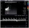
We used an ultrasonography (SSA-660; Toshiba, Japan) with a 10 MHz linear probe. Our technique for color Doppler imaging has been previously described [7]. Briefly, the left eyes and orbits were imaged with the dog lightly restrained in a sitting position. Coupling gel was applied to the region dorsal to the left zygomatic arch, and the transducer was positioned in a horizontal plane (Fig. 1). Baseline measurements were performed in a predetermined order (IOP, RI of mLPCA). Thereafter, one drop of the medication was topically administered into the left eye. Measurements were repeated 120 and 240 min after administration. Single doses of the following drugs were administered topically into the left eye with nasolacrimal occlusion: 0.5% levobunolol (Betagan; Allergan Pharmaceuticals, Ireland), 0.1% dipivefrin (Propine; Allergan Pharmaceuticals, Ireland), and 4% pilocarpine (Samil Pharm, Korea). All dogs (nine left eyes) were used for experiments involving each drug. Withdrawal periods between each drug were greater than 14 days. Tonopen-XL (Mentor, USA) was used to measure IOP. The mean of three IOP measurements was calculated.
Statistical analysis was performed using the SPSS statistical computer program (IBM, USA). A repeated measure ANOVA was used for data analysis. For all tests, p values < 0.05 were considered significant.
Effects of the anti-glaucoma drugs on RI values and IOP are presented in Table 1. All drugs under study relatively reduced IOP although the differences were not statistically significant. Topical administration of pilocarpine had no significant influence on the RI of mLPCA. However, levobunolol (post 4 h; p < 0.05) and dipivefrin (post 2 h; p < 0.05) significantly increased the RI of mLPCA following administration.
Like timolol, levobunolol is a non-selective β-blocker which lowers IOP, probably by decreasing the secretion of aqueous humor by ciliary epithelium [2]. Bloom et al. [2] suggested that levobunolol had a variable effect on human retinal circulation with a tendency to produce a slightly overall increase in flow. Another study has shown that ocular blood flow change was not observed with levobunolol in humans [9]. However, in our study, levobunolol increased mLPCA RI, suggesting a decrease in the velocity of blood flow in the treated eye. It is not certain why these differences were observed.
Dipivefrin directly acts as an α and β agonist. This drug, which is metabolized to epinephrine by corneal estrases, has been shown to reduce blood flow in the ciliary body in humans [8]. The direct vascular effect, together with the increase in uveoscleral outflow, may contribute to the intraocular pressure-reducing capability of dipivefrin [9]. Our data suggests that dipivefrin lowers choroidal blood flow. These results align with the finding of a slight decrease in choroidal blood flow after topical application of epinephrine in rabbits [6].
Theoretically, cholinergic miotics cause contraction of the longitudinal ciliary musculature and sphincter muscle of iris [3]. Ciliary muscle fibers are attached to the corneoscleral trabecular meshwork. Contraction of the ciliary muscle results in opening of the extratrabecular spaces of the corneoscleral trabecular meshwork and then increases drainage of an aqueous humor [4]. In our study, pilocarpine had no effect on ocular blood flow, which concurs with the results from a previous human study [9]. Another investigation reported using a radioactive microsphere technique with similar results in rabbits [6]. This group suggested that although the difference is not statistically significant, pilocarpine decreases blood flow to the iris and ciliary process. In our opinion, these trends may result from indirect compression by muscle contraction.
We found that some anti-glaucoma drugs could affect ophthalmic blood flow, even though it is not known whether these effects are detrimental to the eye. Further studies are under way to investigate which anti-glaucoma treatments lower IOP while increasing ophthalmic blood flow, and whether effects on ophthalmic vascular resistance are detrimental or beneficial to eye.

 XML Download