

 PDF
PDF ePub
ePub Citation
 Print
Print


Introduction
In cattle, the use of assisted reproductive technologies started around 1982 with the development of in vitro fertilization (IVF) [3], followed by the use of embryo transfer. Cloning in mammals began with the birth of a cloned sheep [24], but Bovidae is undoubtedly the most widely studied species and the one in which technology for somatic cell nuclear transfer (SCNT) is the most advanced. The first calves derived from SCNT were born in 1998 [6]. Subsequently, it can be estimated that up to the year 2004 about 1,500 SCNT calves have been produced worldwide by different research groups and companies, mainly in North America, Japan, New Zealand, and Europe but also in other countries including in ones in South America or Asia [8].
Since synthetic oviduct fluid (SOF) culture medium was invented [22], modifications of SOF have supported greater developmental competence of bovine embryos in vitro [5,10,15]. In order to improve developmental competence of blastocysts, protein sources such as bovine serum albumin (BSA) or fetal bovine serum (FBS) are generally added to the culture media but these may be associated with "large offspring syndrome" (LOS) [20,21,25], with potential risk of disease or transmission of pathogens [7]. As a way to overcome these problems and to evaluate the effects of individual culture medium components, chemically defined media were introduced for culturing IVF or SCNT embryos but result in low developmental competence [2]. Recently, developmental competence of IVF embryos cultured in chemically defined medium (CDM) was shown to be similar to that of embryos cultured in SOF media supplemented with BSA or FBS [16]. Accordingly, the objective of this study was to compare developmental competence of SCNT embryos cultured in modified synthetic oviduct fluid (mSOF) or CDM, and to investigate pregnancy rates, length of pregnancy, and body weights of cloned calves following the transfer of embryos cultured in these media.
Materials and Methods
In vitro maturation (IVM) of immature oocytes
An IVM protocol was followed as previously described [9]. Bovine ovaries collected from a local slaughterhouse were transported to the laboratory within 2 h of harvesting the ovaries in a 0.9% NaCl solution at about 35℃. The cumulus oocyte complexes (COCs) were retrieved from small antral follicles 3 to 8 mm in diameter by aspiration with an 18 gauge hypodermic needle attached to a 10 mL syringe and washed 3 or 4 times in HEPES-buffered tissue culture medium (TCM)-199 (Invitrogen, USA) supplemented with 10% FBS, 2 mM NaHCO3, 5 mg/mL BSA (Invitrogen, USA), and a 1% mixture of penicillin and streptomycin. COCs with evenly granulated cytoplasm and enclosed by more than three layers of compact cumulus cells were selected. A group of 30 to 40 COCs were cultured for maturation in one well of a multi-well dish containing 0.5 mL of bicarbonate-buffered TCM-199 supplemented with 10% FBS, 0.005 IU/mL follicle stimulating hormones (FSH; Teikoku Seiyaku, Japan) and 1 µg/mL 17β-estradiol at 39℃ in a humidified atmosphere of 5% CO2.
Donor cell preparation
Fibroblasts were isolated from bovine fetuses on day 45 of gestation. The head of the fetus was removed using iris scissors, and soft tissues such as liver and intestine were discarded after removing with 2 watchmaker's forceps. After washing three times with Dulbecco's phosphate buffered saline (DPBS; Invitrogen, USA), the carcass was minced with a surgical blade in a 100 mm culture dish (Becton Dickinson, USA). The minced fetal tissues were dissociated in Dulbecco's modified Eagle's medium (DMEM; Invitrogen, USA) supplemented with 0.25% trypsin and 1 mM EDTA (Invitrogen, USA) for 1 h at 37℃. Trypsinized cells were washed once in Ca2+- and Mg2+-free DPBS by centrifugation at 43 ×g for 2 min, and subsequently seeded into 100 mm plastic culture dishes. Seeded cells were subsequently cultured for 6 to 8 days in DMEM supplemented with 10% FBS (Invitrogen, USA), 1 mM glutamine (Invitrogen, USA), 25 mM NaHCO3, and a 1% minimal essential medium non-essential amino acid solution (Invitrogen, USA) at 39℃ in a humidified atmosphere of 5% CO2 and 95% air. After the removal of unattached clumps of cells or explants, attached cells were further cultured until confluent, subcultured at intervals of 4 to 6 days by trypsinization for 5 min using 0.1% trypsin and 0.02% EDTA, allocated to three new dishes for further passaging, and then stored in freezing medium in liquid nitrogen at -196℃. The freezing medium consisted of 80% DMEM, 10% dimethyl sulfoxide (DMSO) and 10% FBS. Cultured cells were used at passages 4 to 6 for SCNT. Prior to SCNT, cells were thawed, cultured for 3 to 4 days until 100% confluent for contact inhibition, and retrieved from the monolayer by trypsinization for 30 sec.
SCNT and cell number
After 22 h of maturation culturing, cumulus cells were removed by repeated pipetting in 0.1% hyaluronidase in HEPES-buffered TCM-199, and metaphase and first polar body were removed. The transfer of a fetal fibroblast into an enucleated oocyte was carried out as previously described [9]. Reconstructed embryos were fused, activated for 4 min with ionomycin followed by incubating for 4 h in 1.9 mM 6-dimethylaminopurine. Cloned embryos were cultured in 25 µL microdrops of mSOF or CDM overlaid with mineral oil for 8 days at 39℃ in an atmosphere of 5% O2, 5% CO2, and 90% N2. mSOF and CDM were prepared as previously described [9,16]. Cleaved embryos were observed at 24 h after culture, and on day 8 viable SCNT blastocysts were used for counting cell numbers by nuclear staining with bisbenzimide.
Embryo transfer and diagnosis of pregnancy
Embryo transfer and diagnosis of pregnancy were perfomed as previously described [9]. One or two mid- or hatching blastocysts with normal morphology in phosphate buffered saline supplemented with 20% FBS were transferred to one uterine horn of each recipient cow (n = 34) using a nonsurgical transcervical method on day 7 (estrus = day 0 = day of fusion). In order to determine embryo survival and pregnancy, cows were examined by rectal palpation on day 45 post-estrus. Pregnant cows were monitored by rectal palpation at regular 60 days intervals thereafter.
Microsatellite analysis
Parentage analysis was performed on cloned calves, surrogate cows, and nuclear donor cells to compare the genetic identity of the calves with that of the donor cells used for SCNT as previous report [9]. Genomic DNA from blood and trypsinized donor cells was extracted with the G-spin Genomic DNA Extraction Kit (iNtRON, Korea) according to the manufacturer's protocol. The isolated genomic DNA samples were dissolved in 50 µL TE and used for microsatellite assay with bovine specific markers (BM1824, BM2113, ETH10, ETH225, AGLA293, TALA122, TGLA73, and TGLA126) labeled with a fluorescent dye (FAM, NED or HEX) [9]. Polymerase chain reaction (PCR) amplification was carried out with one cycle for denaturing at 95℃ for 15 min, and 35 subsequent cycles with denaturing at 95℃ for 30 sec, annealing at 55℃ for 30 sec, and extension at 72℃ for 30 sec; and a final extension at 72℃ for 10 min. Ten µL of PCR product were separated on a 2.0% agarose gel and stained with ethidium bromide. Length variations were assayed by PCR amplification with fluorescent-labeled locus-specific primers and PAGE on an automated ABI 373 DNA sequencer (Applied Biosystems, USA). Proprietary software (GeneScan and Genotyper; Applied Biosystems, USA) was used to estimate PCR product size.
Experimental designs
Experiment 1 (Developmental competence and cell numbers of reconstructed embryos cultured in mSOF or CDM): Reconstructed embryos with fetal fibroblasts were randomly divided into two groups and were cultured in mSOF or CDM. On day 7, blastocysts were observed and on day 8 cells of the blastocysts were counted.
Experiment 2 (Production of cloned claves): On day 7, pre-implantation embryos with normal morphology from both media cultures were transferred into the uterine horns of naturally synchronized recipients. Pregnancy was assessed by non-return to estrus and rectal palpation.
Statistical analysis
Data were analyzed using a statistical analysis system program (Version 9.0; SAS, USA). Analysis of interaction among experimental parameters was first performed. Since no interaction was found, the data were subjected to analysis of variance and a protected least significant different test using general linear models to determine differences among experimental groups. When a significant model effect was found for each experimental parameter, data were compared by the least squares method. Statistical significance was determined where the p-value was less than 0.05.
Results
Developmental competence and cell number of reconstructed embryos cultured in mSOF or CDM
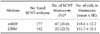
SCNT embryos were cultured in mSOF or CDM, and development to the blastocysts stage was 26.6% vs. 22.5%, respectively. Nuclear staining showed that blastocysts derived in mSOF and CDM had a mean of 118.6 ± 12.2 cells and 111.7 ± 15.1 cells, respectively (Table 1).
Production of cloned claves
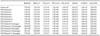
We further investigated the effect of mSOF and CDM on production of cloned calves from transferred SCNT embryos. As shown in Table 2, a total 67 pre-implantation stage embryos derived in mSOF or CDM were transferred into 34 recipient cows. Survival rates on day 60 and term after transfer the embryos cultured in mSOF were 29.6% (to recipients) and 18.5% (to recipients), respectively. In contrast, 28.6% and 42.9% of the 15 embryos transferred after culture in CDM resulted in pregnancy on day 60 and at term, respectively (Table 2). Although a statistical difference was not found, survival of transferred blastocysts on the live cloned calves rates was lower in the mSOF group [18.5% (to recipients), 9.6% (blastocysts)] than in the CDM group [42.9% (to recipients), 20.0% (blastocysts)].
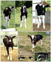
In addition, two pregnant recipient cows carrying mSOF-derived embryos aborted at 4 and 6 months, respectively; the latter was pregnant with twin fetuses. Cloned twin calves derived from SCNT embryos cultured in CDM were prematurely delivered with the assistance of a veterinarian at 264 days post-embryo transfer; however, they died from severe respiratory distress. A total of six cloned calves including a pair of twins were normally born by caesarean section, or assisted or natural delivery (Fig. 1 and Table 3).
Ranges of gestation length and body weight of the cloned calves were 270~280 days and 29~53 kg, respectively (Table 3). Due to severe diarrhea, one twin derived from mSOF embryos died 1 month after birth. Microsatellite DNA analyses examining eight loci confirmed that all the calves were genetically identical to the donor fetal fibroblasts used for SCNT. Additionally, the cloned calves were not genetically related to the respective recipient cows (Table 4).
Discussion
Increased efficiency in cloning cattle will enhance application of technology for assisting large animal production and provide a critical foundation for creating transgenic animals used for cell therapy and biopharmaceuticals [17]. In this study, we demonstrated that cloned embryos produced by SCNT and cultured in mSOF or CDM showed equivalent developmental competence in vitro, and both groups of embryos resulted in the production of viable cloned calves. In an effort to improve the development of bovine blastocysts in vitro, there have been many trials using somatic cell co-culturing, cell-conditioned media, or complex media containing serum [1]. Among these studies, mSOF culture medium containing BSA or FBS is widely used for producing bovine embryos. Our previous studies show that blastocysts cultured in mSOF containing BSA or FBS support the birth of cloned calves after embryo transfer [4,5]. However, due to concerns about LOS or transmission of unknown pathogens, several studies used chemically defined culture media without FBS or BSA but reported low developmental competence, cell numbers, and calving rates [2,5,13,14,19]. In the present study comparing the developmental competence of embryos, no difference was found between ones cultured in mSOF containing BSA or CDM. In addition, blastocyst cell number, which is considered to be an indicator of viability, was not different between blastocyts from mSOF and CDM cultures, indicating that CDM was not inferior to mSOF containing BSA in this study.
Although it is believed that improved in vitro culture conditions by supplementing with vitamins, growth factors, and hormones can support higher embryo developmental competence, cell numbers, or calving rates, this is not always the case. For instance, Choi et al. [5] reported that culture media supporting the highest embryo developmental competence in vitro did not yield the most efficient production of live cloned calves. We thought that the survival of transferred blastocysts until term and the birth of normal live cloned calves are critical factors in improving efficiency of cloning. CDM, which has been used for producing in vitro fertilized embryos that subsequently develop into live calves [16], increased developmental competence of SCNT embryos to a level similar to that obtained with mSOF. Additionally, transferred SCNT embryos derived in CDM survived to term with higher calving rates compared to ones derived in mSOF even though the number of surrogate mothers was limited. Since CDM induces changes in the expression of implantational-related genes in vitro in fertilized embryos results in increased calving rates [16], we can infer the same effect on the SCNT embryos derived from CDM was observed in our study as well.
In contrast to all pregnant cows that received embryos derived in CDM maintained to term and produced live cloned calves, 50% of the pregnant cows that received SCNT embryos from the mSOF group produced live cloned calves. Even though number of recipient cows was low and there was no significant difference, we believe the CDM used in this study is better for producing live cloned calves. Moreover, CDM did not contain any toxic components or unknown pathogens, or a wide variability of unidentified components that are commonly present in BSA and FBS [11,12]. Therefore, this medium may be more useful for generating healthy offspring from cloned embryos or ones produced in vitro. However, we did not observe LOS in cloned calves derived from embryos cultured in either medium.
While microsatellite DNA analyses were consistent in showing that the cloned calves were genetically identical to the donor fetal fibroblasts, there were differences in the coat color patterns among the nine cloned calves. Piebald coat patterning appeared not to be under absolute genetic control. Environmental influences in utero have been found to induce a degree of variability in the multiplication and migration of melanoblasts which form the melanocytes necessary for pigment production in the developing skin of the fetus [18,23].
In conclusion, our study demonstrated that transferred SCNT embryos cultured in CDM had higher survival rates and resulted in more births of cloned calves compared to blastocysts derived from mSOF. Furthermore, our results demonstrated that the CDM system may support the production of healthier cloned calves without BSA and FBS which contain pathogens or unidentified factors. Our findings will help advance research in assisted reproductive technologies and lead to the development of more efficient livestock propagation techniques.


 XML Download