

 PDF
PDF ePub
ePub Citation
 Print
Print


Introduction
Influenza A virus infects different animal species including humans, dogs, pigs, and a wide variety of domesticated and wild birds [10,14]. Influenza virus infections are mediated by specific interactions between the viral hemagglutinin (HA) and sialic acid (SA) residues on the host cell surface. The influenza virus specifically recognizes oligosaccharides of the respiratory epithelial cell receptors containing a terminal SA linked to galactose (gal) either by an α-2,6 linkage (SA α-2,6-gal) or SA α-2,3-gal moieties [12].
Host cell receptors are important determinants of virus entry. Differences in receptor distribution and receptor specificity among hosts could account for variations in susceptibility to infection [10]. It has been generally accepted that the majority of avian influenza viruses bind to receptors with SA α-2,3-gal, while human viruses preferentially bind to SA α-2,6-gal [10]. It is assumed that differences in the ability to bind cell surface receptors will, at least in part, prevent a massive influenza outbreak, which has not occurred.
Genomic mutations of avian influenza viral HA have been known to change the binding preference from SA α-2,3-gal to SA α-2,6-gal-linked receptors, which leads to extending the viral host range [12]. One possible way this may occur is through infection of animals such as pigs and some terrestrial poultry that have both types of receptors [7]. An increasing number of reports showing that several influenza viruses are established in land-based poultry [1,2,11] suggest the potential role of some avian species as "mixing vessels" which provide a proper environment for re-assortment of influenza viruses in birds and mammals. Therefore, clear understanding of the expression and distribution of influenza virus receptors in various avian species has become an important issue that could further explain interspecies transmission of influenza A viruses.
Previous studies have demonstrated the presence or absence of receptors in various poultry including chickens, ducks, and quails by lectin histochemistry [9,13,15]. However, some major discrepancies in receptor distribution were observed among those studies, warranting additional investigation to validate previous results. Moreover, it is of great interest to compare specific receptor densities in the respiratory tract of several intermediate hosts since the presence or absence of receptors may play key roles in generating viruses with pandemic potential. Therefore, we sought to identify the types of sialyloligosaccharides at influenza virus replication sites in representative land-based poultry such as chicken, duck, quail, and pheasant.
Materials and Methods
Specific pathogen-free (SPF) white leghorn chickens (Gallus gallus, 8 weeks old) were purchased from Namduk SPF (Korea). Pekin ducks (Anas platyrhynchos domestica, 7 weeks old), quails (Coturnix coturnix, 8 weeks old), and ring-necked pheasants (Phasianus colchicus, 8 weeks old) were obtained from commercial breeders in Korea, where they were raised for eggs or meat. At least five animals from each species were included in this study. All procedures for animal use were approved by Institutional Animal Care and Use Committee at Konkuk University. Animals were euthanized by cervical dislocation. Fresh upper, middle, and lower regions of the trachea were collected separately and a piece of lung tissue was also taken from the right lung. These samples were fixed in 4% paraformaldehyde immediately after sample collection then subjected to a routine paraffin-embedding process. The blocks were cut into 6 µm-thick sections using a microtome (Leica Biosystems, Germany).
Lectin histochemistry was performed using previously described procedures with some modifications [9,16]. Prior to lectin histochemistry, the sections were quenched with 0.3% hydrogen peroxide for 15 min and blocked with 5% bovine serum albumin (BSA) in phosphate buffered saline (PBS) for 1 h at room temperature. To detect influenza receptors, the sections were incubated with biotinylated Maakia amurensis lectin II (1.3 µg/mL; Vector, USA), which is specific for α-2,3-gal-linked SA, and biotinylated Sambucus nigra lectin (2.6 µg/mL; Vector, USA) specific for α-2,6-gal-linked SA. Each lectin was diluted in 1% BSA/PBS and the sections were incubated in both for 2 h at 37℃. The sections were washed with PBS then subsequently incubated with horseradish peroxidase conjugated streptavidin (0.4 µg/mL; Vector, USA) in PBS for 1 h at room temperature. The lectin signal was detected by using diaminobenzidine and hydrogen peroxide as substrates. Stained sections were further counterstained with methyl green then dehydrated, mounted, and evaluated with a light microscope. Negative control staining was carried out either by omitting the lectin incubation or by pre-incubating the tissue sections with recombinant neuraminidase cloned from Clostridium perfringens (12.5 U/µL; New England Biolabs, USA) at 37℃ for 24~48 h to remove oligosaccharides from the influenza receptors [3]. All the sections from four avian species were processed under identical lectin staining conditions to compare staining intensity. After thorough examination of each section, the relative intensity of receptor expression was categorized into six grades: no signal at all (-), extremely weak signal (+/-), weak signal (+), medium signal (++), slightly strong signal (+++), strongest signal (++++). The intensity grades were assigned to compare receptor expression patterns at a cellular level as well as regional expression differences within the respiratory tract among the various species.
Results
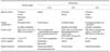
Lectin histochemistry revealed striking differences in the receptor expression patterns of chickens, ducks, pheasants, and quails. Each species showed cell-specific and/or regional differences in receptor densities. When examining receptor expression, we mainly focused on the primary cells that are in contact with influenza viruses. The main observations are summarized in Table 1, and the major differences in lectin staining procedures between our study and previous studies are summarized in Table 2.
When comparing signal intensities among species, pheasants and quails showed a higher expression of SA α-2,3-gal-linked receptors than chickens and ducks. The expression of SA α-2,3-gal-linked receptors was minimal in the lungs of all species examined (Fig. 1). On the other hand, the expression of SA α-2,6-gal-linked receptors was highest in quails, intermediate in chickens and pheasants, and lowest in ducks. Unlike lung expression of SA α-2,3-gal-linked receptors, relatively high expression of SA α-2,6-gal-linked receptors was observed in both pheasant and chicken lung atria (Figs. 1 and 3).
In order to determine the specificity of the lectins used in our study, a control experiment was carried out by pre-incubating tissue sections with neuraminidase, which cleaves both α-2,3-gal and α-2,6-gal residues. Neuraminidase pre-treatment effectively abolished lectin signals for α-2,3-gal-linked receptors in all the species tested (Fig. 2). Neuraminidase pre-treatment also reduced lectin signals for α-2,6-gal linked receptors in chickens, ducks, and pheasants while weak but negligible signal was observed in quails (Fig. 4).
Regional differences in receptor distribution along the respiratory tract were also observed in chickens and pheasants. The number of SA α-2,6-gal-linked receptors increased toward the lower part of the respiratory tract in chickens and pheasants (Fig. 3). Cell-specific variations in receptor types in the trachea were also evident. While chickens, ducks, and pheasants showed more influenza receptors in ciliated tracheal epithelia, quails showed higher receptor expression in non-ciliated mucin-producing cells (Fig. 2).
In chickens, some ciliated epithelial cells were stained positive for SA α-2,3-gal- and SA α-2,6-gal-linked receptors in the upper and middle trachea, whereas considerably strong staining was observed in most of the ciliated epithelial cells in the lower trachea. No signal was found in non-ciliated epithelial cells. An extremely weak signal for SA α-2,3-gallinked receptors was observed in the lung while medium intensity signal for SA α-2,6-gal-linked receptors was also observed (Fig. 4).
In ducks, only a few ciliated epithelial cells stained positive for SA α-2,3-gal- and SA α-2,6-gal-linked receptors in the upper, middle, and lower trachea. No signal was found on non-ciliated epithelial cells. Few cells were positive for both SA α-2,3-gal- and SA α-2,6-gal-linked receptors in the lung.
In pheasants, the majority of ciliated epithelial cells in the entire trachea stained positive for SA α-2,3-gal- and SA α-2,6-gal-linked receptors (Fig. 3). Staining intensities on ciliated cells were somewhat higher than non-ciliated cells throughout the respiratory tract. Extensive staining of SA α-2,6-gal-linked receptors was observed throughout the air sacs in lung (Figs. 3 and 4). While most cells were positive for SA α-2,6-gal-linked receptors in the lung, almost no signal for SA α-2,3-gal-linked receptors was observed.
In quails, strong staining for SA α-2,6-gal-linked receptors was observed in ciliated and non-ciliated epithelial cells in the upper, middle, and lower trachea (Fig. 4) while SA α-2,3-gal-linked receptors were found mainly in a large proportion of non-ciliated epithelial cells (Fig. 2). It is interesting to note that all parts of the pheasant and quail respiratory tract were positive for both SA α-2,3-linked and SA α-2,6-linked receptors in the tracheal epithelia. These findings are in accordance with Wan and Perez [15], who reported the presence of SA α-2,3-gal-linked receptors on non-ciliated cells and SA α-2,6-gal-linked receptors on ciliated cells in the quail trachea.
Discussion
The present study provides information on the influenza virus receptor distribution in the respiratory tract of four avian species, including pheasant which has not been previously studied. Understanding receptor distribution patterns in the respiratory tract is particularly important because initial virus-host interaction takes place on host cell surface receptors. We undertook this study to verify some of the results from previous studies that contradict one another. In addition, our comparative study with four different avian species generated useful information on host sensitivity to influenza viruses. It may also provide insight into the potential roles of these species as mixing vessels for genetic re-assortment of influenza viruses. When we carried out this study, we divided the respiratory tract in four different regions and carefully observed receptor expression in ciliated versus non-ciliated epithelial cells. This is because it is likely that ciliated epithelial cells in the upper trachea may get exposed to influenza virus more frequently than non-ciliated epithelial cells in the lower respiratory tract.
Since chickens and ducks are the major species of poultry raised all over the world, it was meaningful to revisit receptor distribution in these birds. Previously, Wan and Perez [15] showed in chickens that SA α-2,6-gal-linked receptors were observed only in a small number of non-ciliated epithelial cells, whereas high levels of SA α-2,3-gal-linked receptors was found in both ciliated and non-ciliated cells. This pattern was in contrast to the study by Kuchipudi et al. [9] which showed that the SA α-2,6-gal-linked receptor is the dominant receptor type in chicken trachea. Our results support the Kuchipudi study [9] as we also observed greater SA α-2,6-gal-linked receptor expression in the ciliated epithelium throughout the chicken respiratory tract. However, the most recent report by Pillai and Lee [13] demonstrated that SA α-2,3-gal-linked receptors are more frequently observed in chicken trachea. These discrepancies may stem from the fact that different staining conditions were employed in each study in terms of tissue preparation, source of lectins, lectin concentrations, and incubation environment. Thus, it may not be appropriate to directly compare previously published results with ours.
To achieve optimum staining, it was not possible to use same concentration of Maakia amurensis and Sambucus nigra lectin. Thus, direct comparison between SA α-2,3-gal- or SA α-2,6-gal-linked receptors in a given specie may not be accurate unless the same lectin concentrations are used to visualize both types of receptors. Two studies [9,13] used the same lectin concentrations to stain for both types of receptors; therefore, their results may represent true differences in receptor density. It also should be taken into consideration that lectins from different suppliers may show different binding specificities;. In particular, the source of Maakia amurensis has been shown to significantly affect binding specificity [9,12]. Moreover, isotypes of Maakia amurensis lectin may affect staining outcomes in different manners [8]. We used Maakia amurensis II lectin because the signal from this lectin is much more abundant in chickens and ducks [9]. Thus, we chose to use Maakia amurensis II lectin for examining the rest of the species we tested.
It is believed that ducks are relatively resistant to land-based poultry-adapted influenza virus infection via the respiratory tract [4]. This may be due to a scarcity of α-2,6-gal-linked receptors in duck respiratory tract. It was shown that duck trachea lacks SA α-2,6-gal-linked receptors using a virus binding assay [4]. Kuchipudi et al. [9] also reported that SA α-2,6-gal-linked receptor expression is very low in duck trachea. Only a few ciliated epithelial cells stained positive for SA α-2,3-gal- and SA α-2,6-gal-linked receptors in the upper, middle, and lower trachea in ducks. No signal was found on non-ciliated epithelial cells in ducks. In contrast, different results have been reported by Pillai and Lee [13] who demonstrated strong expression of SA α-2,3-gal-linked receptors in duck trachea using Maackia amurensis lectin. They also showed that both SA α-2,3-gal-linked and SA α-2,6-gal-linked receptors are highly expressed in duck respiratory tracts. This contrasting result could be due to the use of different lectin isotypes or lectins supplied by different companies. Different specificity of Maackia amurensis lectins has been previously reported. While Maackia amurensis I is specific for SA α-2,3-galβ(1-4)GlcNAc, Maackia amurensis II is specific for SA α-2,3-galβ(1-3)GlcNAc [8]. The isotype of lectin used by Pillai and Lee [13] was not specified so it is uncertain why different results were obtained for α-2,3-gal-linked receptors in ducks.
One of the most interesting observations made in our study was the abundant receptor expression in both pheasants and quails. In pheasants, we observed the continuous expression of both SA α-2,3-linked and SA α-2,6-linked receptors on ciliated epithelial cells in all parts of the respiratory tract including lungs. Quails also highly expressed both SA α-2,3-linked and SA α-2,6-linked receptors, primarily on non-ciliated epithelial cells. Carrying both types of receptors throughout the entire respiratory tract of these birds increases their chance of being infected with both human and avian influenza viruses. Wan and Perez [15] already pointed out the importance of quails as a potential intermediate host, and here we report that pheasants are another species that provides a proper environment for viral replication of various influenza subtypes. Pheasants are known to be an ideal carrier of influenza viruses and are capable of producing genetic variants of the influenza virus [5,6]. Our findings explain, at least in part, why quails and pheasants can serve as mixing vessels for influenza viruses. Although the portion of pheasants and quails are relatively small in poultry industry in Korea, they are frequently raised in other Asian countries. Therefore, their substantial roles in interspecies transmission of influenza are far greater than what we previously believed.


 XML Download