

 PDF
PDF ePub
ePub Citation
 Print
Print


Introduction
Rabies is an almost invariably fatal viral disease in animals and humans. According to the World Health Organization, rabies infections result in approximately 55,000 human deaths worldwide annually [23]. More than 29,000 human deaths from rabies were reported in Far East Asia including Thailand and China in 2006. However, there were no cases of human rabies reported in some countries such as Japan, Singapore, Korea, Malaysia and Taiwan in 2006 [5]. Rabies is transmitted by vector species such as bats, red foxes, skunks, raccoon dogs, dogs, wolves, and mongooses, depending on the country [9,12]. Dogs were known to be the main vectors for the transmission of rabies virus (RABV) to humans or other animals in Korea prior to 1980. Raccoons such as Nyctereutes procyonoide and Procyon lotor have been involved in RABV circulation in eastern Europe and the eastern United States since the late 1990s [2,13,23].
RABV belongs to the genus Lyssavirus in the family Rhabdoviridae and consists of a non-segmented negative single-stranded RNA genome that encodes five structural proteins: nucleoprotein (N), phosphoprotein, matrix protein, glycoprotein (G), and large protein [12]. Recent advances in technology have contributed to better understanding of the molecular epidemiology and geographic relationships of RABV isolates [20,21,26]. The nucleoprotein, which contains antigenic sites such as NI and NIII, is associated with encapsidation of the genomic RNA and the formation of an active cytoplasmic ribonucleoprotein complex that is essential for viral replication [25]. The N gene or protein has been targeted for diagnosing wild rabies using RT-PCR or indirect fluorescent antibody tests because the gene is highly conserved and the protein is produced in large quantities in infected brain tissue. Moreover, the nucleotide sequence of the N gene has been used extensively as a molecular marker to explain the patterns of the geographic distribution of RABV at the regional and global levels [20]. The glycoprotein plays an important role in attachment of the virus to the host cell surface, pathogenicity, immunogenicity, and neurovirulence [6,14,19]. Additionally, the RABV G gene has been used as another target for studying genetic diversity and antigenic typing because it contains several major antigenic sites designated as I~VI, 'a', and G1 on the ectodomain (1~439 amino acids) and is related to pathogenicity.
In Korea, the first case of rabies was reported in 1907, and a number of rabies cases were identified nationwide until 1945 [8]. After implementing an effective RABV control program using live and inactivated vaccine, the number of rabies cases decreased dramatically to an average of 32 cases per year by 1984, and there were no reports of rabies between 1985 and 1992 [7,11]. However, rabies was identified in a dog in Gyeonggi-do Province in 1993. Since then, a few rabies cases have been reported annually. Although a renewed national RABV control program involving the distribution of bait vaccine was implemented in early 2000, rabies still occurs in some regions of Korea. Korean RABV isolates collected between 1998 and 2004 have been identified as genotype I and found to be closely related to the Arctic lineage virus, but distantly related to other Asian strains including Chinese and Sri Lanka strains [8,16]. Therefore, we analyzed the N and G gene diversity of the recent strain of RABV circulating in Korea as well as the correlation among rabies genes and phylogenetic relationships. The genetic characterization of RABV is considered important for developing diagnostic and preventive measures, including a more effective vaccine. In this study, we cloned and sequenced the N and G genes of 11 RABV isolates obtained between 2008 and 2009 in Korea and analyzed the phylogenetic relationships of the isolates with other available sequences retrieved from GenBank.
Materials and Methods
Samples
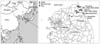
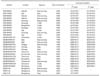
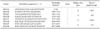
Eleven RABV isolates were obtained from brain samples that had tested positive for rabies using indirect immunofluorescence with monoclonal antibody against the N gene in Gangwon-do Province of Korea between 2008 and 2009. Five isolates (KRVR0801, KRVR0803, KRVR0804, KRVR0901, and KRVR0906) originated from raccoon dogs (Nyctereutes procyonoides koreensis), while another five isolates (KRVB0902, KRVB0903, KRVB0904, KRVB0905, and KRVB0907) were from cattle, and one (KRVC0802) was from a dog. These Korean RABV isolates are described in Fig. 1 and Table 1.
Extraction of viral RNA and RT-PCR
Viral RNA was extracted from the 11 isolates using an RNA extraction kit (Qiagen, Germany) according to the manufacturer's instructions. The extracted RNA was eluted in 50 µL of RNase- and DNase-free water. RT-PCR was conducted using primers specific for the N and G regions of RABV (RVN1F, RVN1R, RVN2F, RVN2R, RVG1F, RVG1R, RVG2F, and RVG2R; Table 2). RT-PCR was conducted in a reaction mixture containing 10 µL of denatured RNA, 1 µL of each primer (50 pmol), 10 µL of 5 × buffer (12.5 mM MgCl2), 2 µL of dNTP mix, 2 µL of enzyme mix (reverse transcriptase and Taq polymerase), and 24 µL of distilled water (Qiagen, Germany). The cycling profile consisted of cDNA synthesis at 42℃ for 30 min, followed by 35 cycles of 95℃ for 45 sec, 50℃ for 45 sec, and 72℃ for 1 min, with a final extension at 72℃ for 5 min. The PCR products were visualized using electrophoresis on 1.8% agarose gels containing ethidium bromide.
Cloning and sequencing
All of the PCR products that were purified using the gel extraction kit were ligated with pGEM-T easy vector (Promega, USA) according to the manufacturer's protocol. Plasmid DNA was isolated from amplified Escherichia coli (DH5α), and recombinant plasmids were identified by EcoRI enzyme digestion (Bioneer, Korea). The sequences of the purified plasmids were analyzed using an MJ Research PTC-225 Peltier Thermal Cycler and ABI PRISM BigDye Terminator Cycle Sequencing kits with AmpliTaq DNA polymerase (FS enzyme; Applied Biosystems, USA) according to the manufacturers' protocols. Single-pass sequencing was conducted for each template using universal primers (e.g., SP6 and T7). The fluorescent-labeled fragments were purified from the unincorporated terminators using an ethanol precipitation protocol. The samples were resuspended in distilled water and subjected to electrophoresis in an ABI 3730xl sequencer (Applied Biosystems, USA). Both DNA strands were sequenced to verify the sequences.
Phylogenetic analysis
The nucleotide sequences, accession numbers, and names of the strains used for the phylogenetic analysis in this study were obtained from the GenBank database. Nucleotide sequence similarity calculations were conducted using the DNASIS (Hitachi Software, Japan) software. Individual sequences were initially aligned using BioEdit and Clustal X 1.81. Phylogenetic reconstructions were generated using the neighbor-joining method by the computer program PHYLIP 3.572c. Phylogenetic trees were reconstructed on aligned nucleotide sequences using ClustalW (version 2.0.12; European Bioinformatics Institute, UK). The robustness of the phylogenetic analysis was determined by bootstrap analysis with 1,000 replications. Graphic output was produced by TreeView (version 1.6.6; Institute of Biomedical and Life Science, UK).
Results
Phylogenetic analysis of the complete N gene sequences
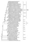
A total of 1,350 bp nucleotide sequences of the complete N gene encoding the nucleoprotein from 11 isolates were determined, and their amino acids sequences were deduced. The N gene sequences of 51 other RABVs obtained from GenBank were compared with those of the 11 Korean isolates to analyze molecular epidemic relationships. The nucleotide sequence analysis showed that all of the Korean isolates were classified into genotype I of Lyssavirus, and the nucleotide similarity among the 27 Korean isolates ranged from 98.1 to 99.8% (Fig. 2). The highest sequence similarity of the 11 Korean isolates with non-Korean RABV corresponded to the Chinese strains NeiMeng1025B and NeiMeng927 (96.1 and 96.7% at the nucleotide level and 98.8 and 99.5% at the amino acid level, respectively). The nucleotide similarity of the Korean isolate (KRVB0902) with Russian strain (857r) and Sri Lanka strain (SRL1145) were 95.7 and 86.8%, respectively [13]. The lowest nucleotide similarity of the Korean isolates with 35 non-Korean RABV strains was with the 99ABV3306 strain ranging from 82.7 to 82.9%. A phylogenetic analysis based on the N gene classified the Korean isolates into four subgroups (Gangwon I, II, III and Gyeonggi) with high similarity (Fig. 2).
Phylogenetic analysis of the complete G gene sequences
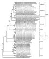
The 1,575 bp nucleotide sequences of the G gene encoding the glycoprotein from the 11 RABV isolates were identified and their amino acids were also deduced. The nucleotide sequences of the 11 Korean isolates were compared with those of 42 RABVs retrieved from GenBank to analyze the evolutionary relationships of RABVs. The phylogenetic tree based on the nucleotide sequence analysis of the G gene revealed that all of the Korean isolates were also divided into four groups (Gangwon I, II, III and Gyeonggi) and were most closely related to the Chinese strains designated as Chinese I in Fig. 3, similar to the results for the N gene. The similarity of the G gene nucleotide sequences of RABV ranged from 97.9 to 99.3% among all Korean isolates and 95.4 to 96.2% between Korean isolates and Chinese strains such as NeiMeng927A. Nucleotide similarity between the Korean isolates and Arctic strain ranged from 89.6 to 90.6% and comparison of Korean isolates with the NY516 and USA7-BT strains showed 78.4 and 78.9% similarity, respectively. Alignment of the deduced amino acid sequences revealed 98.3 to 99.8% similarity among the 24 Korean isolates.
Comparison of the proteins coding the N and G genes
The deduced amino acid sequences of the nucleoprotein and glycoprotein were compared to analyze the genetic variability among Korean RABVs. Antigenic sites NI (374~383) and NIII (313~337) and the phosphorylation site (389) mapping to serine were conserved in the 11 Korean RABVs. Comparison of the nucleoprotein among the 11 Korean RABVs revealed eight amino acid substitutions (data not shown). The deduced amino acid sequence demonstrated that RABV circulating in Korea consisted of the SP (-19~1), Ecto (20~439), TM (440~461), and Endo (462~524) domains. Among the 524 residues in the deduced amino acid sequences of the G gene were 38 substitutions among 24 Korean RABV isolates (data not shown). The arginine at position 333 of the residues within antigenic site III, which is essential for virulence, was not changed in the 11 Korean isolates (data not shown). All 11 Korean RABV isolates had two N-glycosylation sites at amino acid positions 37 and 319 of the ectodomain of the glycoprotein.
Discussion
Since rabies recurred in the northern part of Korea in 1993, many RABV cases have been reported in Gyeonggi-do and Gangwon-do Provinces [8,11]. According to national data regarding rabies, no cases were reported in Gyeonggi-do Province in 2009, whereas 18 rabies cases were reported in Gangwon-do Province. These findings suggest that our efforts to prevent rabies should be focused on limited regions.
Among the 11 Korean RABV isolates used in this study, five were obtained from domestic cattle and one was obtained from a dog infected by contact with rabid raccoon dogs, while five isolates were obtained from naturally infected raccoon dogs. Although various vector species of RABV are known, the raccoon dog has been the main carrier between domestic and wild animals in Korea. Raccoon dogs, which are the only canids known to hibernate in winter, were introduced to Far East Asia from Russia for the production of fur and pelts in 1928 [1]. However, the raccoon dog industry decreased due to the rise in popularity of silver foxes and goats. Raccoon dogs eventually escaped from fur farms and became wild animals in Korea. The Korean raccoon dog population increased until 2007 due to lack of predators. However, in 2008, the population of raccoon dogs decreased in Gyeonggi-do Province due to various parasitic diseases involving Demodex spp., tremadoes, cestodes and rapid urbanization of the region.
As advanced technology expanded, the nucleotide sequences of the RABVs circulating in various regions of the world have been submitted to several genetic information banking systems [8,16,18]. Using recent genetic information, we determined the nucleotide sequences and conducted a phylogenetic analysis of the N and G genes of Korean isolates obtained between 2008 and 2009 to understand the evolution of RABV in Korea. The genetic relationships among various RABV strains have been described elsewhere [10,15,26]. Analysis of the N and G genes provided similar results, suggesting that either gene can be used for phylogenetic analysis. Comparing the nucleotide similarity with non-Korean isolates, the Korean RABV isolates formed a close phylogenetic relationship with the NeiMeng1025B strain, which was isolated from a naturally RABV-infected raccoon dog in Jilin Province of China and with 857r strain, which was isolated from Chabarovsk of Russia and classified into group B [13]. However, it was only distantly related to other Asian RABV strains (8764THA, 9702INDI and SRL1145). The phylogenetic analysis revealed that the Korean isolates belonged to four subgroups (Gangwon I, II, III and Gyeonggi) that shared high homology, indicating that the genetic features of rabies are related more to geographic relationships and isolation year than to the species infected [2]. A previous study showed that Korean isolates obtained from 1998 to 2004 had the closest relationship with Arctic-like virus, such as Canadian strains [8,16]. The results of this study indicate that RABV in Korea was transmitted from the northeastern region of China.
The deduced amino acid sequences of the N and G genes of the 11 RABV isolates were aligned to investigate the antigenic variation in RABV, as reported earlier [17]. The genetic analysis of the N and G genes of the 11 Korean isolates showed that all of the antigenic sites were conserved, and the putative phosphorylation site at amino acid position 389 in the N gene was also conserved. It is well known that the arginine or the lysine at residue 333 on the ectodomain of glycoprotein are essential for neurovirulence within antigenic site III, and that virus variants that substitute glutamine, isoleucine, and glycine at that position show less phathogenic or avirulent symptoms [3,19,22]. Genetic analysis of the compared Korean RABVs showed no substitutions at these positions. Therefore, the Korean isolates recently circulating in Korea are pathogenic in several hosts such as dogs and cattle and have the ability to infect neural cells, such as neuroblastoma NG-108 cells [4]. Wunner et al. [24] reported that glycosylation is essential for complete folding and there are three or four putative glycosylation sites on the glycoprotein depending on the virus strain. The 11 Korean isolates had two putative N-glycosylation sites (Asparagine-X-Serine or Asparagine-X-Threoine) at amino acids 37 and 319 within the ectodomain, indicating the existence of antigenic variants of RABV with altered glycosylation in rabies viruses.
In conclusion, our results indicate that Korean RABV isolates are closely related with northeastern Asian RABV strains based on a phylogenetic analysis. Based on these findings, raccoon dogs play an important role as a vector species for rabies and raccoon dog movement could be responsible for the distribution of RABV. Current strategies to prevent rabies in Korea should be reconsidered and new tactics such as point infection control and trap-vaccinate-release programs should be reviewed to eradicate rabies. Application of bait vaccine for wild animals should also be scaled up around the place of outbreaks and genetic surveillance of RABV circulating in Korea is needed to monitor the epidemiological status of rabies in Korea.
 XML Download