

 PDF
PDF ePub
ePub Citation
 Print
Print


Introduction
Escherichia (E.) coli is a major causative agent of urinary tract infection (UTI); these strains are designated uropathogenic E. coli (UPEC) and may be considered to represent a subgroup of extraintestinal pathogenic E. coli (ExPEC). UTI occurs not only in humans but also in companion animals such as dogs and cats, and has been associated with urogenital disease such as cystitis, nephritis, and prostatitis [25]. The main virulence determinants of UPEC isolated from dogs and cats include type 1 fimbriae (fim), pilus associated with pyelonephritis (pap), S fimbriae (sfa), afimbrial adhesion (afa), α-hemolysin (hly), aerobactin, cytotoxic necrotizing factor 1 (cnf1), and cytolethal distending toxin (cdt) [7]. In particular, the combination of virulence genes that appears with high frequency includes papC, sfa, hlyA, and cnf1 [8,27].
Phylogenetic studies showed that E. coli strains can be assigned to one of four main phylogenetic groups: A, B1, B2, and D [15,24]. Most ExPEC, including those with the most robust virulence factor repertories and those which are best able to infect non-compromised hosts, are derived from phylogenetic group B2 [10]. Group D contains the second highest number of ExPEC; extraintestinal isolates from this group typically have somewhat fewer virulence factors and a different mix of virulence factors than group B2 isolates. E. coli strains belonging to groups A and B1 do not frequently cause extraintestinal infection. These strains that are not highly virulent cause disease only in immunocompromised hosts, and could be pathogenic in healthy hosts only if they were to acquire sufficient extraintestinal virulence factors [10]. The DNA profile of repetitive regions dispersed throughout the prokaryotic genome obtained by polymerase chain reaction (PCR)-based methods, repetitive extragenic palindromic (REP)-PCR or enterobacterial repetitive intergenic consensus (ERIC)-PCR, have been used to identify and characterize E. coli populations and to study the clonal relationship among subgroups within these populations [5,12,19]. In the present study, we assessed the association between virulence genotype and phylogenetic groups within a collection of 40 E. coli isolates obtained from dogs and cats with cystitis in Italy. REP- and ERIC-PCR were used to explore the relationship among these strains.
Materials and Methods
Collection of strains
Forty E. coli strains were isolated from urine samples obtained by cystocentesis from dogs and cats with presumably uncomplicated cystitis at the Veterinary Teaching Hospital of Medicine Veterinary Faculty (Turin, Italy) and private veterinary practices in the Turin area between January 2008 and December 2009. These patients showed urinary symptoms as well as pollakiuria, dysuria, stranguria, and hematuria, but did not exhibit fever or flank pain. Dogs in this study included 22 females and 8 males with a mean age of 7 years (range, 2 to 13 years). There were also eight female and two male cats in our study with mean age of 9 years (range, 2 to 18 years). Urine samples were cultured on MacConkey agar (Oxoid, UK) and lactose-fermenting, indole-positive colonies were evaluated by a BBL Crystal test (Becton Dickinson, USA). All E. coli strains were stored in Luria-Bertani broth (Oxoid, UK) containing 15% glycerol at -80℃ until use.
Virulence genotyping and phylogenetic analysis
E. coli isolates were cultured in MacConkey agar at 37℃ and harvested during late exponential growth phase (OD600nm of 1). The genomic DNA of the bacteria was extracted by using the InstaGene DNA extraction kit (Bio-Rad, USA) as per the instructions of the manufacturer. Individual PCR reactions were used to screen each E. coli isolate for the presence of virulence genes using specific primers (Sigma, USA): afa, papC, sfa, aerobactin receptor (iutA), fimA, cnf1, cdt, and hlyA [11,13,16,22,26]. E. coli strains were classified into four main phylogenetic groups by PCR as described by Clermont et al. [4], and isolates were assigned to phylogenetic groups A, B1, B2, or D according to the amplification of chuA and yjaA genes, and TspE4C2 fragment. The virulence score was calculated for each isolate as the median of all virulence-associated genes detected (afa, papC, sfa, iutA, fimA, cnf1, cdt, and hlyA).
REP- and ERIC-PCR analysis
All strains were typed with REP- and ERIC-PCR as described by Versalovic et al. [23]. The reaction for each strain was performed in three separated experiments to confirm the pattern of the amplified bands. The relative size of the DNA fragments found by electrophoresis of REP- and ERIC-PCR products was ascertained by direct comparison with a 100 bp DNA ladder plus molecular weight markers (Fermentas, USA). Generated fingerprints were analysed with Bionumerics software (Applied Maths, Belgium). The positions of the bands were normalized with respect to the molecular size standards. Bands were assigned manually to correct for suboptimal detection. PCR products ranging from 0.1 to 1.5 kb were considered for analysis in this study. Dice coefficient was used for the calculation of the similarity between fingerprints [14]. The results obtained with the two primer sets were analysed first separately and then combined together. In both instances, dendrograms representing the relatedness among strains were generated by the Unweighted Pair Group Method with Arithmetic Mean algorithm [21]. Since the dendrogram combining the results produced a more comprehensive representation of the genetic variability, it was chosen for cluster analysis.
Statistical analysis
Data were organised into 2 × 2 tables in order to explore associations between each virulence factor and the phylogroups. In both cases, data were analysed using Fisher exact probability test to assess the significance of the associations. p-values below 0.05 were considered statistically significant and p-values < 0.01 were considered to be highly significant. In addition, the distribution of virulence scores between the phylogroups B2 and non-B2 was assessed using Wilcoxon two-sample test, with p < 0.05 considered to be significant.
Results
Virulence gene profiles and phylogenetic distribution
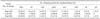
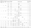
PCR analysis of 40 E. coli strains indicated that at least one of the virulence-associated genes was present in all strains (Tables 1 and 2). The fimA gene was the most commonly found among the 8 target genes and was present in 34 isolates whose hosts included 25 dogs and 9 cats. The sfa gene was detected in 23 isolates from 16 dogs and 7 cats. The cnf1 gene was found in 21 isolates collected from 16 dogs and 5 cats, while the papC and iutA genes were found in 15 animals (11 dogs and 4 cats). The hlyA gene, detected in 11 isolates from 7 dogs and 4 cats, was always found in conjunction with cnf1 and sfa genes, and often with papC and fimA. Six samples carried only one of the target genes and no sample possessed more than six virulence factors.
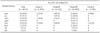
Phylogenetic analysis showed that 65.0% of the isolates (n = 26) belonged to phylogenetic group B2, 10.0% (n = 4) to group D, 15.0% (n = 6) to group B1, and 10.0% (n = 4) to group A (Table 3). In addition, UPEC belonging to group B2 harboured a greater number of virulence factors compared to strains from phylogenetic groups A, B1 and D (p < 0.001; Table 2). FimA and iutA genes were detected in all groups at different percentages (Table 3). PapC, hlyA, cdt, and afa were found in phylogenetic group B2 but were absent in the remaining groups. Additionally, sfa and cnf1 were predominantly detected in group B2.
REP- and ERIC-PCR
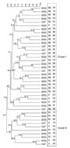
DNA amplification of E. coli strains by REP and ERIC primers was reproducible: identical band patterns were obtained from three different experiments, each of which was performed on three different days, and all comparisons among the strains were performed on the same gel (data not shown). Fingerprinting with REP- and ERIC-PCR primers pairs generated distinct amplification bands ranging in size from 100 bp to 1.5 kb. The 40 strains produced 38 different patterns by REP-PCR ranging from 4 to 12 bands, and 36 different patterns by ERIC-PCR ranging from 3 to 10 bands. The dendrograms produced using REP and ERIC fingerprints separately (data not shown) showed grouping of the strains similar to that of the dendrogram produced by the combination of the results. In both dendrograms, the strains were clearly distinct based on the phylogroup. Strains belonging to phylogroups B2 and D (cluster I) were all clustered together and separated from strains belonging to phylogroups A and B1 (cluster II). In particular, the dendrogram obtained combining the REP and ERIC results (Fig. 1) showed an overall similarity of 53.7%; strains belonging to cluster I showed a similarity of 54.9% and strains belonging to cluster II showed a similarity of 60.0%. Three of the four strains belonging to phylogroup D were clustered together at 63.2% of similarity and showed the same virulence factors. The analysis of virulence factors distribution over the dendrogram indicated that the strains presenting the same pattern of virulence factors were not the nearest neighbours, but rather scattered all along the dendrogram. This finding was observed for all the phylogroups except for group D. Another feature common to the dendrograms was that strains retrieved from cat samples were not genetically distinct from those obtained from dogs. In particular, considering the clustering of the cat samples (n = 10), these shared genetic similarity as high as 94.4% with dog samples. These values were also higher when separately considering REP and ERIC results, both of which showed one cat sample to be 100% similar to one dog sample (data not shown).
Statistical analysis
For the analysis of 2 × 2 tables, strains were divided into two groups: one joining strains belonging to phylogroups A and B1, and the other assembling strains belonging to phylogroups B2 and D. Cdt and afa were removed from the analysis given their low detection frequency. In addition, since the hlyA gene was used as a genetic marker for E. coli virulence, the association between hlyA alone and the presence of other factors (sfa, papC and cnf1) was evaluated. This combination was chosen because of its high frequency of occurrence (Table 2). The associations between hlyA and the combination of sfa, papC and cnf1 revealed highly significant results (p < 0.001), with all the hlyA-positive strains also being positive for sfa and cnf1, whereas one strain was positive for hlyA but negative for papC. The association between phylogroups and virulence factors revealed that papC and hlyA were only associated with cluster I (phylogroups B2 and D; p < 0.05). The results of Wilcoxon two-sample test showed a highly significant difference in score distribution between phylogroups B2 and the other phylogroups (median virulence score of 3.5 vs. 2.0; p < 0.001).
Discussion
In the present study, we analyzed E. coli isolates from dogs and cats with cystitis in order to obtain evidence of an association between virulence genotype and phylogenetic background, and the clonal relationship as determined by REP- and ERIC-PCR. In agreement with other studies, the presence of several types of virulence factors was a common characteristic among dog and cat UPEC strains. A survey of fimA, sfa, cnf1, papC, iutA, hlyA, and cdt genes yielded results similar to those obtained in other studies [2,8,13,27] of human and animals strains. The afa gene was not frequently found, indicating that this virulence factor is uncommon among urophatogenic bacterial strains [2,19,20]. However, in contrast to these results, we found one strain having an afa gene associated with sfa, fimA, iutA, and cdt sequences. The results provided evidence that two virulence factors (papC and hlyA) were present only in cluster I. In particular, repeated observation of hlyA only in the B2 phylogroup indicated that the presence of hly is associated with higher pathogenicity of the strains, not only because of the toxin produced but also because the strains harbouring this gene may also be carriers of numerous other virulence factors. In fact, the association of hlyA with papC, sfa, and cnf1 observed in the present research indicated that when hlyA is present in the genome, these other genes are present as well. This may be explained by the presence of a direct chromosomal linkage among these operons within particular DNA units on the chromosome, termed pathogenicity islands (PAIs), which carry virulence-associated genes coding for cnf1, hlyA, and papC [3].
In concurrence with several authors [1,9,18], we observed a link between phylogroups and extraintestinal strains because the majority of the strains isolated from urine belong predominantly to phylogenetic group B2 and, to a lesser extent, to group D, whereas they were sparsely represented within groups A and B1. Strains belonging to group B2 harboured a greater number of virulence factors compared to strains from other phylogenetic groups isolated in extraintestinal infections, suggesting a putative association between virulence factors and pathogenic potential [6]. In the same way, a link between phylogroups and intestinal E. coli was observed in works by other groups [6,10] showing that intestinal pathogenic E. coli belong mainly to groups A and B1 and are rarely derived from phylogenetic group B2, while commensal strains are distributed in an homogeneous manner all over the phylogroups. In this study, all isolates harbouring four or more virulence genes belonged to phylogroup B2, suggesting these strains were more virulent that the others. Similar results were observed in a recent study conducted in northern Italy that found an association between virulence factors and phylogroups in urinary E. coli strains isolated in human [17], suggesting the presence of strains responsible for UTI that are common to animals and humans.
Our analysis also included two methods for assessing relatedness of the strains: REP- and ERIC-PCR. The generated dendrograms showed that the majority of isolates had a distinct profile, indicating their genomic heterogeneity, while strains belonging to phylogroup D showed a low degree of polymorphism. The isolates were grouped into two main clusters: a large cluster (cluster I) that included groups B2 and D, and a smaller cluster (cluster II) that included isolates belonging to groups A and B1. UPEC strains belonging to cluster I harboured a greater number of UPEC virulence factors compared to strains from cluster II. In particular, we observed a frequent association of hlyA, papC, sfa, and cnf1 genes that indicate the presence of PAIs in E. coli belonging to cluster I, suggesting a possible correlation between PAIs and the clonal origin of UPEC strains. These data could indicate a selective receptivity of phylogroups B2 and D, included in cluster I, to acquire virulence determinants responsible of urinary infections. Moreover, the dendrogram showed that strains were not grouped according to their virulence factor pattern or on the animal species of origin. The finding that feline strains did not cluster separately from canine strains indicated that the strains circulate between these two hosts and that no niche adaptation occurs. However, in order to further explore this hypothesis, a studies with a stratified random sample of patients needs to be performed, ideally with a large number of samples of each species.
In summary, among the E. coli isolated from dogs and cats with cystitis we found that a high frequency of virulence factors such as fimA, sfa and cnf1 was significantly associated with phylogenetic group B2. Our results indicate a strong association between hlyA and papC, sfa, and cnf1 operons related to the presence of PAIs in these strains. In addition, we observed that REP- and ERIC-PCR had high discriminatory power. Our results demonstrated high genetic variability among the UPEC strains studied, probably indicating that cystitis may be caused by E. coli with a variety of different genotypes.
 XML Download