Introduction
Foot and mouth disease (FMD) is an economically important disease of cloven-hoofed livestock such as cattle, sheep, and other domestic animals in addition to several wild-life species. The causative agent of the FMD is the foot and mouth disease virus (FMDV) [9,11,18]. The disease is considered one of the most important barriers to the worldwide trade of livestock and animal products [12]. FMDV isolates sampled from the world have been categorized into seven different serotypes, A, O, C, Asia1, and South African Territories 1 (SAT1), SAT2, and SAT3, according to antigenic levels; and a large number of variants have appeared within each serotype. Out of the three serotypes (O, A, and Asia1) found to be prevalent in Iran in recent years, serotype A has been found to be more antigenically and genetically diverse than the others [6,10,14].
The FMDV possesses a single-stranded RNA molecule consisting of about 8,200 nucleotides within an icosahedral capsid composed of structural proteins [12]. The open reading frame encodes a single polyprotein which can be cleaved into four structural proteins (VP4, VP2, VP3, and VP1) and eight non-structural proteins (L, 2A, 2B, 2C, 3A, 3B, 3C, and 3D polymerase [3Dpol]). In general, structural proteins are more variable than non-structural proteins. Mutations or deletions in structural proteins may help the FMDV to evade immune responses of the host whereas mutations or deletions in non-structural proteins can inhibit viral replication and protein processing [5].
The FMDV usually infects cells by binding to integrin receptors via a long flexible loop (G-H loop) of VP1 (1D). The sequence of this loop contains a conserved arginineglycine-aspartic acid (RGD) tripeptide motif which is characteristic of ligands that bind to integrin receptors [16]. The VP1 protein is encoded by the VP1 coding region of viral RNA; this coding region is 627~639 bp long and produces a protein containing 209~213 amino acid residues depending on the serotype [6]. The 3Dpol gene sequence is 1,410 nucleotides in length. This gene contains a TAA stop codon and is responsible for encoding a protein containing 470 amino acid residues [4]. Similar to other picornaviruses, the FMDV 3Dpol protein is a viral-encoded RNA polymerase. Among the different FMDV serotypes and subtypes, both the nucleotide and amino acid sequences of 3Dpol are highly conserved [14]. Uninterrupted co-circulation of the virus in the environment and the absence of viral polymerase proofreading activity during the replication process lead to the appearance of various genetic isolates [2]. Analysis of the viral genome sequence is a crucially important approach for monitoring field isolates in areas where the disease is endemic.
Previous studies have shown that Iran has one of the highest reported rates of FMD cases per year [17]. In Iran, FMD is largely controlled by vaccinating cattle and sheep with vaccines prepared against isolates that are likely to be encountered in the region. Spread of the disease is promoted by the presence of large populations of susceptible animals, low vaccination rates, prevalence of multiple serotypes (including serotypes A, Asia1, and O), and unlimited movement of susceptible animals in the country. Furthermore, there is endemic co-circulation of multiple genotypes of type A virus [19]. The high incidence of FMD has allowed the identification of new variants of the virus over the last 7 years [17]. The general objective of this study was to determine the nucleotide sequences of genes encoding VP1 and 3Dpol proteins of a type A FMDV (A/Iran87) highly adapted to cell culturing, and to compare them to other corresponding sequences available in the GenBank database.
Materials and Methods
The Razi Vaccine and Serum Research Institute (Iran) generously provided samples of the FMDV used for our study. Clinical specimens of FMDV which included samples in tongue epithelium tissue were collected in 1987 from infected calves displaying clinical symptoms of FMD in an Iranian field located in Tehran. The A/Iran87 strain was initially detected and isolated nearly more than two decades ago. Thus, the highly passaged virus investigated in this study was not virus isolated from the field. The tissue samples were used to infect baby hamster kidney 21 (BHK-21) cells. The virulent isolate was passaged in BA (cell line derived from pig kidney) and BHK-21 cell monolayers. Propagation steps included six passages in BA cells and then four passages in BHK-21 cells. Thereafter, it has been used for the development of FMD vaccines during the past years as a vaccine seed. The infected cell culture supernatant from the ~150th passage was clarified and stored at -70℃ before use.
RNA extraction and reverse transcription-polymerase chain reaction (RT-PCR)
Total RNA was extracted from FMDV-infected BHK-21 cells using an RNX plus kit (Cinnagen, Iran) according to the manufacturer's instructions. Briefly, the cell culture was centrifuged at 85,000× g at 4℃ for 2 h. The pellet was resuspended in 200 µL of PBS, mixed well with 400 µL of RNX reagent, and incubated at room temperature for 5 min. RNA was extracted with 0.2 µL chloroform : isoamylalcohol (24 : 1). RNA in the aqueous solution was precipitated by adding an equal volume of isopropanol. The mixture was then centrifuged at 10,000 × g for 20 min. The pellet was washed with 75% ethanol and dissolved in 20 µL of RNase-free water.
RT-PCR was carried out in 50 µL of reaction mixture containing 10 µL of 5× reaction buffer, 4 µL of mixed dNTPs (2.5 mM each), 1 µL of AMV enzyme (Titan One Tube RT-PCR system kit; Roche Diagnostic, Germany), 1 µL of each primer (10 pmol each), 4 µL of RNA template, 2.5 µL DTT, 3 µL 25mM MgCl2, and 23.5 µL of H2O. The following conditions were used for amplification: 42℃ for 30 min, 94℃ for 3 min; 30 cycles of 94℃ for 30 sec, 52℃ for 30 sec, 72℃ for 40 sec; then followed by 72℃ for 5 min. The VP1 and 3Dpol coding regions of FMDV (621 and 690 bp, respectively) were amplified using standard methods with a one-step RT-PCR system and specific primer combinations. Primers sequences were as follows: forward: 3FMG6 5'-ctctggtaccatcaacctgcac-3' and reverse: Nk61 5'-gacatgtcctggtgcatctg-3' for 3Dpol, and forward: FMG13 5'-accaggatgatgattggcag-3' and reverse: FM15 5'-tttcactcctacggtgtcgc-3' for VP1. Amplified PCR products of the expected length were separated by electrophoresis in a 1% agarose gel, stained with ethidium bromide, and visualized under a UV transilluminator.
Cloning of the PCR product, DNA sequencing, and sequence analysis
After successful amplification of the target DNA sequences, fragments were purified using a gel extraction kit (Roche, Germany) following the recommendations of the supplier. Ligation was performed with plasmid vector pTZ57R/T (Fermentas, Germany) in 0.165 µg, 0.18 pmol ends and 0.54 pmol ends purified PCR fragment in 1× ligation buffer, polyethylene glycol (PEG 4000), 5 units of T4 DNA ligase, and up to 30 µL of deionized water at 22℃ for 16 h. The ligated products were used to transform chemically competent cells (XL1-Blue cells) and white colonies grown on LB plates were randomly selected. The cloned PCR products were purified using plasmid purification kit (Roche Diagnostics, Germany) according to the manufacturer's instructions and sequencing was carried out in both directions using a T7 promoter primer (MWG Biotech, Germany).
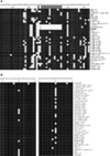
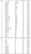
The published sequences of 66 FMDV type A isolates recovered from different parts of the world were included in this analysis and compared to the corresponding sequence of the A/Iran87 isolate. Reference FMDV sequences were obtained from the National Center for Biotechnology Information (NCBI, USA). The sequences used for each gene were first examined to exclude ambiguous sequences which were incomplete or frame-shifted. To determine the degree to which genetic diversity was observed in the VP1 and 3Dpol proteins, multiple alignments and comparisons of the predicted amino acid sequences of isolates were carried out (Fig. 1). Nucleotide sequence homology/divergence was calculated using the MegAlign project of the DNAStar software package (ver 5.1; DNAStar, USA; data not shown). The histories of the type A FMDV field isolates, including year of isolation, accession number, and geographical distribution, are presented in Table 1. The nucleotide sequences of the VP1 and 3Dpol coding regions of A/Iran87 have been submitted to GenBank (accession No. AY370770 and AY248743, respectively).
Results
Genetic comparison of the field isolates to the vaccine strain is of significant importance for testing the usefulness of the existing vaccine strain as well as selecting new vaccine strain(s). During virus circulation, amino acid substitutions accumulate at the different positions of the protein sequence. In the present study, a highly passaged field isolate of FMDV (A/Iran87) named Mardabad or A87 isolate was selected for genetic analysis. Sequences of the protein coding regions (VP1 and 3Dpol) from the high-passage cell-adapted vaccine strain were determined by PCR amplification and sequencing. Comparison of the amino acid sequences revealed amino acid changes in both structural and non-structural proteins. The number of sequence differences exhibited by each of the isolates showed that A/Iran87 contains four amino acid substitutions at positions 17, 26, 50, and 57 in the 3Dpol coding region (Fig. 1). There is clear evidence of novel amino acid substitutions (Ala→Ser, Asp→Glu, Glu→Asn, and Ala→Gly) in two domains of the 3Dpol protein. Comparison of the VP1 protein among different variants showed that A-Iran-vaccinal contained a change at position 179. At this position, Ala was replaced with Val. Fig. 1A shows that amino acid changes among the VP1 genes of the field isolates consist of numerous nucleotide substitutions and deletions compared to the consensus sequence. Comparison of the predicted amino acid sequences of VP1 region in the field isolates revealed amino acid deletions in three Iranian vaccine isolates (A-Iran-vaccinal, A Iran04, and A/Iran87). As shown in Fig. 1A, deletions occurred within the 13 amino acid positions (168 to 180) of the VP1 region. Three-dimensional analysis of 3Dpol protein showed that the Asp→Glu substitution occurred in a beta sheet located in a small groove of the protein.
The variations were not distributed uniformly along the genes; there were areas of high and low incidence of nucleotide sequence variation (data not shown). The region of the VP1 gene between amino acids 157 and 183 contained a high degree of variation and encoded an important immunogenic site on the viral surface. Consequently, nucleotide changes in this region are most likely involved in the appearance of new antigenic variants. The VP1 and 3Dpol nucleotide sequences of the selected FMDV-A subtypes isolated from outbreaks in Iran and other countries were used to construct a FMDV-A VP1 and 3Dpol-based sequence similarity tree. Fig. 2 shows a phylogenetic tree that was constructed based on the sequence alignment of 35 genomes of the VP1 region and 42 genomes in the 3Dpol region which are distinctly divided into different lineages. As depicted in Fig. 2A, the A/Iran87 isolate clustered with a Saudi Arabian and five Iranian isolates into a branch separate from other type A isolates.
Fig. 2B demonstrates that A/Iran87 and all isolates examined in the present study originated from different geographical areas and did not cluster in relatively similar lineages based on 3Dpol sequences. All the field isolates shared comparatively lower homology (90~93%) with the A/Iran87 isolate in the 3Dpol coding region (data not shown). The topology of the phylogenetic tree indicated that there was no remarkable similarity between A/Iran87 and all other isolates in the 3Dpol coding region. Space-filling and worm styles of the three-dimensional structures of the 3Dpol and VP1 proteins from the A/Iran87 isolate were observed. Analysis of the 3Dpol protein structure showed that the Asp→Glu substitution occurred in a beta sheet located within a small groove of the protein. In order to examine the long amino acid deletion in the three-dimensional structure of the VP1 protein, homology-based modeling of the protein domains was carried out (Fig. 3). It indicated that, 13 amino acid deletions in the VP1 protein of A/Iran87 isolate caused a change in three-dimensional structure of protein in the G-H loop region.
Discussion
Detailed knowledge of the molecular characteristics of the major FMDV immunogenic components would be useful for monitoring various processes like evolution, genetic diversity, and virus origin, and for developing protective vaccines. Among the FMDV serotypes, serotype A has been found to include the greatest number of recently developed variants. Extreme genetic heterogeneity is largely due to the absence of viral polymerase proofreading activity [2]. FMDV variants also appear during continuous infection of animals on farms or proliferation in cell cultures. Furthermore, genetic diversification among FMDV subtypes can be caused by events occurring during recombination.
Along with above mechanisms, there is continuous co-circulation of multiple genotypes of FMDV type A which may lead to recombination; this progresses gradually between closely related isolates when multiple viral genotypes co-infect the host. Exchange of different genomic regions between FMDVs by recombination is directly linked to FMDV diversification. Recombination, particularly in the structural protein-coding region, may offer selective advantages to the virus. This can be great concern to areas where multiple FMDV genotypes co-circulate [19]. The presence of extensive antigenic variation among different FMDV isolates can hinder vaccination against FMDV [1,13].
Genetic polymorphisms amongst different type A subtypes demonstrated that four nucleotides in the 3Dpol coding region of isolate A/Iran87 (49, 78, 148, and 170) were changed from G to T, C to A, G to A, and C to G, respectively, resulting in amino acid substitutions Ala17→Ser, Asp26→Glu, Glu50→Asn, and Ala57→Gly, respectively. In contrast to the A/Iran87 isolate, the remaining 41 isolates showed no corresponding changes at these positions. Analysis of the sequence data revealed that the A/Iran87 isolate also contained 30 nucleotide deletions in region 501~540 of the VP1 nucleotide sequence, which led to long amino acid deletions (RGDLGSLAARVAA) in the G-H loop. It should be noted that the RGD sequence was deleted from the G-H loop. The three-dimensional and antigenic structures of many different serotypes of the FMDV have been examined [8]. In spite of losing the RGD sequence and acquiring a Asp26→Glu substitution in a beta sheet located in a small groove of the 3Dpol protein, the virus grew in a BHK-21 suspension cell culture and exerted cytopathic effects after 16~18 h. Since this strain is used as a vaccine strain, it may be inferred that the RGD does not have a key role in the virus binding to cells during infection. This virus subtype can probably use other pathways for cell attachment. As RGD sequence was found to be deleted, such a natural deletion in VP1 gene of FMDV is a novel phenomenon and has not been previously reported in compared isolates.
The VP1 capsid protein contains a mobile loop between the βG and βH strands on the virus surface; this protein not only contains the major immunodominant epitopes of the virion but also a highly conserved RGD amino acid sequence motif. RGD participates in binding FMDV to susceptible cells. Several studies using different approaches have indicated that naturally developed FMDV isolates attach to cells via the highly conserved RGD motif. FMDVs have been shown to use multiple RGD-dependent integrins of the αv subgroup to initiate infection; these include αvβ3, αvβ6, αvβ1, and αvβ8. On the other hand, these viruses are capable of entering cells via non-integrin pathways. Tissue culture-adapted FMDVs are able to utilize heparan sulfate (a glycosaminoglycan found on the cell surface) as a receptor for entering the cells. It has recently been found that FMDV can utilize unknown receptors except for integrin and heparan sulfate. This indicates the presence of other receptors and possible alternative mechanisms for viral particle entrance into the cells. This conclusion is also supported by the finding that destruction of the βG-βH loop RGD motif by site-directed mutagenesis created viruses which were able to replicate and assemble, but were not capable of initiating further infection cycles because of their inability to interact with cell receptors [18].
Previous findings from studies of synthetic peptides suggested that the carboxy-terminus of the VP1 protein situated in the vicinity of the RGD motif is required for RGD-mediated cell binding [3]. The G-H loop of the VP1 protein on the viral particle surface is considered to be the dominant epitope for several FMDV types and the most variable part of the particle. Antibody-based responses against the loop have the ability to neutralize FMDVs in vitro and are protective in vivo. For example, replacing the whole or partial G-H loop leads to the doubling of antibody titers against type A virus. New findings in this field are in complete agreement with a previous report documenting that the only mutation which did not increase heterologous responses was deletion of the RGD-motif (3A) [8]. In another study, the RGD sequence was deleted from a genetically engineered FMDV (Type A12), thereby rendering the virus particles incapable of binding to cell surfaces. Protective immunity in cattle has been obtained by utilizing particles from which the receptor binding site was deleted as a vaccine, revealing that this deletion has no significant effect on the antigenic properties of viruses with the RGD sequence [15].
Many reports [21] have suggested that FMDV subtypes can be obtained in the field by immune selection during outbreaks of FMDV which have not been effectively neutralized in animal populations with incomplete immunization. Thus, key changes in protein sequences that give rise to differences in immunogenicity and virulence can be seen among viruses isolated from partially immunized animals. In addition, it is possible that other significant changes take place when the virus moves from one host cell type to another. For instance, this may occur when FMDV spreads from one species to another (from cattle to swine and then back to cattle) or when FMDV cultured in BHK cells infects animal herds. For a number of years, it has been observed that FMDV easily adapts to many different tissue culture cell types. A recent report on a single passage of subtype A12 (AVR1) in BHK cells showed that some viral variants selected from this passage contained amino acid changes at residues 148 and 153 of the VP1, including ones that have a limited capability for eliciting cross-neutralizing antibodies [21].
The rapid generation of immunogenically distinguishable variants among FMDV subtype A viruses may affect the large-scale production of vaccines in BHK cells. Adaptation for growth in tissue culture can be used to select viruses that have antigenic and cell attachment site(s) different from those of the parental virus. Amino acid residues 134~158 form a βG-βH loop structure which contains the main FMDV immunogenic epitope. In FMDV type C, mutation of residues 138~140 and 148~150 in the VP1 region has been shown to affect FMDV antigenicity. Mutation frequency in the VP1 gene sequence of the FMDV has been estimated to be 1.6 × 10⁻³~6.4 × 10⁻³ substitutions for each nucleotide per year [6]. G-H loop-specific antibody responses are known to play a major role in immunity induced by the current FMDV vaccines. Considering the high sequence variability of this loop within and among the serotypes, it will be valuable to direct immune responses against other protective epitopes that are not as variable. Residues 140~160 of VP1 have been previously shown to induce neutralizing antibodies against FMDV types O and A [20]. Due to a novel deletion of the 13 amino acid region including the RGD motif in the VPI capsid protein of A/Iran87, the effect of this motif in receptor binding and cell infection must be determined.
The FMDV-3D protein is a viral-encoded RNA polymerase. This protein was initially called the FMDV infection-associated antigen since antibodies to this antigen are be detected in serum from recovery animals [22]. The 3Dpol position is located at the C-terminus of the polypeptide next to the 3C gene product. The 3Dpol coding region produces a 470-amino acid protein with a molecular weight of ~55 kDa. The pivotal role of 3Dpol in the viral replication process is attributed to the fact that the 3Dpol gene is highly conserved, particularly in the functional motifs. Since RNA polymerase is necessary for viral replication, the degree of sequence identity among the 3Dpol genes is much greater than that of VP1 gene sequences [4].
A wealth of three-dimensional structural information is currently available for a large number of RNA-dependent RNA polymerases from different families of positive- and double-stranded RNA viruses, including three different members of the Picornaviridae family [7]. Considering the fact that this strain can infect BHK-21 cells and has been used as a vaccine strain for nearly two decades, it may be concluded that deletion of RGD has no critical role in virus binding to cells during the initiation of infection. It is probable that this FMDV subtype can utilize other pathways for cell binding. The findings of the current study demonstrated that the cell-adapted vaccine strain A/Iran87 containing 13-amino acid deletions in the VP1 protein can be considered as a novel variant of FMDV type-A. This variant serves as a potent vaccine strain in Iran.


 PDF
PDF ePub
ePub Citation
 Print
Print


 XML Download