Journal List > J Vet Sci > v.12(3) > 1041103

Youn, Koo, Ahn, Lim, and Park: Determination of staphylococcal exotoxins, SCCmec types, and genetic relatedness of Staphylococcus intermedius group isolates from veterinary staff, companion animals, and hospital environments in Korea

Abstract

The Staphylococcus (S.) intermedius group (SIG) has been a main research subject in recent years. S. pseudintermedius causes pyoderma and otitis in companion animals as well as foodborne diseases. To prevent SIG-associated infection and disease outbreaks, identification of both staphylococcal exotoxins and staphylococcal cassette chromosome mec (SCCmec) types among SIG isolates may be helpful. In this study, it was found that a single isolate (one out of 178 SIG isolates examined) harbored the canine enterotoxin SEC gene. However, the S. intermedius exfoliative toxin gene was found in 166 SIG isolates although the S. aureus-derived exfoliative toxin genes, such as eta, etb and etd, were not detected. SCCmec typing resulted in classifying one isolate as SCCmec type IV, 41 isolates as type V (including three S. intermedius isolates), and 10 isolates as non-classifiable. Genetic relatedness of all S. pseudintermedius isolates recovered from veterinary staff, companion animals, and hospital environments was determined by pulsed-field gel electrophoresis. Strains having the same band patterns were detected in S. pseudintermedius isolates collected at 13 and 18 months, suggesting possible colonization and/or expansion of a specific S. pseudintermedius strain in a veterinary hospital.

Introduction

Staphylococcus (S.) intermedius and S. pseudintermedius are both Staphylococcus intermedius group (SIG) members. These microorganisms are opportunistic pathogens that cause skin and nosocomial infections in dogs and cats [32]. S. pseudintermedius is the predominant SIG species in Korea and until recently was misclassified as S. intermedius [30,33]. These pathogens rarely cause human disease although they do have zoonotic potential [5,6,26]. Increased identification of methicillin-resistant SIG (MRSIG) isolates has emphasized the importance of public health precautions for veterinary staff and pet owners [7,11,26].
Many current studies have evaluated methicillin-resistant characteristics and the staphylococcal cassette chromosome mec (SCCmec) of SIG isolates, particularly S. pseudintermedius. Although type I to type VIII SCCmec types have been identified, there is no significant studies have been conducted to examine SCCmec types isolated from veterinary hospitals located nationwide in Korea. SCCmec types may produce staphylococcal exotoxins including staphylococcal enterotoxins (SEs), exfoliative toxins (ETs) including S. intermedius exfoliative toxin (SIET), and toxic shock syndrome toxin 1 (TSST 1); these toxins are associated with atopic dermatitis, mastitis, food poisoning, canine pyoderma, and chronic otits [1,3,10,14,29]. SIET (encoded by the siet gene), first described by Terauchi et al. [27], plays a potential role in the pathogenesis of canine pyoderma and chronic otitis [31]. There may also be a greater chance of SIG isolates to colonization and/or expand in veterinary staff, companion animals, and hospital environments. Therefore, this study is focused on the genetic identification of staphylococcal exotoxins, SCCmec types, and genetic relatedness by pulsed-field gel electrophoresis (PFGE) of the SIG isolates in Korea.

Materials and Methods

Samples of S. pseudintermedius (n = 167) and S. intermedius (n = 11) were isolated; their identity was confirmed by Gram-staining and biochemical testing such as coagulase, DNase production and hydrolysis [33]. Polymerase chain reaction (PCR) was carried out with primers targeting S. intermedius nuclease and 16S rRNA genes [2]. To differentiate S. pseudintermedius from S. intermedius and S. delphini, PCR-restriction fragment length polymorphism analysis was performed using pta gene-specific primers (pta_f1:5'-AAA GAC AAA CTT TCA GGT AA-3', and pta_r1: 5'-GCA TAA ACA AGC ATT GTA CCG-3'), and the restriction enzyme MboI (New England Biolabs, USA) [3]. Samples were collected from eight veterinary hospitals from four different regions in Korea between November 2006 and January 2010 as previously described [33]. PCR analysis to detect the methicillin-resistance gene (mecA) was performed with gene-specific primers as previously described [32]. PCR was also performed to amplify genes encoding SEA (sea), SEB (seb), SEC (sec), SED (sed), SEE (see), SEG (seg), SEH (seh), SEI (sei), TSST 1(tsst-1) and ETs (eta, etb, etd, and siet) using primers and PCR conditions used in other previous studies [12,16,19,20,24,25]. FRI913, FRI 361, FRI 472, FRI 569, MNHOCH, and RN4220 strains were used as either positive or negative controls for superantigen genes. A superantigen gene obtained in this study was further analyzed by DNA sequencing using the Vector NTI Align X program (Invitrogen, USA).
All MRSIG isolates including 49 methicillin-resistant S. pseudintermedius (MRSP) and three methicillin-resistant S. intermedius (MRSI) were subsequently classified as SCCmec type I to VIII by either single or multiplex PCR assays using protocols in other previous studies including primers and PCR conditions [4,9,13,15,17,18]. Genomic DNA plug kits (Bio-Rad, USA) with SmaI (New England Biolabs, USA) digestion were used for the PFGE analysis of 39 S. pseudintermedius isolates from a private referral veterinary hospital collected at different times (eight isolates from November 2006, 24 isolates from April 2008, four isolates from June 2009, and three isolates from October 2009) and six S. intermedius isolates (all collected during April 2008). PFGE was performed according to protocol used in previous study [8] and the manufacture's instruction (Bio-Rad, USA) except for an initial pulse time of 5 sec and final time of 40 sec for 21 h [8].

Results

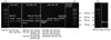
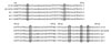
Analysis of the SIG isolates (n = 178) for SEs and TSST 1 genes showed that only a single S. pseudintermedius isolate from a veterinary staff member (H1-23) harbored the enterotoxin C (sec) gene (Fig. 1). Subsequent DNA sequencing analysis (Fig. 2) revealed that the amplified sec gene the canine type C SE gene (seccaine). A total of 166 out of 178 SIG isolates (155 S. pseudintermedius and 11 S. intermedius) isolated from veterinary staff members (n = 38, 95%), companion animals (n = 107, 93%), and veterinary hospitals (n = 21, 91.3%) harbored siet originally detected in S. intermedius. However, other ET genes such as eta, etb, and etd, known to originate from S. aureus, were not identified.
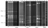
SCCmec typing of the 49 MRSP isolates identified 38 type V isolates, one type IV isolate, and 10 non-identifiable isolates. Three MRSI isolates were characterized as type V isolates (Table 1). PFGE analysis showed that SIG isolates from a private referral animal hospital (collected between 2006 and 2009) recovered from veterinary staff, companion animals, and the environment had the same band patterns (Fig. 3). Six S. pseudintermedius isolates (two from companion animals collected in September 2006, three from veterinary staff members, and one from companion animals collected in April 2008) and two S. pseudintermedius isolates (one from a veterinary hospital environment isolated in April 2008, and one from a companion animal isolated in June 2009) showed the same band patterns (Fig. 3).

Discussion

Since SIG is closely related to S. aureus, studies have been performed to determine whether SIG has adapted the eta, etb and etd toxins from S. aureus by PCR test with specific primers [16,24,25]. None of these toxins were detected. However, 166 out of 178 (93.3%) SIG isolates harbored the siet toxin originating from S. intermedius [27]. This result and those of other studies [22,31] imply that the majority of SIG isolates harbor the siet gene. Although the siet gene was present in 93.3% of the SIG isolates (from 108 dogs and two cats) in this study, only 14 dogs had a history of various skin disease including allergy and prolonged inflammation lesion in skin (n = 13) or otitis (n = 1) (data not shown). Therefore, other factors such as the general health of an animal and existence of other SIG virulence factors may play an important role in outbreaks of various kinds of skin disease or otitis.
The sec gene was detected in a single isolate in the present study, which was identified as seccanine by DNA sequencing. However, this isolate was isolated from a veterinary staff member, and no additional SECcanine isolates were identified in the veterinary hospital where this individual worked. This suggested that the isolate might be transmitted from an in- or outgoing companion animal with which the veterinary staff was in contact. The low incidence of toxins in this study could be secondary to the small number of isolates collected from companion animals that a history of skin disease or otitis. Only 11.3% of S. pseudintermedius in a previous study had exotoxins although all samples were taken from patients diagnosed with pyoderma or chronic otitis and referred to a veterinary teaching hospital [30]. This result suggested that SE toxins may be associated with pyoderma and chronic otitis.
A total of 52 out of 178 SIG isolates harbored the mecA gene. Although the majority of SCCmec types were type V (78.9%), one isolate was type IV. In a previous study [10], 23 isolates (85.2%), three isolates (11.1%) and one isolate (3.7%) from veterinarians, staff, students, companion animals and environment in the veterinary hospitals were determined to be an MRSP hybrid SCCmec type I~II, type V, or non-identifiable, respectively. A previous European and North American study [21] identified 75 hybrid SCCmec type II~III isolates (72.8%), two type III isolates (1.9%), six type IV isolates (5.8%), 14 type V isolates (13.6%), four type VII isolates (3.9%), and two non-identifiable isolates (1.9%) from diseased and healthy dogs in veterinary diagnostic laboratories of different countries. This demonstrated that the majority of MRSIG isolates in Korea harbor the SCCmec type V whereas the hybrid type II~III is the main SCCmec type found in veterinary hospitals in Japan, Europe, and North America.
In the present study, PFGE analysis of the 39 S. pseudintermedius and six S. intermedius isolates from a private referral veterinary hospital (collected during November 2006, April 2008, June 2009, and October 2009) showed that 20 isolates (lines 1~6, lines 7~9, lines 10~15, lines 16~18, and lines 19~20) had the same band patterns. Moreover, some isolates obtained on different sampling dates showed the same band patterns (six isolates from lines 12~17, and two isolates from lines 21~22). These results suggest potential contamination or expansion of S. pseudintermedius and S. intermedus isolates among veterinary staff, companion animals, and veterinary hospital environments, and colonization by these specific strains for more than 13 or 18 months in the same hospital.
In conclusion, eta, etb, etd genes were not detected but siet toxin was found in 166 isolates in the current study. PFGE analysis results of isolated from H animal hospital showed that S. pseudintermedius isolates collected in over a period of 13 and 18 months from veterinary staff, companion animals, and the hospital environment had the same band patterns. S. pseudintermedius infections in humans [5,26,28], spread of MRSP populations [6,23,29], and association of SIG with canine pyoderma and chronic otitis [3,14,29] has been previously reported. Therefore, SIG, especially MRSIG, may have significant clinical implications for companion animals with skin infections or chronic otitis that is of concern for veterinary staff, companion animal owners, and healthy companion animals.

Figures and Tables

Fig. 1
Staphylococcal enterotoxins A (sea), B (seb), C (sec), D (sed), E (see), G (seg), H (she), and I (sei), and the toxic shock syndrome toxin 1 (tsst-1) were detected by single or multiplex polymerase chain reaction (PCR) in 178 SIG isolates collected from veterinary staff, companion animals, and veterinary hospital environments in Korea. Genomic DNA from FRI913, FRI 361, FRI 472, FRI 569, MNHOCH, and RN4220 were used as controls. Multiplex PCR for sec, tsst-1/see, seg, sei/seb, sed, and seh and uniplex PCR for sea were performed. Only a single isolate from a veterinary staff (H1-23, hand) was positive for sec.
jvs-12-221-g001
Fig. 2
The PCR-amplified sec gene sequence from the Staphylococcus (S.) pseudindermedius isolate H1-23 collected from a veterinary staff member was aligned with that of sec genes encoding S. intermedius SECcanine from the S. intermedius gene. Nucleotide sequences with GenBank numbers U91526, X05815, DQ192646, and X51661 were used for the alignment analysis of seccanine, sec1, sec2, and sec3, respectively. Aligment was peformed using the Vector NTI Align X program.
jvs-12-221-g002
Fig. 3
Comparison of S. intermedius group isolates from the H veterinary hospital between September 2006 and October 2009 using pulsed-field gel electrophoresis (PFGE) to detect identical PFGE patterns between isolates from veterinary staff, companion animals, and veterinary hospital environments collected on the same and different sampling dates. Lane M: Lambda Ladder PFG Marker (50~1,000 kb); Lanes 1~3 (SI) from single CA#1 (dog: anus, nasal mucosa, skin); Lane 4 (SI), VS#1 (nasal mucosa); Lanes 5~6 (SI) from single VS#2 (nasal mucosa, hand); Lanes 7~8 (SP) from a single CA#3 (dog: ear canal, ear canal); Lane 9 (SP), VS#3 (nasal mucosa); Lanes 10~11 (SP) from a single CA#4 (dog: anus, nasal mucosa); Lane 12 (SP), CA#5 (anus); Lane 13 (SP) VS#4 (nasal mucosa); Lane 14 (SP) VS#5 (nasal mucosa); Lane 15 (SP) VS#6 (nasal mucosa); Lane 16 (SP) CA#6 (anus); Lane 17 (SP) VS#7 (nasal mucosa); Lane 18 (SP) VS#8 (nasal mucosa); Lane 19 (SP) VHE#1 (floor); Lane 20 (SP) CA#7 (anus). CA: companion animal, VS: veterinary staff, VHE: veterinary hospital environment, SI: Staphylococcus intermedius, SP: Staphylococcus pseudintermedius.
jvs-12-221-g003
Table 1
Staphylococcal cassette chromosome mec (SCCmec) typing of Staphylococcus intermedius group (SIG) isolates from Korean veterinary hospitals (veterinary staff, companion animal, and the veterinary hospital environment)
jvs-12-221-i001

*NT: non-typeable. Included 3 Staphylococcus (S.) intermedius isolates and the other SIG isolates were S. pseudintermedius.

Acknowledgments

This study was supported by the National Veterinary Research and Quarantine Service (B-AD 3-2006-14-04) and Basic Science Research Program through the National Research Foundation of Korea (NRF) funded by the Ministry of Education, Science and Technology (2010-0003357). Additional support was provided by the Research Institute of Veterinary Science, Department of Veterinary Microbiology, College of Veterinary Medicine, and the BK21 Program for Veterinary Science, Seoul National University, Korea.
The authors thank Dr. Sa-Im, Dr. Sun Young, Dr. Bo Youn, Dr. Ka Hee, Dr. Min Ki, Dr. Ki Yeon, and Dr. Young Kyung for their excellent technical support.

References

1. Balaban N, Rasooly A. Staphylococcal enterotoxins. Int J Food Microbiol. 2000. 61:1–10.
crossref
2. Bannoehr J, Franco A, Iurescia M, Battisti A, Fitzgerald JR. Molecular diagnostic identification of Staphylococcus pseudintermedius. J Clin Microbiol. 2009. 47:469–471.
crossref
3. Baron F, Cochet MF, Pellerin JL, Ben Zakour N, Lebon A, Navarro A, Proudy I, Le Loir Y, Gautier M. Development of a PCR test to differentiate between Staphylococcus aureus and Staphylococcus intermedius. J Food Prot. 2004. 67:2302–2305.
crossref
4. Boye K, Bartels MD, Andersen IS, Møller JA, Westh H. A new multiplex PCR for easy screening of methicillin-resistant Staphylococcus aureus SCCmec types I-V. Clin Microbiol Infect. 2007. 13:725–727.
crossref
5. Chuang CY, Yang YL, Hsueh PR, Lee PI. Catheter-related bacteremia caused by Staphylococcus pseudintermedius refractory to antibiotic-lock therapy in a hemophilic child with dog exposure. J Clin Microbiol. 2010. 48:1497–1498.
crossref
6. Frank LA, Kania SA, Kirzeder EM, Eberlein LC, Bemis DA. Risk of colonization or gene transfer to owners of dogs with meticillin-resistant Staphylococcus pseudintermedius. Vet Dermatol. 2009. 20:496–501.
crossref
7. Futagawa-Saito K, Makino S, Sunaga F, Kato Y, Sakurai-Komada N, Ba-Thein W, Fukuyasu T. Identification of first exfoliative toxin in Staphylococcus pseudintermedius. FEMS Microbiol Lett. 2009. 301:176–180.
crossref
8. Herschleb J, Ananiev G, Schwartz DC. Pulsed-field gel electrophoresis. Nat Protoc. 2007. 2:677–684.
crossref
9. Higuchi W, Takano T, Teng LJ, Yamamoto T. Structure and specific detection of staphylococcal cassette chromosome mec type VII. Biochem Biophys Res Commun. 2008. 377:752–756.
crossref
10. Ishihara K, Shimokubo N, Sakagami A, Ueno H, Muramatsu Y, Kadosawa T, Yanagisawa C, Hanaki H, Nakajima C, Suzuki Y, Tamura Y. Occurrence and molecular characteristics of methicillin-resistant Staphylococcus aureus and methicillin-resistant Staphylococcus pseudintermedius in an academic veterinary hospital. Appl Environ Microbiol. 2010. 76:5165–5174.
crossref
11. Kadlec K, Schwarz S, Perreten V, Andersson UG, Finn M, Greko C, Moodley A, Kania SA, Frank LA, Bemis DA, Franco A, Iurescia M, Battisti A, Duim B, Wagenaar JA, van Duijkeren E, Weese JS, Fitzgerald JR, Rossano A, Guardabassi L. Molecular analysis of methicillin-resistant Staphylococcus pseudintermedius of feline origin from different European countries and North America. J Antimicrob Chemother. 2010. 65:1826–1828.
crossref
12. Lautz S, Kanbar T, Alber J, Lämmler C, Weiss R, Prenger-Berninghoff E, Zschöck M. Dissemination of the gene encoding exfoliative toxin of Staphylococcus intermedius among strains isolated from dogs during routine microbiological diagnostics. J Vet Med B Infect Dis Vet Public Health. 2006. 53:434–438.
crossref
13. Lim SK, Nam HM, Park HJ, Lee HS, Choi MJ, Jung SC, Lee JY, Kim YC, Song SW, Wee SH. Prevalence and characterization of methicillin-resistant Staphylococcus aureus in raw meat in Korea. J Microbiol Biotechnol. 2010. 20:775–778.
14. Loeffler A, Linek M, Moodley A, Guardabassi L, Sung JML, Winkler M, Weiss R, Lloyd DH. First report of multiresistant, mecA-positive Staphylococcus intermedius in Europe: 12 cases from a veterinary dermatology referral clinic in Germany. Vet Dermatol. 2007. 18:412–421.
crossref
15. McClure JA, Conly JM, Elsayed S, Zhang K. Multiplex PCR assay to facilitate identification of the recently described staphylococcal cassette chromosome mec type VIII. Mol Cell Probes. 2010. 24:229–232.
crossref
16. Nakaminami H, Noguchi N, Ikeda M, Hasui M, Sato M, Yamamoto S, Yoshida T, Asano T, Senoue M, Sasatsu M. Molecular epidemiology and antimicrobial susceptibilities of 273 exfoliative toxin-encoding-gene-positive Staphylococcus aureus isolates from patients with impetigo in Japan. J Med Microbiol. 2008. 57:1251–1258.
crossref
17. Okuma K, Iwakawa K, Turnidge JD, Grubb WB, Bell JM, O'Brien FG, Coombs GW, Pearman JW, Tenover FC, Kapi M, Tiensasitorn C, Ito T, Hiramatsu K. Dissemination of new methicillin-resistant Staphylococcus aureus clones in the community. J Clin Microbiol. 2002. 40:4289–4294.
crossref
18. Oliveira DC, de Lencastre H. Multiplex PCR strategy for rapid identification of structural types and variants of the mec element in methicillin-resistant Staphylococcus aureus. Antimicrob Agents Chemother. 2002. 46:2155–2161.
crossref
19. Omoe K, Ishikawa M, Shimoda Y, Hu DL, Ueda S, Shinagawa K. Detection of seg, seh, and sei genes in Staphylococcus aureus isolates and determination of the enterotoxin productivities of S. aureus isolates harboring seg, seh, or sei genes. J Clin Microbiol. 2002. 40:857–862.
20. Omoe K, Hu DL, Takahashi-Omoe H, Nakane A, Shinagawa K. Comprehensive analysis of classical and newly described staphylococcal superantigenic toxin genes in Staphylococcus aureus isolates. FEMS Microbiol Lett. 2005. 246:191–198.
crossref
21. Perreten V, Kadlec K, Schwarz S, Grönlund Andersson U, Finn M, Greko C, Moodley A, Kania SA, Frank LA, Bemis DA, Franco A, Iurescia M, Battisti A, Duim B, Wagenaar JA, van Duijkeren E, Weese JS, Fitzgerald JR, Rossano A, Guardabassi L. Clonal spread of methicillin-resistant Staphylococcus pseudintermedius in Europe and North America: an international multicentre study. J Antimicrob Chemother. 2010. 65:1145–1154.
crossref
22. Ruscher C, Lübke-Becker A, Semmler T, Wleklinski CG, Paasch A, Šoba A, Stamm I, Kopp P, Wieler LH, Walther B. Widespread rapid emergence of a distinct methicillin- and multidrug-resistant Staphylococcus pseudintermedius (MRSP) genetic lineage in Europe. Vet Microbiol. 2010. 144:340–346.
crossref
23. Sasaki T, Kikuchi K, Tanaka Y, Takahashi N, Kamata S, Hiramatsu K. Methicillin-resistant Staphylococcus pseudintermedius in a veterinary teaching hospital. J Clin Microbiol. 2007. 45:1118–1125.
crossref
24. Sato H, Matsumori Y, Tanabe T, Saito H, Shimizu A, Kawano J. A new type of staphylococcal exfoliative toxin from a Staphylococcus aureus strain isolated from a horse with phlegmon. Infect Immun. 1994. 62:3780–3785.
crossref
25. Sila J, Sauer P, Kolar M. Comparison of the prevalence of genes coding for enterotoxins, exfoliatins, panton-valentine leukocidin and tsst-1 between methicillin-resistant and methicillin-susceptible isolates of Staphylococcus aureus at the university hospital in Olomouc. Biomed Pap Med Fac Univ Palacky Olomouc Czech Repub. 2009. 153:215–218.
crossref
26. Stegmann R, Burnens A, Maranta CA, Perreten V. Human infection associated with methicillin-resistant Staphylococcus pseudintermedius ST71. J Antimicrob Chemother. 2010. 65:2047–2048.
crossref
27. Terauchi R, Sato H, Hasegawa T, Yamaguchi T, Aizawa C, Maehara N. Isolation of exfoliative toxin from Staphylococcus intermedius and its local toxicity in dogs. Vet Microbiol. 2003. 94:19–29.
crossref
28. Van Hoovels L, Vankeerberghen A, Boel A, Van Vaerenbergh K, De Beenhouwer H. First case of Staphylococcus pseudintermedius infection in a human. J Clin Microbiol. 2006. 44:4609–4612.
crossref
29. Weese JS, van Duijkeren E. Methicillin-resistant Staphylococcus aureus and Staphylococcus pseudintermedius in veterinary medicine. Vet Microbiol. 2010. 140:418–429.
crossref
30. Yoo JH, Yoon JW, Lee SY, Park HM. High prevalence of Fluoroquinolone- and Methicillin-resistant Staphylococcus pseudintermedius isolates from canine pyoderma and otitis externa in veterinary teaching hospital. J Microbiol Biotechnol. 2010. 20:798–802.
31. Yoon JW, Lee GJ, Lee SY, Park C, Yoo JH, Park HM. Prevalence of genes for enterotoxins, toxic shock syndrome toxin 1 and exfoliative toxin among clinical isolates of Staphylococcus pseudintermedius from canine origin. Vet Dermatol. 2010. 21:484–489.
crossref
32. Youn JH, Hwang SY, Kim SH, Koo HC, Shin S, Lim SK, Park YH. mecA gene transferrability and antibiogram of zoonotic Staphylococcus intermedius from animals, staff and the environment in animal hospitals in Korea. J Microbiol Biotechnol. 2010. 20:425–432.
33. Youn JH, Yoon JW, Koo HC, Lim SK, Park YH. Prevalence and antimicrogram of Staphylococcus intermedius group isolates from veterinary staff, companion animals, and the environment in veterinary hospitals in Korea. J Vet Diagn Invest. 2011. 23:268–274.
crossref
TOOLS
Similar articles