

 PDF
PDF ePub
ePub Citation
 Print
Print


Introduction
Organ transplantation is considered the preferred solution for the treatment of terminal organ failure [28]. However, there has always been a serious shortage of suitable human donors [13]. The constant shortage of donor organs led to research into xenotransplantation, which was first reported in 1906 [14]. Pigs are currently considered the most appropriate source of organs for xenotransplantation to humans based on several advantages, including physiological/anatomical organ similarities, reproductive characteristics, the possibility of controlled breeding, and ethical considerations [1,3,42]. Furthermore, recently, research to avoid the rejection of grafted organs has involved the production of genetically-modified pigs such as the α 1,3-galactosyltransferase gene knock-out pig, expression of human complement regulatory proteins (CD46, CD55, and/or CD59), and reducing risk of endogenous porcine retrovirus infection [6,8,20,26,27,32]. To resolve existing hurdles prior to the clinical application of pig organs, an appropriate method for evaluating the vascular system of micropigs must be established and immunological barriers must be addressed.
To transplant micropig solid organs into humans, evaluation of the vascular system and anatomical comparisons are essential for selection of a suitable organ as well as to gather sufficient pre-clinical data [5,18]. Previously, the standard method for preoperative angiographic evaluation of the donor vascular system was conventional angiography, whose disadvantages include being invasive and time-consuming, as well as the fact that it requires the use of ionizing radiation and large amounts of contrast agents. In contrast, the multidetector row computed tomographic angiography (MDCTA) process using doses of nonionic contrast media and ionizing radiation exposure that are less than conventional angiography [38]. Furthermore, venography is needed to obtain additional information regarding the venous system prior to organ transplantation. With remarkable advancements in spatial and temporal resolution, MDCTA is now routinely performed to evaluate human donors for solid organ transplant. This technology has been confirmed as a valuable method that can provide a road map for surgical planning as well as to assist in donor selection [17,33]. In addition, MDCTA has several advantages over traditional angiography; it is less invasive and permits visualization of organ structures and possible pathology [38].
The goal of this study was to confirm the feasibility of using MDCTA to evaluate the vascular system of micropigs and establish standard reference values for the vascular diameter and anatomy, which would be useful for selection of suitable donor organs in the future.
Materials and Methods
Animals
All experimental protocols were approved by the Ethics Committee of Chonnam National University, Korea (CNU IACUC-YB-2008-29). Physiologically and genetically intact male micropigs (n = 6) were purchased from PWG Genetics Korea (Korea). The animals were kept in individual cages at the university's central animal facility and received a standard pig diet and water ad libitum. The mean age and weight of the animals was 360 days and 30.50 ± 1.24 kg, respectively. Prior to undergoing MDCTA, all animals were fasted for 24 h. The animals were premedicated with an intramuscular injection of azaperone (0.5 mg/kg) and xylazine (8 mg/kg) and anesthetized with an intramuscular injection of a combination of zolazepam/tiletamine (4.4 mg/kg).
MDCTA protocol
The examinations were performed using a 64-channel multi-detector row helical CT scanner (LightSpeed VCT; GE Healthcare, USA) according to the following parameters: 0.5 sec per rotation, 5 mm collimation, 1.0 pitch, and a tube current of 120 kV per 140~200 milliamperes. The MDCTA images were acquired with spatial resolution of 0.35 × 0.35 × 0.8 mm. The CT angiographic scan was obtained in the craniocaudal direction, and reconstruction thickness and reconstruction increment were 1 mm and 0.5 mm, respectively.
For administration of intravenous contrast material, a 20-gauge peripheral line was placed in an ear vein. After a scout CT image was obtained, arterial phase volumetric image data sets were acquired following initiation of an intravenous injection of 60 mL of nonionic contrast media (Ultravist 370; Schering AG, Germany) at the rate of 3 mL/sec using an automated injector (LF CT 9000; Liebel-Flarsheim, USA). An automatic bolus triggering software program was systematically applied, with a circular region of interest positioned at the level of the superior vena cava (SVC) and a threshold for triggering data acquisition preset at 100 Hounsfield units to obtain arterial phase images. All image acquisitions were obtained in the craniocaudal direction and supine position. Imaging extended from the C1 cervical vertebrae to the knee joint including both pelvis and thigh. Volumetric data sets were transferred to an Advantage Workstation 4.3 (GE Healthcare, USA) equipped with Volume Viewer Plus three-dimensional (3D) software for subsequent review. Transverse 0.625-mm-thick sections were reformatted into maximum intensity projection images and volume rendered images.
Image analysis
A single radiologist reviewed all CT images at a workstation which permitted editing of CT volume data sets to create optimal 3D CTA images. Source images as well as 3D display images were evaluated. For 3D CTA, volume-rendering techniques were typically employed, but maximum-intensity-projection rendering was also used as an adjunct display. The 3D images were reviewed by scrolling the acquisition displayed on a workstation monitor in conjunction with the assessment of conventional 2D axial images.
The reviewer measured and recorded the diameter of the aorta and major branches. The aorta was divided into four sections: ascending, arch, thoracic, and abdominal. Major aortic branches measured were right and left common carotids, celiac trunk, superior mesenteric, splenic, external iliac, and superficial femoral. The diameter of the main arteries was assessed from the most appropriate point of the segment, 1~1.5 cm from the ostium, using the workstation electronic cursor. The presence of any anatomic variations or intrinsic vascular disease such as atherosclerosis and/or calcification was also recorded.
In addition, both a morphological evaluation and measurement of the diameter of the SVC and inferior vena cava (IVC) were also performed. The diameter of the SVC was measured at the point just proximal to the SVC-right atrium junction. The diameter of the IVC was measured at three segments: hepatic, suprarenal, and infrarenal. The values presented in this study are expressed as mean ± SD. The data obtained from the micropigs was compared to pertinent human data published in the literature.
Results
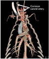
CT examinations were successfully performed in all six micropigs. There was no evidence of vascular malformation, arterial stenosis, aneurysm, atherosclerosis, or calcification found in any animal. In the present study, we measured the diameters of the major systemic vessels and compared those data to previously published human data (Table 1). The mean diameters of the right and left common carotid arteries measured were 0.57 ± 0.08 cm and 0.55 ± 0.05 cm, respectively (Fig. 1). There were no significant differences between micropigs and humans with regard to anatomy or diameter of the common carotid arteries.
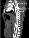
The mean diameters of the micropig ascending and descending thoracic aorta, aortic arch, and SVC were 1.69 ± 0.12 cm, 1.23 ± 0.11 cm, 1.50 ± 0.07 cm, and 1.93 ± 0.33 cm, respectively. The anatomic structure of the thoracic aorta and aortic arch of the micropigs was similar to that of humans (Fig. 2), but the diameters of these vessels were considerably smaller than those in humans. In addition, the significant anatomical differences in SVC of micropig compared with human were not observed.
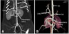
In the abdominal region, we evaluated the abdominal aorta, celiac trunk, superior mesenteric artery, splenic artery, and hepatic/suprarenal/infrarenal IVC. The mean diameters of these vessels were 0.85 ± 0.06 cm, 0.52 ± 0.08 cm, 0.68 ± 0.05 cm, 0.38 ± 0.05 cm, and 1.65 ± 0.20/1.59 ± 0.21/1.26 ± 0.07 cm, respectively. There were no anatomical variations in the micropigs in relation to humans; however, the diameter of the abdominal aorta was significantly smaller than in humans (Fig. 3). In addition, there were no significant differences between micropigs and humans with regards to anatomy or diameter of the IVC.
In the pelvic region, the diameters of the external iliac artery and superficial femoral artery were 0.52 ± 0.05 cm, and 0.48 ± 0.05 cm, respectively which were 42.4% and 46.3% comparable to human vessels, respectively.
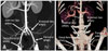
In all six micropigs examined, the external and internal iliac arteries arose directly from the aorta. There was no discernable common iliac artery in the micropigs. These findings were clearly different from the human vasculature (Fig. 4).
Discussion
Solid-organ transplantation is currently the definitive solution for end-stage organ failure. Accurate preoperative imaging of donor vasculature is of great importance because vascular variations, such as accessory arteries and early branching, are particularly important when determining optimal organ extraction procedures and the type of anastomosis [7,24,37]. Furthermore, imaging evaluation of vascular systems using MDCTA plays a critical role in solid-organ transplantation to facilitate the selection of suitable donors, planning the surgical procedure, and revealing any co-existing pathology [17,33]. The gold standard technique for preoperative donor evaluation is conventional angiography, but this procedure has the drawback of being invasive [4]. Angiography using MDCT is fast, safe, minimally invasive, and now is routinely used in the preoperative evaluation of potential human donors for renal and liver transplantation [12,35,40,41]. In this study, we performed anatomical evaluations and diameter measurements of the major systemic vessels in micropigs using 64-channel MDCTA. The morphology and branching patterns of the major vessels were constant between the micropigs and there were no anatomical variations found during this study. In addition, the morphology of the major micropig vessels did not reveal significant differences when compared to those of humans, except for in the case of the iliac artery. In all micropigs evaluated, the external and internal iliac arteries arose directly from the aorta. The external artery detached one branch, the deep femoral artery, which continued as the femoral artery. There was no common iliac artery corresponding to that of humans, which arises from the aorta and branches off into the external and internal iliac arteries. Although differences in vascular diameter, morphology, and branching pattern between micropigs and human [2,19,25,30,39] can be overcome with modern surgical techniques at the time of transplantation, there is the possibility that the function of the related micropig organs could be compromised in human systems following transplantation. Thus, further studies are needed to evaluate and compare micropig organ function with that of humans.
In addition, the smaller diameter of micropig arteries compared to human vessels [11,16,31] may be problematic in terms of perioperative complications. It has been suggested that a smaller diameter donor artery may contribute to an increased incidence of post-transplantation complications. For example, hepatic arteries with diameters less than 3 mm are considered to present a high surgical risk for liver transplantation [15]; thus, accurate preoperative evaluation of the arterial diameter is essential for successful organ transplantation. Previous studies reported that CTA can replace conventional angiography traditionally used for preoperative evaluation of potential organ donors [4,21,22]. Along with the rapid evolution in technique, the number of detectors has gradually increased, allowing shorter scan rotation times, submillimeter slice acquisition parameters, and isotropic datasets [9,17,18,30]. MDCTA appears to be an ideal method to evaluate hepatic arteries and venous anatomy, as well as detect potential hepatic transplant complications such as hepatic artery and/or portal vein stenosis or thrombosis [10]. In addition, MDCTA has been reported to be as accurate as renal angiography for evaluating the arterial anatomy [29,34] and more sensitive for detecting venous and parenchymal structures [23]. Therefore, MDCTA is a suitable method to evaluate the anatomy of vascular structures of potential xenotransplantation donors as well as human recipients.
In conclusion, we present CTA data for the major systemic vessels in micropigs, which can be used as standard reference values for xenotransplantation studies. We have determined that 64-channel MDCTA allows accurate evaluation of the major systemic vasculature in micropigs.

 XML Download