

 PDF
PDF ePub
ePub Citation
 Print
Print


Caudolateral curvilinear osteophyte (CCO) [6], which is a thickened osteophyte at the site of the joint capsule attachment on the caudal aspect of the femoral neck, has been advocated as one of the radiographic criteria for hip subluxation [7]. Occasionally referred to as 'Morgan's line', CCO is considered to be an important finding for the early detection of canine hip dysplasia (CHD) [10] and for diagnosing CHD and hip subluxation [8,11]. However, its diagnostic significance remains debatable because of past reports that do not consider CCO as a diagnostic criterion in the absence of subluxation [1,2] and suggest that longitudinal research need to be performed to evaluate its diagnostic utility and significance [6].
The purpose of this study was to assess the relationship between CCO size on three-dimensional computed tomographic (CT) images to the presence of CCO on radiographs.
Data obtained from 22 clinically healthy Border Collies (age, 7-120 months; weight, 11.1-22.0 kg; gender, 7 males and 15 females) were analyzed. Gait abnormalities and the Ortolani sign were absent in all the dogs, and the radiographs revealed no evidence of osteoarthritis. Radiographic and CT procedures were performed under general anesthesia. Anesthesia was induced with an intravenous injection of 4.0 mg/kg propofol (Rapinovet; Schering-Plough Animal Health, Japan). All experiments were approved by the animal experiments committee of the Obihiro University of Agriculture and Veterinary Medicine.
All CT images were acquired from the wing of the ilium to the ischial tuberosity by using a multidetector-row CT scanner (Asteion Super 4; Toshiba, Japan) with the following technical parameters: 0.5 mm slice thickness, 120 kV, 150 mA, 1.0 sec/rotation. The dogs were placed in the weight-bearing position on the CT table [3,4].
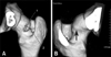
The presence or absence of CCO on the radiographs (radiographic-CCO) in 44 femurs was reviewed by veterinarians. Cross-sectional CT images, including those of the acetabulum and femoral head, were constructed into a three-dimensional image by an image processing workstation (Virtual Place Advance PLUS; AZE, Japan). Next, the lengths (mm) of the minor and major axes of the CCO in the three-dimensional images (3D-CCO) were measured, and its multiplier was applied to the index of the CCO size (CCO index, Fig. 1A). Then the coxofemoral joint evaluation was performed using the transverse CT images. The dorsal acetabular rim angle (DARA) [9], the dorsolateral subluxation score (DLS score) [3], the center distance index (CD index) [4], and the lateral center edge angle (LCEA) [4] were calculated using the measurements obtained from the transverse CT images, which included the largest diameter of the femoral head [5]. In addition, correlation coefficients were calculated to assess the relationships between radiographic-CCO, 3D-CCO, age, gender, body weight, and the CT parameters (DARA, DLS score, CD index, and LCEA). All correlations were analyzed using Spearman rank correlation. p values <0.05 were considered statistically significant.
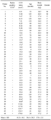
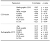
Data regarding the radiographic-CCO, CCO index, age, body weight, and gender of the 44 femurs are shown in Table 1. Radiographic-CCO was detected in 14 of the 44 femurs (31.8%). From the three-dimensional images, 3D-CCO was detected in all the 44 femurs. The mean CCO index radiograph positive CCO femurs (14 femurs) was 81.0 and was 23.5 for the femurs that did not have CCOs appear on radiographs (30 femurs). Radiographic-CCO was detected in 2 femurs with a CCO index lower than 23.5 (femurs No. 13 and 15). In contrast, there were femurs for which CCO could not be detected even when the CCO index was greater than 81.0 (femurs No. 38 and 39). The correlation coefficients and p value of each parameter are shown in Table 2. The CCO index correlated positively with radiographic-CCO, and also with DARA and the CD index. In addition, there was a negative correlation between the CCO index and age. Radiographic-CCO correlated with DARA and the CD index. Both the CCO index and radiographic-CCO did not correlate with body weight or gender.
In this study, there was a positive correlation between the CCO index and radiographic-CCO. The result suggested that a larger CCO is more detectable on radiographs. The CD index reflects the laxity of the coxofemoral joint, and DARA reflects the reduction of the dorsal acetabular rim. Hence, the correlation between the CCO index, radiographic-CCO, CD index, and DARA suggests the possibility that the CCO enlarges with the progression of coxofemoral subluxation and becomes detectable on radiographs. However, a larger CCO is not necessarily detected on radiographs; in this study, there were femurs with a small CCO index (lower than 23.5) for which radiographic-CCO was detected, and femurs with a large CCO index (greater than 81.0) for which radiographic-CCO was not detected. Hence, it is likely that the mass density, thickness of the CCO, and direction of the radiographic beam play a role in the detection of radiographic-CCO [7].
In addition, there was no correlation between radiographic-CCO and age, while there was a negative correlation between the CCO index and age. A possible cause for this observation is the age-related relative decrease in 'remarkable projection' of the CCO. A three-dimensional CT image of the CCO of the oldest dog in this study is seen in Fig. 1B. The femoral neck has thickened around the joint capsule, but compared with Fig. 1A, there is no 'remarkable projection'. The osteophytes have to form the 'remarkable projection', which is clearly differentiated from the surrounding area and is detected as a 'line' on radiographs. It is possible that if the width of the 'remarkable projection' of the CCO increases with age and forms a 'gently raised region', it may not be detected on radiographs. Therefore, radiographic CCO cannot be considered as highly reliable, especially for the evaluation of dogs with severe subluxation or older dogs.
In conclusion, it was determined that radiographs do not necessarily depict CCO accurately. Therefore, radiographic-CCO findings should be applied cautiously in evaluation of the hip in Border Collies. Future studies should be performed with various breeds of dogs.
 XML Download