

 PDF
PDF ePub
ePub Citation
 Print
Print


Introduction
In general, atopic dermatitis occurs mainly from dysregulation in helper T (Th) cell reactivities [1,4]. The Th cell is classified into two types, types 1 and 2, depending on type of cytokine secreted by the cell and immunoregulatory functions driven by the secreted cytokine [11,22,29]. Any disruption of the balance between these two types of Th cells may cause a variety of immunological diseases, such as allergic asthma and allergic rhinitis. Atopic dermatitis is an immunologic disease induced from an imbalance favoring type-2 helper T (Th2) cell [3].
Interleukin-4 (IL-4) production from Th2 cells, mast cells, or other immune components cells is known to trigger isotype-switching to IgE and IgG1 in B cells [18]. Evaluation of IL-4 production from splenic T cells is a tool for investigating the predominance of Th2 reactivity [13]. Hyper IgE production is a hallmark of atopic dermatitis in humans [1,4], and well documented in transgenic mice or NC/Nga mice suffering from atopic dermatitis-like skin lesions [8,26,33]. IFNγ from type-1 helper T (Th1) cells, natural killer cells, other immune cells induces isotype-switching to IgG2a in mice [17,31]. Therefore, analysis of the serum IgG1 or IgG2a levels could reflect the alteration of homeostasis between type-1 and type-2 immune responses. Since histamine is immediately released following allergen exposure, increase in serum histamine level is often considered as a parameter for diagnosing the onset of allergic diseases. Previous studies also showed that eosinophils play an important role in atopic dermatitis, characterized by elevated blood eosinophil counts [17,30].
Atopic dermatitis is a chronic skin disorder and has been medically treated using steroids, antihistamines, immunosuppressive agents, and other medications. But many studies have reported that long-term use or even abuse of these agents may cause various side effects, so relevant recent studies have focused upon complementary therapies based on alternative medicine [9,28].
Aromatic oils are natural essential oils consisting of volatile organic compounds containing original scents and therapeutic substances. It is well known for positive effects in insomnia, relief from anxiety and stress, recovery from fatigue, muscular relaxation, anti-histaminic or anti-allergic actions [5,7,27]. German chamomile (GC) oil, an aromatic oil, has been reported to help relieve physical or mental stress or fatigue through its sedative or alleviating effects along with fruity scent, is also known to be effective in the treatment of dry and itchy skin [23]. Historically, GC oil has been used for the treatment of skin disorders such as eczema, as it contains three major sesquiterpene constituents (azulene, bisabolol, farnesene) with anti-inflammatory or anti-histaminic effects [6,32]. Particularly, it is known that these effects of GC oil come from α-bisabolol, a substance that has strong anti- inflammatory effects of all. However, there have been no scientific studies to date on the possible effects of GC oil on alleviating atopic dermatitis. Therefore, this study tested the application of GC oil on BALB/c mice demonstrating atopic dermatitis-like immune alteration and skin lesions. The 2,4-dinitrochlorobenzene (DNCB) skin application model was adopted to induce atopic dermatitis-like phenomena in mice, which has been reported as a reliable model demonstrating similar immunologic and skin alteration as human atopic dermatitis [24].
Materials and Methods
Composition of GC oil
For the experiment, this study used GC oil (Sanoflore, France) as 100% pure and natural essential oil tested by organic product certification organization (ECOCERT, France) as well as jojoba oil (Desert Whale, USA). Jojoba oil was used as a control due to its clinical application.
Animals
Forty BALB/c mice (SPF male, 7 weeks old) were purchased from Daehan Biolink (Korea). Both animal care and protocol for this study were in accordance with Institutional Animal Care and Use Committee (USA) and OECD guideline. Animals fed on unlimited amount of water and feed throughout whole experiment duration. The animals were divided into four groups (ten mice each) and were housed separately in plastic cages per each group. The normal group was applied with saline throughout atopic dermatitis induction stage and oil treatment period. The control group was applied with saline following induction of atopic dermatitis. The vehicle group was applied with jojoba oil and the experimental group with 3% GC oil following atopic dermatitis induction (Fig. 1). Three % GC oil was selected in accordance to the clinical application.
Induction of atopic dermatitis-like immunologic and skin disorders
For induction of atopic dermatitis-like immunologic and skin disorders, DNCB was applied onto mice skin. After complete removal of dorsal hairs in area of approximately 8 cm2, 100 µL of 1% DNCB was applied on their dorsal skin twice every 3 days for sensitization. In the next week, 100 µL of 0.2% DNCB was applied on their dorsal skin twice every 3 days for challenge. DNCB was dissolved in a 4 : 1 mixture of acetone and olive oil. As soon as the challenge was completed, 3% GC oil (diluted in jojoba oil) was applied once a day (70 µL, 6 times week) on the dorsal skin for 4 weeks. Saline or jojoba oil was applied onto skin of the control mice. Three hundreds µL of blood was sampled at the completion of second challenge and 2 weeks after initiating oil application via periorbital sinus. After the 4-week oil application period, under ether anesthesia blood sampled from posterior vena cava was used for immunologic or hematologic analysis, and skin tissue was sampled and fixed with 10% neutral buffered formalin solution for histopathologic examinations, splenic cells were sampled by separation from the extracted spleen and stored in a freezer after freezing in liquid nitrogen for cytokine analysis.
Immunologic observation
Measurement of serologic parameters
Serum IgE levels were determined using a sandwich ELISA method, as previously described [15]. The levels of serum IgG1 and IgG2a were also determined using a sandwich ELISA method, using goat anti-mouse IgG1 and IgG2a capture antibodies and a peroxidase conjugated anti-mouse IgG detection antibody [16]. In addition, serum histamine levels were determined using the IBL histamine ELISA kit (IBL, Germany) according to the quantification methodology of Duda et al. [10].
Measurement of IL-4 in splenic cell culture solution
Splenic cells (5 × 10⁵ cells) were stimulated with immobilized anti-CD3 mAb (5 µg) for 48 h in a CO2 incubator. The level of IL-4 in culture supernatants was determined using a sandwich ELISA method prepared by BD Pharmingen (BD, USA) [15]. The lower limit of detection was 5 pg/mL for IL-4.
Behavioral observation
Behaviors of mice were monitored according to observation methodology of Kobayashi et al. [20]. The frequency of scratching occurring on facial or dorsal skins was counted with a 30-min visual observation on the first day and on the 21st day after GC oil application.
Results
GC oil-mediated suppression of IgE hyperproduction
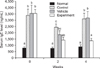
Mice at the completion of 0.2% DNCB second challenge demonstrated significantly higher levels of serum IgE (approximately 4.4 times) than the normal group (Fig. 2), implying a successful induction of atopic dermatitis-like immune alteration. Serum IgE levels were significantly downregulated in the experimental group after application of GC oil for 4-weeks, while the saline-applied control or the jojoba oil-applied vehicle groups still demonstrated significantly upregulated IgE levels compared to the normal mice.
Modulation of serum IgG1 level following GC oil application
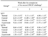
Levels of IgG1 and IgG2a were substantially increased following DNCB sensitization and challenge compared to the normal mice (Table 1). Application of GC oil for 4 weeks resulted in a reduction of approximately 31% (13.75 mg/mL) in IgG1 levels compared to 19.80 mg/mL after 2-weeks of GC oil application. There were no significant differences in the levels of IgG1 in saline-applied control or jojoba oil-applied vehicle groups between at 2 week and at 4 week following atopic dermatitis induction. Meanwhile, persistent application of GC or jojoba oil did not lead to any changes in IgG2a levels when compared between 2 and 4 weeks following the induction of atopic dermatitis.
Effect of GC oil application on modulation of serum histamine level
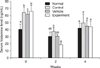
Histamine levels were significantly higher in all mice groups exposed to DNCB compared to the normal mice (Fig. 3), indicating a successful induction of atopic dermatitis-like alterations of the immune system. Two weeks after GC oil application, the GC oil group (18.45 ng/mL) showed significantly lower (approximately 51%) serum histamine level than the saline-applied control (37.43 ng/mL), and also lower (approximately 40%) than the jojoba oil-applied vehicle (30.60 ng/mL) group. However, no further downregulation of histamine levels was found 4 weeks after the start of treatment.
Effect of GC oil application on production of IL-4 from splenic T cells
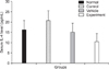
Four weeks of GC oil application (10.4 pg/mL) resulted in lower (approximately 50%) IL-4 production with no statistical significance (p > 0.05) in splenic cell culture supernatants compared to the saline treated control mice (20.7 pg/mL) (Fig. 4). These findings imply that GC oil controls the production of IL-4 from Th2 cells, contributing to controlling IgE and IgG1 generation from B cells.
Decreased scratching frequency in GC oil-applied mice
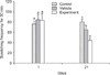
The frequency of scratching on facial and back skin was observed for 30-min on one day after the second DNCB challenge (day 1) and on the 21st day after initiating GC oil application (day 21). It was found that the GC oil group (44 times) and jojoba oil group (65 times) showed significantly lower (approximately 45% and 19%, respectively) scratching frequency than the saline treated control group (80 times) at day 21 (Fig. 5). Furthermore, scratching frequency of the GC oil group was significantly lower (approximately 32%) than the jojoba oil vehicle group.
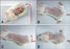
Decreased inflammatory cell infiltration in GC oil-applied mice skin
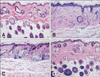
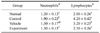
Numerous leukocytes were infiltrated into the dermis and epidermis was thickened in the control mouse skin compared to the normal mouse skin, while inflammatory cell infiltration, such as neutrophils and lymphocytes, and hyperplasia of the epidermis were significantly (p < 0.05) reduced in the experimental mouse skin (Table 2 and Fig. 6). In this study, it was found that application of GC oil for 4 weeks resulted in significantly lower leukocytes in the peripheral blood than the saline treated control mice, and showed similar values to the normal mice (Table 3). The number of neutrophils, lymphocytes, monocytes, eosinophils and basophils of GC oil application for 4 weeks showed significantly (p < 0.05) lower levels than the saline treated control mice by 31, 33, 42, 50 and 50%, respectively (Table 3).
Discussion
The present study was undertaken to evaluate the efficacy of GC oil alleviating atopic dermatitis-like immune alterations in mice. Even though GC oil has been historically used for the treatment of pruritic skin disorder [2,6,12,32], there has been no scientific study investigating GC oil on the treatment or prevention of atopic dermatitis. Our study is the first to demonstrate GC oil's immunoregulatory potential for alleviating atopic dermatitis through influencing of Th2 cell activation. Overall, we found that GC oil possessed an ability to influence Th2 cell activation involved with the onset or progression of atopic dermatitis, in that dermal application of GC oil resulted in the suppression of IgE or IgG1 over-production, downregulation of IL-4 production from Th2 cells, and demonstrated a reduction in histamine release. In addition, frequent scratching due to itchy sensations was alleviated following GC oil application onto the damaged skin involved with the pathogenesis of atopic dermatitis.
Histamine release from activated mast cells or basophils is a hallmark of the occurrence of allergic diseases such as atopic dermatitis, and cause itching, increased vascular permeability, and the wheal and flare response of immediate hypersensitivity [1]. Pruritus is a symptom found in a variety of skin disorders like atopic dermatitis and contact allergic dermatitis. For atopic dermatitis cases, it has been reported that scratching due to itchy sensations causes skin damage, promotes inflammation and thereby further aggravates pruritus [1,4,21,34,35]. Therefore, it is important to reduce itchy sensations and scratching frequency to prevent aggravation of skin lesions due to pruritic disorders and improving quality of life.
Kobayashi et al. [19,20] reported that the oral administration of GC extract combined with histamine receptor antagonists was more effective for the suppression of scratching behavior in mice than administration of histamine receptor antagonists alone. GC oil may inhibit the binding of histamine to its receptor, which will eventually control histamine mediated clinico-pathologic effects such as itching [20]. Concerning our results showing the downregulation of serum histamine levels 2 weeks after the start of GC oil application following atopic dermatitis induction, this compound may also exhibit its anti-histamine effect by directly suppressing histamine release from mast cells through downregulation of IL-4 production from Th2 cells and following by suppression of IgE or IgG1 over-production.
The most important aspect to be discussed is GC oil's systemic effect on the modulation of the in vivo type-2 response. The type-2 response becomes predominant when differentiation or activation of Th2 cells is preferred, but development or stimulation of Th1 cells is suppressed [13,14]. Considering that the skewedness toward type-2 responses is a background mechanism for the occurrence of atopic dermatitis [1,4,25], dermal application of GC oil could systematically block the pathogenesis of atopic dermatitis through correcting the immune homeostasis skewed in favor of Th2. Considering our experimental results, it could be concluded that the application of GC oil on the skin of atopic dermatitis cases could alter production of IL-4 from Th2 cells and also alter IgE, IgG1 and histamine production, performing a series of immunoregulatory functions to alleviate the occurrence or progression of atopic dermatitis. Hence, it is believed that the application of GC oil on atopic dermatitis cases will be significantly meaningful for public health and alternative medicine.
Although it is known that α-bisabolol has the strongest anti-inflammatory effects of all the constituents of GC oil, it is not clear which constituent(s) of GC oil contribute to the GC oil-mediated alleviation of atopic dermatitis-like immunologic and skin alterations in mice at the moment. Future studies will be directed towards identifying which components of GC oil are responsible for its immunomodulatory effects. Furthermore, whether GC oil exerts its immunomodulatory effects independently, sequentially, or concomitantly on each of the immune component cells involved in the pathogenesis of atopic dermatitis such as Th, B, and mast cells needs to be answered. Further investigations are necessary to identify target immune cells for GC oil-mediated alleviation of atopic dermatitis-like immunologic and skin alterations.

 XML Download