

 PDF
PDF ePub
ePub Citation
 Print
Print


Tendon and ligament injuries are among the most common musculoskeletal injuries and contribute to a considerable economic loss for the equine industry [5,8,10,14]. New regenerative therapies are thought to harness the animal's ability to repair the injury site and to reduce scar tissue formation [6].
Seven horses of different breeds - 3 mares, 4 geldings, aged 4 to 12 years, used as sport and/or pleasure riding horses - were included in this study. The horses were presented at the clinic and met the following criteria: acute severe tendinitis of the superficial digital flexor tendon (SDFT) or deep digital flexor tendon (DDFT), or acute desmitis of the inferior check ligament (ICL) of one or both forelimbs; no treatment prior to admission; and otherwise normal results of the clinical examination. All horses were subjected to a general examination with whole blood analysis and a lameness examination. Tendons and ligaments were evaluated ultrasonographically and the damaged tendon/ligament areas were documented, with evaluation of the following parameters: the cross sectional area (CSA; cm2), the percentage of the maximum injury zone compared to the size of a healthy tendon/ligament (%MIZ), the echogenicity score (ES) of the lesion (range: 0 to 4), and the fibre alignment score (FAS: range 0 to 4). The FAS was determined in the longitudinal view of the lesion [12]. Autologous conditioned plasma (ACP) therapy was performed in each horse (n = 7) at the day of presentation at the clinic (D0), and also after a 2-week interval (D1) in horse SDFT 2. Autologous conditioned plasma was prepared using a 15 mL ACP double syringe (Arthrex GmbH, Germany) according to the manufacturer's instructions. An average of 5 mL of ACP was obtained from every 15 mL syringe. At D0, a 1 mL fraction of the ACP was analyzed for hematocrit (Hct), platelets and white blood cell (WBC) count. After preparation the ACP was injected into the tendon/ligament-defect through a 21 gauge needle under ultrasonographic control. The injected volume (2 mL to 4 mL) depended on size and type of the lesion. The horses were subjected to stable confinement for 28 days. Thereafter they were hand-walked for 15 min each day, with the time progressively extended up to one hour in the coming months. Follow-up for each horse was obtained twice by clinical and ultrasonographic examination in 2 to 3 week intervals (D1 and D2). Follow-up at 7 to 9 months as well as at 10 to 13 months (D3) was obtained by phone through questioning of the owners, trainers and/or referring veterinarians.
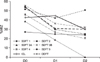
Five horses had acute SDFT lesions (zone 1A-3B) with one of them showing bilateral lesions (n = 6 limbs). One horse had an ICL injury (zone 2A; n = 1) and one horse presented with a core lesion of the DDFT (zone 2A; n = 1). The %MIZ of the SDFT lesions (n = 6) ranged from 27.23% to 54.20% at D0 and decreased at D1 and D2 in all horses except for one horse (SDFT 2) which showed a higher %MIZ at D2 compared to D1 (Fig. 1). The CSA decreased continuously in the SDFT lesions from D0 to D2 in all horses except for one horse (SDFT 2) in which it increased between D1 and D2 (Fig. 2). The ES and FAS decreased continuously from D0 (ES: 2 to 3; FAS: 1 to 3) to D2 (ES: 0 to 1; FAS: 0 to 1) in all horses except for horse SDFT 2, which showed an increase in ES between D1 (ES:1) and D2 (ES:2), and horse SDFT 3B which had an unchanged FAS between D0 and D2. The %MIZ for the DDFT lesion (n = 1) was 53.10% at D0. The %MIZ for the ICL lesion (n = 1) was 42.80% at D0. The %MIZ of the DDFT- and ICL-lesions decreased till D2 (Fig. 1). The CSA decreased continuously in DDFT- and ICL-lesions from D0 to D2 (Fig. 2). The ES improved from D0 (ES:2) to D2 (ES:1). The FAS improved for the DDFT lesion from D0 (FAS:3) to D2 (FAS:2) and the ICL-lesion from D0 (FAS:1) to D2 (FAS:0). Follow-up of all horses at 7 to 9 months post injury and at D3 revealed a positive outcome with all horses either performing at their previous work-load or were back in full training. No horse experienced re-injury. Whole blood and ACP analysis (all horses) revealed that platelet counts were on average 1.3-fold higher in ACP compared to whole blood. The platelet count in ACP ranged from 160 × 109 platelets/L to 197 × 109 platelets/L, compared to the platelet blood values ranging from 115 × 109 platelets/L to 173 × 109 platelets/L. On average, the WBC count was 10.92-fold lower in ACP compared to whole blood. The WBC count in ACP ranged from 0.44 × 10⁹ cells/L to 1.58 × 10⁹ cells/L compared to WBC values in whole blood which ranged from 6.41 × 10⁹ cells/L to 11 × 109 cells/L. The Hct was 3.26 × 10³-fold lower in ACP compared to whole blood. The Hct count in ACP ranged from 0% to 0.01% compared to that of whole blood which ranged from 30% to 36%.
Tendinitis and desmitis result in a considerable economic loss to the equine industry due to decreased performance, prolonged rehabilitation, recurrent injuries and early retirement. Treatment methods that induce the regeneration of healthy tendon tissue, i.e. restoration of the tendon's normal structural architecture and biomechanical function rather than formation of scar tissue should therefore decrease the risk of re-injury [6]. New regenerative treatment options are thought to recapitulate processes taking place during embryonic development, with temporal and spatial interactions between scaffold, growth factors and cells. Growth factors are molecules which regulate cellular metabolism. They have an anabolic effect on tendon and ligament tissues through stimulation of cell proliferation, increase of extracellular matrix synthesis and promotion of vascular invasion. They also down-regulate catabolic matrix degradation cytokines e.g. matrix-metalloproteinases and interleukins [3]. Platelet rich plasma (PRP), an established growth factor enhanced therapy strategy, is a concentrate of platelets which release growth factors, including platelet-derived growth factor, transforming growth factor-β, insulin-like growth factor I, epidermal growth factor, vascular endothelial growth factor and fibroblast growth factor from the platelet α-granules [4,11,15].
The data obtained in this clinical case study are based on a small number of horses used for various disciplines (racing, pleasure riding, dressage or show jumping) referred for various tendon and ligament lesions. The regenerative therapy method of intralesional injection of ACP was chosen based on the encouraging results with PRP and bone marrow-ACP treatments in equine orthopaedics [2,7]. The data obtained in this case-study are encouraging and reveal that there could be remarkable positive changes in the early stages of injury healing, expressed by %MIZ, CSA, ES and FAS, of both tendinitis and desmitis if defect areas are directly treated with ACP. Partly, these effects could be explained with a coincidence with conditions and phases of normal tendon healing. Nevertheless, our data reflect cases of severe tendon lesions with %MIZ values greater than 25%; such values are associated with a poor prognosis [13]. All seven horses, with lesions of the SDFT, DDFT and ICL, had %MIZ values far higher than this critical value but nevertheless underwent good recoveries. Horse SDFT 2 showed sufficient healing of the SDFT lesion at D1. The horse received a second ACP-injection at D1 and had a subsequent deterioration of tendon structure and texture at D2 (Figs. 1 and 2). Nevertheless the horse recovered completely within five months. The described case underlines the fact that a repetitive intralesional ACP-injection has to be evaluated critically. Follow-up data of the horses described in this clinical case report - performed by interviewing the owners, trainers and/or referring veterinarians - revealed a positive outcome at 7 to 9 months as well as in an intermediate time range (10 to 13 months post injury, D3). All horses treated with ACP were either performing at their previous work-load or were back in full training. None of the horses treated with ACP showed signs of re-injury. These findings are consistent with those of another study in which 40% of bone marrow-ACP treated horses were sound and 60% of treated forelimbs showed an ultrasonographic improvement within four to six weeks [7]. Recent investigations into the biochemical, biomechanical and histological properties of tendons treated intralesionally with PRP in a placebo-controlled experimental study reflected a positive outcome of PRP-therapy after 24 weeks [3].
The ACP platelet count was 1.3-fold higher than that of whole blood. In contrast, PRP platelet counts have been found to be equal to or higher (range: 1.3- to 13.1-fold) than that of whole blood [9,11,16]. The ACP preparation used in this study had a mean 10.92-fold decrease in WBC counts compared to whole blood. In comparison, PRP preparations have shown either a lower decrease (mean 1.85-fold) or an increase in mononuclear cell concentration compared to whole blood [1,9,15,16]. Autologous conditioned plasma - in contrast to PRP - might therefore be less likely to increase disturbing inflammatory tissue reactions due to WBCs during regenerative therapy.
In conclusion, the limitations of this intermediate-term clinical study are the small number of horses included, their heterogenic use and breed and the variability of the tendon and ligament lesions treated with ACP. Further studies with a homogenous horse population and injury profiles, as well as long-term follow-up, need to be performed to strengthen these clinical observations.

 XML Download