

 PDF
PDF ePub
ePub Citation
 Print
Print


Introduction
Cyclic guanosine monophosphate (cGMP) has become increasingly important in the understanding of antinociceptive mechanisms in the central nervous system [1,13,16,21]. The concentration of intracellular cGMP is regulated by the action of guanylyl cyclase, as well as by its rate of degradation by cGMP-specific phosphodiesterase [2,14]. The increased cGMP may activate protein kinase G (PKG), which may regulate calcium channels [18]. It has been known that calcium ions are widely recognized as having a fundamental role in the central nervous system by modulating neuronal transduction in the spinal cord [9]. There are four types of calcium channels (L-, N-, T- and P-types) in the central nervous system [12,19]. These findings suggest that the increased cGMP may alter the activity of the calcium channels through the activation of PKG, thereby leading to a modulation of nociceptive processing.
Sildenafil (Viagra) is clinically used to treat impotence as a cGMP-specific phosphodiesterase 5 inhibitor [4,10] and produces antinociception in animal pain model, which is mediated through the nitric oxide-cGMP-PKG-potassium channel pathway or opioid receptors [1,13,21]. It has been recently reported that sildenafil inhibits cerebroarterial vasoconstriction by blocking L-type calcium channels [15]. Furthermore, the blocking of L-type calcium channels produced antinociceptive effects at the spinal level [8,11]. The aforementioned findings jointly suggest that activation of PKG followed by L-type calcium channel inhibition may be involved in the effect of sildenafil.
Therefore, the aim of this study was to assess the possible role of PKG and L-type calcium channels on the activity of sildenafil on formalin-induced nociception at the spinal level.
Materials and Methods
Intrathecal cannulation
This study was approved by The Institutional Animal Care Committee, Research Institute of Medical Sciences, Chonnam National University. Adult male Sprague-Dawley rats weighing 250~300 g were used in all experiments. Rats were allowed free access to a standard rat diet and tap water. Room temperature was maintained at 20~23℃ with a 12 : 12 h light/dark cycle. Intrathecal catheters (PE-10) were implanted via the cisterna magna in enflurane-anesthetized rats [20]. The catheter was carefully advanced caudally by 8.5 cm through an incision in the atlantooccipital membrane to the lumbar enlargement. The exterior part of the catheter was tunneled under the skin, externalized on the top of the head, and plugged with a stainless steel wire. The skin was closed with 3~0 silk sutures. Rats showing postoperative neurological deficits were killed immediately with an overdose of volatile anesthetics. Normal rats were kept in individual cages and the formalin test started approximately 7 days after intrathecal catheterization.
Drugs
The following drugs were used in this study: sildenafil (Pfizer Korea, Korea), (9S,10R,12R)-2,3,9,10,11,12-Hexahydro-10-methoxy-2,9-dimethyl-1-oxo-9,12-epoxy-1H-diindolo[1,2,3-fg:3,2,1-kl]pyrrolo[3,4-i][1,6]benzodi azocine-10-carboxylic acid, methyl ester (KT 5823; Tocris, UK), and 2,5-Dimethyl-4-[2-(phentylmethyl)benzoyl]-1H-pyrrole-3-carboxylic acid methyl ester (FPL 64176; Tocris, UK). Sildenafil and FPL 64176 were dissolved in 20% dimethylsulfoxide (DMSO; Sigma-Aldrich, USA), and KT 5823 was dissolved in normal saline. All the drugs were delivered as a 10 µL solution.
Nociceptive test
Nociception was induced by subcutaneous injection of formalin (5%, 50 µL) into the plantar surface of the rat hind paw [21]. Immediately after formalin injection, the rats showed a specific nociceptive behavior (flinching), which was characterized as a rapid and brief withdrawal or flexing of the affected paw. Such pain behavior is quantified by periodically counting the number of flinches of the injected paw. The flinching response was counted for 60 second periods from 1 to 2 min and 5 to 6 min, and then at 5 min intervals during the interval from 10 to 60 min. As a biphasic flinching response was observed after the formalin injection, a 0~10 min interval was defined as phase 1 of the formalin test, whilst the interval from 10~60 min was defined as phase 2. The rats were killed using an overdose of volatile anesthetics directly after the formalin test to minimize suffering.
Experimental design
The effect of intrathecal sildenafil (1.5, 4.5, 15, 45 nmol/L), administered 10 min before the formalin injection, was examined. Rats were pretreated with KT 5823 (PKG inhibitor), or FPL 64176 (L-type calcium channel activator) to assess the involvement of the PKG and L-type calcium channel activity on the antinociceptive effect of sildenafil. These two drugs were administered intrathecally 10 min before the delivery of intrathecal sildenafil (45 nmol/L). The maximum doses of KT 5823 (0.02 nmol/L) and FPL 64176 (0.9 nmol/L) were selected based on the lack of any significant effect on the controlling of the formalin response from the pilot experiments of a previous study [1]. The control study was performed using intrathecal saline or DMSO depending on the solvent for the experimental drug. Animals were experimentally naïve and tested only once in this study. In total, 88 rats were tested in this study and the number of rats per group was 5~8. The investigator was unaware of the drug given to the experimental animals.
Statistical analysis
Data are expressed as mean ± SE. The dose-response data is shown as the sum of the number of flinches in two phases. The dose-response data of sildenafil and the antagonism for the effect of sildenafil were analyzed by one-way analysis of variance with Fisher's PLSD for the post hoc test, with p < 0.05 being considered statistically significant.
Results
Antinociceptive characteristics of intrathecal sildenafil
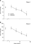
A subcutaneous injection of formalin into the hindpaw resulted in a biphasic flinching response of the injected paw. Intrathecal sildenafil, administered 10 min before the formalin injection produced a dose-dependent suppression of the flinching response during phase 1 and phase 2 in the formalin test (p < 0.05, p < 0.01, p < 0.001; Figs. 1A and B).
PKG-L-type calcium channel to the activity of sildenafil
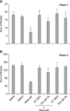
Neither intrathecal KT 5823 nor FPL 64176 when given alone modify the flinching response in control animals at the doses used in this study. Intrathecal KT 5823 and FPL 64176 reversed the antinociceptive effect of intrathecal sildenafil in both phases (p < 0.05, p < 0.01; Figs. 2A and 2B).
Discussion
In the present study, the flinching response decreased in a dose-dependent manner both in the first and second phases after treatment with intrathecal sildenafil. This observation suggests that there is a significant participation of spinal phosphodiesterase 5 in the formalin-induced nociception, and that the inhibition of this enzyme is effective in attenuating the facilitated state pain as well as acute pain in the spinal cord. The antinociceptive action of intrathecal sildenafil on the formalin-induced nociception was consistent with previous findings [1,13,21].
Phosphodiesterase enzymes exist extensively in biological systems [3]. It is an enzyme involved in the hydrolysis of cGMP. Eleven families of phosphodiesterase isoenzymes have been identified, all of which have different physical characteristics, cellular distribution, and selective sensitivity of inhibitors [17]. An in situ hybridization study demonstrated the expression of phosphodiesterases 2, 5, and 9 in the spinal cord [7]. Among these, types 5, 6, and 9 have specificity concerning cGMP hydrolysis, type 5 exerting the most significant effects [14]. It has been suggested that cGMP is involved in central antinociception. This proposal was based on the observation that intrathecal 8-bromo-cGMP reduced the mechanical allodynia in neuropathic rats [16]. This indicates that the cGMP level might be increased by inhibiting this enzyme, thereby producing antinociception. Therefore, it is conceivable that sildenafil, a cGMP-specific phosphodiesterase 5 inhibitor, may exert an antinociceptive effect by inhibiting phosphodiesterase 5 and increasing cGMP concentration at the spinal level.
Here, intrathecal KT 5823 attenuated the antinociceptive effect of intrathecal sildenafil, suggesting that the increased cGMP by the inhibition of phosphodiesterase 5 may activate PKG in the spinal cord, similar to a previous study showing that a PKG inhibitor blocked the activity of sildenafil [1]. A primary action of elevated cGMP levels is the stimulation of cGMP-dependent protein kinase, the major intracellular receptor protein for cGMP. The activation of PKG would lead to phosphorylation and regulation of ion channels to exert its actions [18]. Intrathecal FPL 64176 also reduced the antinociceptive effect of sildenafil. These observations suggest that sildenafil may exert its antinociceptive effect by modulating L-type calcium channels in the spinal cord.
Calcium ions play an important role in the modulation of spinal pain processing [9]. The increase of intracellular calcium by means of intrathecal calcium ionophores or calcium agonists increased the flinching response to the formalin test [6]. Calcium channels are categorized into L-, N-, T- and P-types by their electrophysiological characteristics and pharmacological sensitivities [12]. Moreover, an immunohistochemical study demonstrated the localization of these four types of calcium channels in the spinal cord [19]. Therefore, it is assumed that the blocking of spinal calcium channels may afford antinociception. Indeed, spinal L-type calcium channel antagonists produced antinociception in an animal model of pain [8,11]. The above-mentioned findings jointly suggest the PKG-L-type calcium channel is sequentially involved in the antinociceptive action of sildenafil in the spinal cord.
Since we have not examined the role of N-, T- and P-type calcium channels on the effect of sildenafil in this study, the effect antinociceptive mechanisms related to other calcium channels on the activity of sildenafil can not be excluded. It is known that the modulation of N-, T- and P-type calcium channels are also responsible for antinociception. Intrathecal N and P-type calcium channel blockers produced antinociception [5,8], and cGMP inhibited the activation of N-type calcium channels in dorsal root ganglion neurons [22]. However, unfortunately, other calcium channel activators have not been commercially available. Future studies will be needed with selective calcium channels activators to further clarify the antinociceptive mechanisms of sildenafil with respect to all of the calcium channels.
Seen as a whole, the inhibition of phosphodiesterase 5 by sildenafil increases the cGMP levels in the spinal cord, which in turn decreases the activity of the L-type calcium channel through PKG activation, thereby attenuating the facilitated pain state as well as acute pain evoked by the formalin injection.
 XML Download