

 PDF
PDF ePub
ePub Citation
 Print
Print


Introduction
Endogenous retroviruses (ERVs) are integrated into the germ-line of vertebrates and transmitted to their offspring by Mendelian genetics. The genomic structures of ERVs include group specific antigen (gag), polymerase (pol), and envelope (env) genes which are flanked with 5' and 3' long terminal repeat (LTR) possessing regulatory elements [15]. The expression of ERV genes and their adjacent genes is controlled by transcription regulatory elements placed on the LTR [13]. The majority of ERVs are transcriptionally inactive because deletions and point mutations interrupt the coding potential of the gag, pol, and env genes [14].
The pig also harbors endogenous retroviruses, called porcine ERV (PERV). The first report on PERV was reported in the 1970 [4]. PERVs are the members of family Retroviridae, genus Gamma retrovirus and vertically transmitted like other ERVs [19]. PERV inserted its gene in the pig genome in approximately 30 to 50 sites [1] and 3 classes, PERV-A, PERV-B, and PERV-C, are known [17]. These classes display high sequence similarity in the genes coding for the gag and the pol but differ in the genes encoding the env proteins [21].
In xenotransplantation research, every cell or tissue from a porcine xenograft was thought to carry PERV and could act as a potential source of retrovirus, which could not be eliminated by keeping pigs under specific-pathogen-free conditions or by simple outcross-breeding protocols. Fortunately, there were no trans-species infections of PERV in many in vivo porcine cell or organ transplantation trials [11]. However, PERV-A and -B could successfully infect human originated cell lines in vitro [9,21]. In addition, the state of immunosuppressed patients cannot be excluded in xenotransplantation. Recently, an in vivo study of PERV infection into human cells in a nude mouse suggests the possibility of indirect human PERV infections [25].
In previous studies, many techniques including conventional polymerase chain reaction (PCR), reverse transcription (RT)-PCR [3,16,20], real time PCR, real time RT-PCR [2], and monoclonal antibodies [5,10] were developed to analyze the risk of PERV transmission. Comparisons of PERV mRNA expression patterns were conducted to determine the viral load in various porcine tissues. In that study, the kidney showed the highest expression levels and the pancreas showed the lowest. The assessment of viral load could potentially reduce the risk of PERV transmission [8], and help select appropriate donor pigs expressing low levels of PERV mRNA.
The objective of this study was to establish sensitive duplex RT-PCR protocols for detecting PERV gag mRNA and porcine glyceraldehydes 3-phosphate dehydrogenase (GAPDH) mRNA. In addition, this technique was used to compare the age-related expression levels of various tissues in commercial pigs.
Materials and Methods
Pigs
Twenty pure breed Duroc pigs were allocated into 4 different age (10, 40, 70, 110 days) groups. All animal experiments were in compliance with the current laws of Korea. Care and treatment of animals were conducted in accordance with the protocols and guidelines of the Seoul National University Institutional Animal Care and Use Committee, Korea.
RNA extraction
The organs used for this study were the lung, liver, spleen, kidney, heart, and pancreas. Each organ from a pig was collected and picked up 0.1 g of piece, separately. Collected tissue was minced and suspended in 1 mL of Dulbecco's modified Eagle media without fetal bovine serum. RNA was extracted using TRIzol (Invitrogen, USA) according to manufacturer's recommendations. Briefly, 250 µL of the homogenated samples was mixed with 750 µL of TRIzol and incubated for 15 min at room temperature. Following the addition of 200 µL of chloroform, the mixture was centrifuged at 12,000 × g at 4℃ for 15 min. After adding an equivalent volume of 2-propanol to the supernatants for RNA precipitation followed by 15 min incubation at room temperature, further centrifugation was performed at 12,000 × g at 4℃ for 10 min. RNA pellets were washed with 1 mL of 75% ethanol, and centrifuged at 12,000 × g at 4℃ for 5 min. Pellets were resuspended in 30 µL of diethylpyrocarbonate (DEPC)-treated deionized water after drying.
RT and PCR
Prepared RNA was treated with DNase (Promega, USA) for 30 min at 37℃ according to manufacturer's protocol. Reverse transcription was performed using a random hexamer primer (TaKaRa, Japan) and M-MLV reverse transcriptase (Invitrogen, USA). The random primer (100 pmol) and 1 µg of DNase-treated RNA were mixed, heated at 95℃ for 5 min, and then immediately chilled on ice. The remaining reagents, including ×5 first strand buffer (50 mM Tris HCl, 75 mM KCl, 3 mM MgCl2), 10 mM DTT, 0.3 mM each dNTP, and 100 units of reverse transcriptase, were added, making a final volume of 20 µL. The mixture was incubated at 37℃ for 1 h.
Amplification was performed using the GeneAmp PCR systems (Model 2700; Applied Biosystems, USA). For PERV gag RNA and pig GAPDH detection, 1 µL of cDNA obtained by reverse transcription described above, 0.5 µM of each primers and 16 µL of i-StarMaster mix solution [0.25 mM each of dNTP mixture, 10 mM of Tris-HCl (pH 9.0), 2 mM Mg2+ solution and ×1 chemical stabilizer II] were mixed and adjusted to 20 µL with DEPC treated DW using i-StarMaster mix PCR kit (iNtRon Biotechnology, Korea) containing 2.5 units of i-StarTaq DNA polymerase and ×1 chemical stabilizer I and ×1 loading buffer.
The amplification procedures were as follows: 35 thermal cycles consisting of denaturation at 95℃ for 30 sec, annealing at 60℃ for 30 sec, and extension at 72℃ for 30 sec. Upon completion of the cycles, samples were maintained at 72℃ for 5 min, prior to cooling. PCR products were analyzed by electrophoresis on a 2.0% agarose gel containing ethidium bromide. Loading was executed three times with 5 µL of PCR products in order to obtain statistical analyses of the band densities. Pictures were taken of each electrophoresis using Gel Doc XR (Bio-Rad Laboratories, USA).
Sensitivity test
To determine the sensitivity of duplex RT-PCR, RNA was extracted from PK-15 cells, and treated with DNase (Promega, USA) according to manufacturer's guides. DNase-treated RNA concentrations were measured with a spectrophotometer (Eppendorff, Germany) at 260 nm. RNA was measured as 60 ng, and serially diluted to a concentration of 6 pg, followed by reverse transcription. The corresponding cDNA samples were used to amplify gag, and pig GAPDH.
Density analysis
In order to confirm that there were no differences in the expression ratio between duplex RT-PCR and separate tube RT-PCR with each primer set, the two methodologies were compared. Reactions were conducted in three categories: one containing only PERV gag primer, another containing only pig GAPDH, and the last mixing both PERV gag and pig GAPDH together with cDNA from the PK-15 cell. The ratio between the separate reaction and duplex reaction of RT-PCR was compared.
Image of the gel electrophoresis was obtained and the densities of the amplified DNA bands were measured with a Quantity One quantitation analysis software package (Gel Doc XR; Bio-Rad Laboratories, USA) according to the user manuals. The ratio of the PERV gag / pig GAPDH was expressed so the density of the lower band was divided by the density of upper band which represents PERV gag and pig GAPDH, respectively. The mean ratio of the individual pigs was calculated using the density from three pictures which were from three different duplex RT-PCRs from the same sample. Individual pig expression ratios were was compared with different ages to investigate the relationship between expression levels and ages using ANOVA with Tukey test.
Results
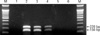
Sensitivity of the developed duplex RT-PCR
The detection limit of the designed duplex RT-PCR was 620 pg for both of PERV gag and pig GAPDH from the PK-15 cell RNA (Fig. 1). The expected product could be successfully distinguished on agarose gels.
Comparison of the PERV gag and pig GAPDH RNA expression
The mean expression levels from separate tube RT-PCR and single tube duplex RT-PCR were 0.559 and 0.561, respectively, with no statistical difference between the two reactions (p > 0.05).
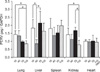
The standard deviations of the expression ratio in each pig after three trials of RT-PCR were between 0.01 and 0.16 in all of the tested pigs. This was presented together with the mean expression ratio for each pig in a bar chart.
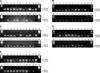
As shown in Fig. 2, established duplex RT-PCR could differentiate two expected products from pig tissues. However, there were no successful amplifications in any pancreas tissue samples. The density of DNA bands which were displayed was measured and the PERV gag / pig GAPDH ratio was calculated, and these data grouped vertical bars plot (Fig. 3). The PERV expression patterns were statistically different among each group in lung, liver, and kidney (p < 0.05). However, there was no statistical difference among each group in the spleen and heart. In the lung and kidney, the PERV gag expression level of the 110 day group was statistically lower than the 10 and 40 days groups, and differences between 40 and 70 day groups were not statistically significant (p < 0.05). In the liver, the 40 day group was significantly lower than the 10 and 70 day groups (p < 0.05).
Discussion
A sensitive duplex RT-PCR protocol, which could detect PERV gag RNA and pig GAPDH mRNA simultaneously, was established in this study. This technique enabled the simultaneous comparison of the PERV gag mRNA and pig GAPDH mRNA expression levels using multiplex RT-PCR. The single tube duplex RT-PCR might be the reasonable for this research, as no differences in PERV gag to pig GAPDH ratio was shown in comparison between separate tube RT-PCR each tube containing only one set of primers for PERV gag or pig GAPDH and single tube duplex RT-PCR. The expression ratio of the PERV gag and GAPDH was not age-related, but the patterns of expression levels in each age were different with each organ. A previous study [8] reported that the PERV mRNA expression level was highest in the kidney among the various tissues tested, and lowest in the pancreas. In addition, more detailed comparisons were performed according to pig breeds or between pig organs. Age-related PERV mRNA expression patterns in the kidney were similar with previous studies [8,23] on retroviral loads in viremia. The viral titer of the pigs was the highest at 2 to 6 months and lowest at 6 months [23]. Moreover, pigs in the low health status showed elevated levels of viremia compared to those of high health status [23].
Compared to a previous study, this study was conducted in four different age groups, 10, 40, 70, and 110 days-old, which could represent the stage of suckling, nursery, grower, and finisher, respectively. In a similar study performed by Tucker et al. [23], age groups were classified into three groups, < 2 months-old, 2 to 6 months-old, and > 6 months-old and then the endpoint of age was above 6 months-old. This classification included various age stages of corresponding groups. However, animals of the same age were included in this study with the endpoint at 110 days-old, which was less than 4 months.
The increasing and decreasing tendencies of PERV between the two studies were similar in the kidney, but the timing of dropping and the patterns in other organs were different. Furthermore, the patterns of PERV mRNA expression with respect to age varied in the organs. In spleen and heart, there was no significant difference in expression levels. The differences among the each age group were observed in the lung, liver, and kidney. However, the patterns of PERV mRNA according to age were dissimilar among the three organs. It was difficult to determine the reason behind the differences. The physical and/or physiological or environmental differences might be one of the reasons. The regulatory signals for ERV were reported as cell and tissue types, and processes were related to differentiation and aging, cytokines and steroids [22]. In addition, various stress signals including injury, infection, oxidative stresses and psychological stresses could modulate their transcription [7]. As a lot of factors can affect PERV expression, more elaborate experiments considering various controlled factors should be done to obtain more clarity on this subject.
Unfortunately, even though the pancreas was tested, there were no amplifications. This could be due to the large amount of ribonuclease A as well as other digestive enzymes in the pancreas that may have degraded RNA [6]. The errors in tissue transport and temperature variations during tissue handling may have affected the RNA in tissue samples. Also, the technical errors in the RNA extraction process or storage of tissue homogenate might accelerate the breakdown of RNA. Moreover, since the pancreas contains more ribonuclease than other tissues, it could be affected more by the inadequate treatment of samples.
To compare PERV expression levels, it is better to estimate the expression level of the all three genes, gag, pol, and env. Moreover, in envelope genes, the estimation of the envA, B, and C mRNA expression levels would be better. Unfortunately, only the gag gene was employed in the current study to investigate the expression of PERV regardless of subtypes because gag is highly conserved in PERV and the expression of gag gene is essential for viron production [18].
This study would be better if all the pigs bred in Korea were included. However, this research was focused on comparing PERV expression to age in pigs. For that reason, pig species was limited to Duroc. And in order to control for the species, the use of pure bred might be beneficial for research. The study about PERV expression in different pig species describing expression of envelope gene common mRNA and envelope A, B, and C was conducted in previously [12]. Though it is difficult to compare with this study because this study only dealt with gag mRNA, expression of envelope genes in previous study [12] presented various phages. Especially, envA and envB were not expressed in Duroc pig which was used in current study.
Even though lymph nodes are good samples for PERV expression, only the major organs were tested in this study because the focus was on the organs which might be used for transplantations. In addition, the PERV expression in embryonic stages might be covered in further studies.
In conclusion, even though consistent age related patterns in expression of PERV mRNA was not observed, the comparison method using duplex RT-PCR with GAPDH was established to be effective in this study. Since elimination of PERV is nearly impossible, the best way might be to focus on reducing the risk of PERV transmission. Finding a suitable donor expressing a lower level of PERV mRNA than the others could diminish the potential risk of PERV transmission.
Even though the physiologic identification was important, the microbiologic safety should be regarded in the concept of public health. Furthermore, established techniques might be helpful in decreasing the infection risk.

 XML Download