Journal List > J Vet Sci > v.10(2) > 1040973

Chang, Lee, Hwang, Hong, Jung, Lee, and Park: A novel β-glucan produced by Paenibacillus polymyxa JB115 induces nitric oxide production in RAW264.7 macrophages

Abstract

The effect of extracellular β-(1→3), (1→6)-glucan, produced by Paenibacillus polymyxa JB115, on nitric oxide (NO) production in RAW264.7 macrophages was investigated. β-glucan induced the production of NO by RAW264.7 macrophages in a concentration- and time-dependent manner. Moreover, β-glucan stimulation increased the mRNA expression of iNOS, COX-2 and IL-6 in RAW264.7 macrophages in a concentration-dependent manner.

Introduction

NO is induced during macrophage activation and thereby contributes to controlling the replication or neutralizing intracellular microbial pathogens [13]. Various studies indicated that NO is an important messenger in diverse biological functions, including neuronal transmission, vascular relaxation, immune modulation, and cytotoxicity against tumor cells [13,14].
β-glucans are heterogeneous groups of glucose polymers usually found in the cell walls of fungi [17], plants [11] and some bacteria [7]. They consist of linear β-1, 3-linked D-glucose molecules with β-1,6-linked side chains of varying length occurring at different intervals along the backbone, and can form complex tertiary structures stabilized by inter-chain hydrogen bonds [2,3].
Some animal studies addressed the beneficial effects of β-glucans on the growth performance of pigs [5,19], on the survival rate of mice challenged with Staphylococcus aureus or Candida albicans [16], and on the somatotropic axis and immune function in weaned piglets challenged with lipopolysaccharide (LPS) [12].
The problems associated with conventional methods of β-glucans extraction from mushrooms and plants, such as low purity and yield, high cost of production, as well as the adverse effects associated with intravenous administration β-glucans, such as inflammation, granuloma formation, and microembolization [18] prompted us to develop a more efficient method for extraction of extracellular β-(1→3), (1→6)-glucan from the soil based Paenibacillus (P.) polymyxa JB115 [7]. This study investigated the effects of β-glucans extracted from P. polymyxa JB115 on NO production in RAW264.7 murine macrophages.
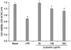
In order to investigate the cytotoxicity of β-glucan on RAW264.7 macrophages, RAW264.7 cells (5 × 10⁴ cells/ml) were incubated in a medium containing either β-glucan 30, 100 or 300 µg/ml or LPS (0.5 µg/ml) for 24 h. The viability of cells was then determined by MTT assay [8]. β-glucan decreased the viability of cells in a concentration-dependent manner (Fig. 1), with a statistically significant decrease (p < 0.05) being observed at a concentration of 300 µg/ml. LPS at 0.5 µg/ml also showed a significant decrease (p < 0.05) of approximately 60% relative to the control.
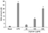
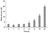
The effect of β-glucan on NO production in RAW264.7 macrophages was examined using a Griess reaction [4]. After 24 h of β-glucan exposure (30, 100 or 300 µg/ml), RAW264.7 cells showed a concentration-dependent production of NO (Fig. 2). This effect was also time dependent (Fig. 3).
Polysaccharides isolated form Phellinus linteus [8], Lentinus edodes [10] and Hericium erinaceum [20] are effective inducers of NO in macrophages. However, there have been other studies that demonstrated the inhibitory effect of β-glucans on macrophages stimulated by LPS or other factors [4,15]. In the present study, β-glucan from P. polymyxa JB115 activated RAW264.7 macrophages and induced the production of NO in a concentration- and time-dependent manner. However, this effect was not as potent as that of LPS (Figs. 2 and 3).
The cytotoxic effect of LPS in different cells including macrophages [21] and endothelial cells [6] has been well documented, and one of the most important factors associated with cell death is induction of NO [1,9]. These may also hold true in this study as the cytotoxicity of β-glucan may possibly be due to the NO production during macrophage activation.
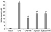
Polymyxin B has shown inhibitory effects on the lethal endotoxic activity of LPS in vivo and on the in vitro mitogenic activity of LPS by forming a stable molecular complex with the lipid A of LPS [21]. Therefore, this study also investigated the effects of polymyxin B on the activity of β-glucan and LPS in order to exclude any possible contamination due to endotoxins during the preparation process. Polymyxin B significantly (p < 0.05) inhibited NO production by LPS actvation. Nevertheless, polymyxin B had no significant effect on NO production induced by β-glucan (Fig. 4).
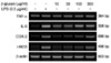
Finally, the mRNA expression of various cytokines was investigated in RAW264.7 macrophages which were exposed to β-glucan or LPS. P. polymyxa JB115 β-glucan induced mRNA expressions of i-NOS in a concentration-dependent manner, which might play a key role in NO production. A similar result was also observed for the mRNA expression of COX-2 and IL-6 (Fig. 5).
Based on our findings, we suggest further studies to be conducted to examine the potential use of the novel β-glucan purified from P. polymyxa JB115 as an immunostimulant or as an adjuvant of some animal vaccines.

Figures and Tables

Fig. 1
Effects of β-glucan and lipopolysaccharide (LPS) on the viability of RAW264.7 macrophages. Data represents the mean ± SD. *Significant difference (p < 0.05) compared to the control group.
jvs-10-165-g001
Fig. 2
β-glucan induced nitric oxide production in RAW264.7 macrophages. RAW264.7 cells were treated with either LPS (0.5 µg/ml) or β-glucan. Data represents the mean ± SD. *Significant difference (p < 0.05) compared to the control group.
jvs-10-165-g002
Fig. 3
β-glucan induced nitric oxide production in RAW264.7 macrophages. RAW264.7 cells were treated with β-glucan (300 µg/ml) for (0, 1, 2, 4, 6, 8, 12 or 24 h). Data represents the mean ± SD. *Significant difference (p < 0.05) compared to the control group.
jvs-10-165-g003
Fig. 4
Role of polymyxin B (PB) on nitric oxide production in RAW264.7 macrophages treated with either LPS or β-glucan. RAW264.7 cells were pretreated with 50 µg/ml of PB for 30 min and then activated with either LPS (0.2 µg/ml) or β-glucan (300 µg/ml). Data represents the mean ± SD. *Significant difference (p < 0.05) compared to the control group, #Significant difference (p < 0.05) compared to the LPS group.
jvs-10-165-g004
Fig. 5
β-glucan induced mRNA expression of cytokines in RAW264.7 macrophages. RAW264.7 cells were exposed to β-glucan at various concentrations, or LPS. After an 8 h incubation, i-NOS, COX-2, IL-6 and TNF-α mRNA were assessed by semi-quantitative RT-PCR.
jvs-10-165-g005

Acknowledgments

This study was supported in part by the Ministry of Knowledge Economy (MKE) through the Center for Traditional Microorganism Resources (TMR) at Keimyung University and in part by the Korea Research Foundation Grant funded by the Korean Government (KRF-2008-521-E00146).

References

1. Albina JE, Cui S, Mateo RB, Reichner JS. Nitric oxide-mediated apoptosis in murine peritoneal macrophages. J Immunol. 1993. 150:5080–5085.
2. Bohn JA, BeMiller JN. (1→3)-beta-D-glucans as biological response modifiers: a review of structure-functional activity relationships. Carbohydr Polymers. 1995. 28:3–14.
crossref
3. Brown GD, Gordon S. Immune recognition of fungal β-glucans. Cell Microbiol. 2005. 7:471–479.
crossref
4. Chang ZQ, Oh BC, Rhee MH, Kim JC, Lee SP, Park SC. Polysaccharides isolated from Phellinus baumii stimulate murine splenocyte proliferation and inhibit the lipopolysaccharide-induced nitric oxide production in RAW264.7 murine macrophages. World J Microbiol Biotechnol. 2007. 23:723–727.
crossref
5. Dritz SS, Shi J, Kielian TL, Goodband RD, Nelssen JL, Tokach MD, Chengappa MM, Smith JE, Blecha F. Influence of dietary beta-glucan on growth performance, nonspecific immunity, and resistance to Streptococcus suis infection in weanling pigs. J Anim Sci. 1995. 73:3341–3350.
crossref
6. Johnson BA, Pitt BR, Davies P. Pulmonary arterial smooth muscle cells modulate cytokine- and LPS-induced cytotoxicity in endothelial cells. Am J Physiol Lung Cell Mol Physiol. 2000. 278:L460–L468.
crossref
7. Jung HK, Hong JH, Park SC, Park BK, Nam DH, Kim SD. Production and physicochemical characterization of β-glucan produced by Paenibacillus polymyxa JB115. Biotechonol Bioprocess Eng. 2007. 12:713–719.
crossref
8. Kim GY, Choi GS, Lee SH, Park YM. Acidic polysaccharide isolated from Phellinus linteus enhances through the up-regulation of nitric oxide and tumor necrosis factor-alpha from peritoneal macrophages. J Ethnopharmacol. 2004. 95:69–76.
crossref
9. Kim YM, Bombeck CA, Billiar TR. Nitric oxide as a bifunctional regulator of apoptosis. Circ Res. 1999. 84:253–256.
crossref
10. Lee JY, Kim JY, Lee YG, Rhee MH, Hong EK, Cho JY. Molecular mechanism of macrophage activation by exopolysaccharides from liquid culture of Lentinus edodes. J Microbiol Biotechnol. 2008. 18:355–364.
11. Lee KY, Lee MH, Chang IY, Yoon SP, Lim DY, Jeon YJ. Macrophage activation by polysaccharide fraction isolated from Salicornia herbacea. J Ethnopharmacol. 2006. 103:372–378.
crossref
12. Li J, Li DF, Xing JJ, Cheng ZB, Lai CH. Effects of beta-glucan extracted from Saccharomyces cerevisiae on growth performance, and immunological and somatotropic responses of pigs challenged with Escherichia coli lipopolysaccharide. J Anim Sci. 2006. 84:2374–2381.
crossref
13. Lowenstein CJ, Dinerman JL, Snyder SH. Nitric oxide: a physiologic messenger. Ann Intern Med. 1994. 120:227–237.
crossref
14. Moncada S, Palmer RMJ, Higgs EA. Nitric oxide: physiology, pathophysiology, and pharmacology. Pharmacol Rev. 1991. 43:109–142.
15. Pacheco-Sánchez M, Boutin Y, Angers P, Gosselin A, Tweddell RJ. Inhibitory effect of CDP, a polysaccharide extracted from the mushroom Collybia dryophila, on nitric oxide synthase expression and nitric oxide production in macrophages. Eur J Pharmacol. 2007. 555:61–66.
crossref
16. Rice PJ, Adams EL, Ozment-Skelton T, Gonzalez AJ, Goldman MP, Lockhart BE, Barker LA, Breuel KF, Deponti WK, Kalbfleisch JH, Ensley HE, Brown GD, Gordon S, Williams DL. Oral delivery and gastrointestinal absorption of soluble glucans stimulate increased resistance to infectious challenge. J Pharmacol Exp Ther. 2005. 314:1079–1086.
crossref
17. Ross GD, Vetvicka V, Yan J, Xia Y, Vetvicková J. Therapeutic intervention with complement and beta-glucan in cancer. Immunopharmacology. 1999. 42:61–74.
crossref
18. Sandula J, Kogan G, Kacurakova M, Machova E. Microbial (1→3)-β-D-glucans, their preparation, physicochemical characterization and immonomodulatory activity. Carbohydr Polym. 1999. 38:247–253.
19. Schoenherr WD, Pollmann DS, Coalson JA. Titration of MacroGard-S on growth performance of nursery pigs. J Anim Sci. 1994. 72:Suppl 2. 57.
20. Son CG, Shin JW, Cho JH, Cho CK, Yun CH, Chung W, Han SH. Macrophage activation and nitric oxide production by water soluble components of Hericium erinaceum. Int Immunopharmacol. 2006. 6:1363–1369.
crossref
21. Vogel SN, Marshall ST, Rosenstreich DL. Analysis of the effects of lipopolysaccharide on macrophages: differential phagocytic responses of C3H/HeN and C3H/HeJ macrophages in vitro. Infect Immun. 1979. 25:328–336.
crossref
TOOLS
Similar articles