Journal List > J Vet Sci > v.10(2) > 1040965

Kim, Yang, Moon, Park, Luo, Lee, Song, Kang, Ann, Jun, and Park: Genetic analysis of ORF5 of recent Korean porcine reproductive and respiratory syndrome viruses (PRRSVs) in viremic sera collected from MLV-vaccinating or non-vaccinating farms

Abstract

The 23 open reading frame (ORF) 5 sequences of Korean type II porcine reproductive and respiratory syndrome virus (PRRSV) were collected from viremic sera from the (modified live vaccine) MLV-vaccinating and non-vaccinating farms from 2007 to 2008. The samples were phylogenetically analyzed with previous ORF5 sequences, including type I Korean PRRSV, and previously reported or collected sequences from 1997 to 2008. A MN184-like subgroup of type II Korean PRRSV was newly identified in the viremic sera collected from 2007 to 2008. And of the type I PRRSVs, one subgroup had 87.2~88.9% similarity with the Lelystad virus, showing a close relationship with the 27~2003 strain of Spain. The maximum parsimony tree of type II PRRSV from 1997 to 2008 showed that they had evolved to four lineages, subgroups 1, 2, 3 and 4. Most of the recently collected type II PRRSVs belonged to subgroup 4 (48%). The region of three B-cell epitopes and two T-cell epitopes of ORF5 amino acids sequences was considerably different from the MLV in subgroups 3 and 4. In conclusion, the existence of type I PRRSV, which was genetically different from Lelystad virus (Prototype of type I PRRSV), and heterologous type II PRRSVs of viremic pigs detected even in the MLV-vaccinating farms indicated the need for new vaccine approaches for the control of PRRSV in Korea.

Introduction

Porcine reproductive and respiratory syndrome virus (PRRSV) is a small, enveloped RNA virus [4,39] that is classified as a member of the genus Arterivirus of family Arteriviridae in the order Nidovirales [6]. PRRSV was first isolated almost simultaneously in Europe [40] and the USA [9].
The PRRSV genome is a polyadenylated, single-stranded, non-segmented, positive-sense RNA approximately 15 kb in size and consisting of at least 9 open reading frames (ORFs) [10,27,30,35,41]. ORF1a and 1b are located immediately downstream of the 5' untranslated region (UTR) and involved in virus transcription and replication. ORFs 2 to 7 are located at the 3' end of the genome and are thought to encode for the PRRSV structural protein. Among them, ORF5 encodes an unglycosylated membrane of 17 kD or a glycosylated protein of 25 kD [22,26]. Furthermore, the ORF5 of PRRSV is in an immunologically crucial region which is mainly related with neutralizing antibody formation. In the case of type II PRRSV, ORF5 has been known to contain three B-cell epitopes and two T-cell epitopes [11,31,32,38].
Remarkable genetic differences have been reported among PRRSV. The major two genotypes are European (Type I, Lelystad as prototype) and North American (Type II, VR-2332 as prototype). The similarity between these two genotypes among the ORFs were shown to be 57~59% (ORF7), 70~81% (ORF6), 51~59% (ORF5), 68% (ORF4), 58% (ORF3) and 63% (ORF2) [3,14,23,24,25,28]. Even within the same genotype, high genetic variation among field viruses has been reported all over the world. Type II PRRSV was first isolated in Korea in 1994 [21], with the molecular characterization of Korean PRRSV from 1996 to 2006 based on ORF5 and ORF7 [7,8,43]. These studies suggested that the Korean PRRSVs belonged to the Type II PRRSV and had geographically evolved with their own genetic clusters. However, one group also reported that type I PRRSV also existed in Korea [18].
To control PRRSV, the modified "test and removal method" was successful in the seed stock breeding farm and a supplying boar studs in Korea [42]. Since this method is laborious and time-consuming, most swine farms with continuous flow systems have applied a modified live vaccine (MLV) .
With the widespread use of MLV, the actual efficacy of the vaccine came into question as the sequence analysis based on the ORF5 sequences of Korean PRRSV showed that the genetic divergence was ranged from 1.3 to 12.9% compared to the MLV [7]. Although MLV had an effect to reduce clinical signs and enhance weight gain in the PRRSV-infected farms, the virus shedding of challenged heterologous viruses was not prevented [5].
With these concerns, this study focused on analyzing the patterns of viremia detected post-vaccination, which could represent the active replication of PRRSV, in the either MLV vaccinated or non-vaccinated farms. Furthermore, the full ORF5 sequences acquired from viremic pigs were compared with the reported [7,21] or previously collected data in the laboratory including type I and II PRRSV from 1997 to 2008.

Materials and Methods

Study design

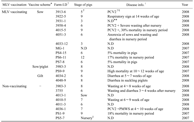
Blood samples were collected from commercial swine farms with information about MLV vaccinating and disease status. In the case of MLV vaccinating farms, blood samples were collected at least 6 months after the first vaccination. If the main clinical signs and PCV2-specific PCR results were available, they were also documented (Table 1).
At first, pilot study was performed to preview the viremic patterns in the MLV vaccinating and non-vaccinating farms. The vaccine schedules used in the MLV-vaccinating farms were one of the sow-only or sow/piglet vaccinations. Among 25 farms, the number of farms with viremic pigs at 3, 6, 9 and 12 weeks of age, including sows, was investigated using a reverse transcriptase nested polymerase chain reaction (RT-nested PCR) method. Serum antibody titer was also evaluated with a commercial enzyme-linked immunosorbent assay (ELISA) kit (Herdchek 2XR, PRRS; IDEXX, USA). The serum IgG titers were expressed as S/P ratio: (Sample O.D - Negative control O.D) / (Positive control O.D - Negative control O.D).
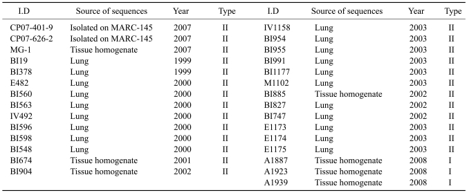
After the pilot study, to know the genetic properties of PRRSV in the viremic sera between MLV-vaccinating or non-vaccinating farms, the viremic samples collected from 2007 to 2008 were further sequenced based on ORF5. A total of 23 PRRSV ORF5 sequences in the viremic sera were obtained. The sequenced data was analyzed with reference and previous laboratory sequence data including type I and II PRRSV from 1997 to 2008, to observe the phylogenetic relationship. The detailed sequence information was presented in Tables 1 and 2.
Except for previously-published reference sequences, the sequence data of type I PRRSV and the other type II PRRSV (not collected from viremic sera) used in this study were collected from 1999 to 2008 in the virus-infected tissues of commercial pigs in Korea. The ORF5 sequences used in this study are available in GenBank accession numbers, from FJ972714 to FJ972766.

Virus isolation

Two Korean field isolates (CP07-401-9 and CP07-626-2, both in 2007) were adapted on MARC-145 cell which was known to be permissive to the PRRSV, especially type II PPRSV [15] and used for genetic analysis. Briefly, the tissue homogenates suspended 10% by volume in Dulbecco's minimum essential medium (DMEM) and centrifuged. The supernatant was filtered with syringe filter (0.45 µm; ChmLab, Spain) and used to inoculate the MARC-145 cells grown in DMEM containing penicillin (100 units/ml), streptomycin (100 µg/ml) and amphotericin B (0.25 µg/ml) with 10% fetal bovine serum (FBS). After 2 h incubation at 37.5℃ and washing with phosphate buffered saline (PBS, pH 7.4), maintenance medium (DMEM with 5% FBS) was added. The suspicious virus candidates were in continuous passage with the same manner after 5~7 days of incubation until the cytopathic effect (CPE) was observed. When the CPE was observed, the PRRSV was identified by PRRSV-specific RT-nested PCR [20] and immunofluorescence assay (IFA). The IFA kit was kindly given from National Veterinary Research Quarantine Service (Korea).

RNA extraction from the PRRSV isolates and collected sera

In this study, the all collected sera were made to a pooled sample with 2 or 3 individual sera in the same age group. The pooled samples of 250 µl and culture medium of isolated virus (passage 3, both CP07-401-9 and CP07-626-2) was mixed with 750 µl of Trizol LS (Invitrogen, USA). In the case of virus infected tissues, 20% tissue suspension with PBS was made, and 250 µl of suspension was reacted with Trizol (Invitrogen, USA). After 15 min incubation, 200 µl of cold chloroform was added, followed by vortexing. The vortexed mixture was centrifuged at 12,000 g, 4℃ for 15 min and 450 µl of the supernatant was mixed with same volume of cold isopropyl alcohol. After precipitating overnight at -20℃, the solution was centrifuged at 12,000 g, 4℃ for 10 min and the final RNA pellet was acquired by washing with 75% ethanol and centrifugation. The RNA pellet was dissolved with 30 µl of 0.1% diethyl pyrocarbonate-treated distilled water.

cDNA synthesis and RT-PCR

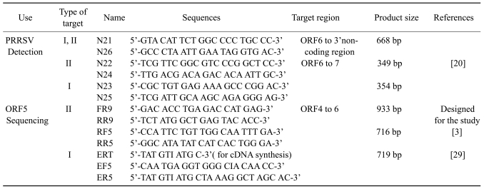
The primers used in this study were presented in Table 3. The cDNA was synthesized with a commercial M-MLV reverse transcriptase kit (Invitrogen, USA) following the manufacturer's instructions. The N26, RR9, ERT primers were used in the cDNA synthesis for virus detection and sequencing. To detect the viremic sera, a previously published RT-nested PCR method, which amplifies a part of ORF6 to ORF7 region, was used [20]. For the ORF5 sequencing of type II PRRSV, a modified RT-nested PCR was used based on the published RT-PCR method [3]. Briefly, FR9 and RR9 primers were newly designed based on the ORF4 and ORF6 regions to generate a 933 bp fragment to include the complete ORF5 sequence. These PCR products were further amplified with two published primers, RF5 and RR5, as a nested PCR which produces 716 bp fragment containing full ORF5 sequence.
The PCR reaction was performed with i-StarMaster mix PCR kit (iNtRON Biotechnology, Korea). The total mixture (20 µl) included 1 µl of each primer at a concentration of 10 pmol/µl, 2 µl of cDNA and 16 µl of master mix containing 250 mM dNTP, 2 mM Mg2+, K+ and NH4+ were added as salts along with 10 mM Tris-HCl (pH 9.0) to the PCR tube, which was coated with 2.5 Unit of i-star Taq DNA polymerase. The first PCR was performed at 95℃ for 5 min followed by 30 cycles of 95℃ 1 min, 55℃ 1 min, 72℃ 1 min, with a final extension at 72℃ or 10 min, and then held at 4℃.
The nested PCR was performed using 2 µl of first PCR products as a template along with FR5 and RR5 primers. The protocol was as follows: 95℃ for 5 min followed by 30 cycles of 95℃ 45 sec, 55℃ 45 sec, 72℃ 45 sec, with a final extension at 72℃ for 10 min, and then held at 4℃. For the ORF5 sequencing of type I PRRSV from the infected tissues, RT-PCR was performed as described in a previous paper [29]. The PCR products were electrophoresed in a 1% agarose gel containing ethidium bromide. The target bands were visualized using an ultraviolet lamp.

Sequencing and data analysis

For sequencing ORF5, a 716 bp fragment of type II PRRSV and a 719 bp fragment of type I PRRSV were purified using the QIAquick Gel Extraction kit (Qiagen Korea, Korea). Both strands of purified products were sequenced by Genotech (Korea). The full ORF5 sequences were analyzed using CLUSTALX v1.83 program and MegAlign software (DNAStar, USA) to determine the phylogenetic relationships and nucleotide similarity. The Neighbor-joining tree and was drawn using Kimura-two parameter as a distance estimation and percent frequencies of the groupings were determined after 1,000 bootstrap evaluation. The Maximum parsimony tree was also drawn as a consensus tree after 1,000 bootstrap evaluations.

Results

Pilot study about MLV vaccination and viremic status in 2007

The viremia of type II PRRSV was detected in both MLV-vaccinating and non-vaccinating farms (Table 4). In the non-vaccinating farms, most viremic pigs were found at 6 to 12 weeks of age. Even in the farms with MLV vaccination, PRRSV viremia was also detected regardless of vaccine schedule. The mean S/P ratio was relatively lower in the non-vaccinating farms than MLV-vaccinating farms at 9 and 12 weeks of age. However, mean S/P ratios were above 0.4 in all age groups, which meant that they were seroconverted for PRRSV.

Phylogenetic relationship of total Korean PRRSV ORF5 sequences with those of other countries

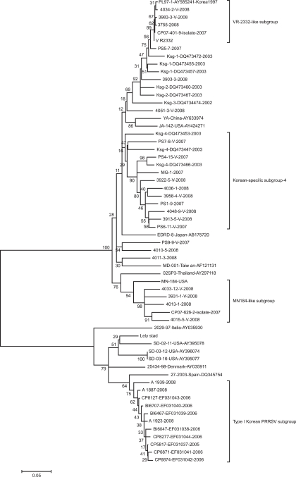
The neighbor-joining tree was presented in Fig. 1. Korean specific subgroups (Ksg)-1, 2 and subgroup 3 were clustered near the VR-2332-like subgroup. The majority of the PRRSV samples collected from 2007 to 2008 were shown to belong to Ksg-4, which was previously reported by Cha et al. [7]. There was no subgroup-specific differences between viruses from MLV-vaccinating or non-vaccinating farms. The viruses of the Ksg-4 subgroup had 87.8-89.0% of nucleotide similarity with VR-2332 in the ORF5 region. The recent Korean PRRSVs from 2007 to 2008 made a novel subgroup which was not reported by Cha et al. [7] and were in the same cluster with MN184 strain from USA. The PRRSVs in this subgroup (MN184-like) had a similarity of 90.3~90.5% compared to MN184 and 84.9~87.2% compared to the MLV on nucleotides of the ORF5 region. The type I Korean PRRSVs were clustered with a 27-2003 strain from Spain with 89.1~91.7% of similarity. The Korean type I PRRSV had 84.9~88.4% of similarity in ORF5 when compared with a European-like PRRSV strain (SD-02-11, Genbank Accession no. AY395078) isolated from USA.

Evolution of type II Korean PRRSV from 1997 to 2008

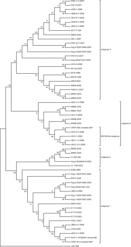
To observe the evolutionary pattern of type II PRRSV from 1997 to 2008, a Maximum parsimony tree was drawn (Fig. 2). The type II Korean PRRSVs were shown to have been evolved into four subgroups, subgroups 1, 2, 3 and 4. Subgroup 1 included a PL97-1, a prototype of type II Korean PRRSV. The previously reported Ksg-1 and 2 subgroups were shown to belong to subgroup 1 and 24% (6/25) of recent type II Korean PRRSVs from 2007 to 2008, were also included in this group. Subgroup 2 containing Ksg-3 had only PRRSVs from samples taken as of 2000 to 2003. The subgroup 3 formed a novel cluster which was not reported before in Korea and had a MN184-like cluster. 28% (7/25) of recent PRRSVs belonged to this subgroup. Subgroup 4 consisted of 48% (12/25) of recent PRRSVs and was consistent with previously reports of the Ksg-4 subgroup.

Analysis of deduced amino acid sequences of type II PRRSV ORF5 in the viremic sera

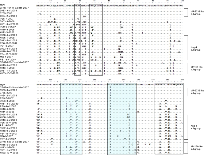
The amino acid sequences of 23 type II PRRSV ORF5 in the viremic sera taken from 2007 to 2008 were aligned to compare several epitope regions (Fig. 3). The Ksg-4-containing subgroup had at least one mutated sequences at H38, L39, L41 and N44 (K/Q38, F/I39, S41 and K44). In the case of the 3rd B-cell epitope (182~200), I189, R191 and Q196 were frequently replaced by V189, K191 and R196 in those groups. The MN184-like subgroup showed the replacement of V185 and R191 to A185 and K191. The first T-cell epitope (117~131) was also a variable region among Korean type II PRRSVs obtained from viremic sera. Most common was V124 and A128 being dominantly replaced by T/I124 and V/T128 in the Ksg-4-containing subgroup. Notably, the ORF5s in MN184-like subgroup had common mutations (FA127-128 → LT127-128) which were also observed in the isolate CP07-626-2 belonging to this subgroup. The second T-cell epitope (149~163) was more conserved than the first T-cell epitope (127~131) in the ORF5 sequences used in this study. The obvious differences of epitope regions were not observed between MLV-vaccinating or non-vaccinating farms, comparing the viruses in same subgroup.

Discussion

The viremia of heterologous virus after MLV vaccination had been reported in several papers. In previous studies, although MLV vaccination could contribute to improve the clinical outcome, sporadic viremia was as observed as ever after challenge with heterologous viruses [5,34]. Although clinical improvement in MLV-vaccinating farms was proved in those studies, those results could not reflect the actual situation in the field. Since the porcine circovirus type 2 (PCV2) became the one of the important pathogens causing the postweaning multisystemic wasting syndrome (PMWS, now one of the PCV-associated diseases), coinfection of other pathogens was the important factor for the disease appearance of PMWS. PRRSV was reported as the crucial factor of PMWS or severe wasting diseases when co-infected with PCV2 [1,12,33]. PCV2 was frequently detected in the wasting, diarrheic pigs and aborted fetuses in Korea [2,16,17]. Most farms in this study suffered from wasting during the nursery period (around 4-9 weeks of age) and several farms were shown to be infected with PCV2. Therefore, the viremia of vaccine-like virus or heterologous PRRSV observed even after MLV-vaccination might be the risk of wasting diseases when PCV2 was endemic in the farms, as viremia indicated the active replication of PRRSV [13].
In the pilot study, we could also observe sporadic viremia even in the seropositive herds of MLV-vaccinating farms applying vaccine schedules, such as sow-only and sow-piglet scheme. From this observation, this study focused on analyzing the PRRSVs obtained from the sera of viremic pigs in MLV-vaccinating or non-vaccinating farms. The PRRSV viremic pigs were frequently detected in both farms. Although high genetic variation was observed among the ORF5 of these viruses, there was no considerable differences between viruses from MLV-vaccinating or non-vaccinating farms. Since Korean swine farms were located not so far from the other farms and pigs were transferred on a national basis, frequent viral transmission 'from farm to farm' may be a factor for this observation. To understand vaccine pressure on PRRSV evolution, a long-term chronological study in the evolution of prevailing PRRSV after MLV vaccination should be performed on a farm to farm basis to reduce interfering factors such as transmission of foreign viruses.
There were some different phylogenetic patterns in this study which included the recent type II PRRSVs from 2007 to 2008. A new subgroup with 84.9~87.2% similarity of ORF5 compared to the MLV was identified. This subgroup consisted of only isolates collected from 2007 to 2008. In the neighbor-joining tree, they were shown to be closely related with MN184 strain from USA, showing 90.3~90.5% of similarity. Since USA is the major trade partner for the swine industry, these newly identified MN184-like viruses might have been introduced from USA one day and evolved in Korea independently.
The successful adaptation the CP07-626-2 strain, belonging to the MN184-like subgroup, on MARC-145 cells (passage 10), could be used to develop a new vaccine to control these newly emerging strains in Korea.
A Maximum parsimony tree of type II PRRSVs from 1997 to 2008 showed that the viruses had evolved after first introduction of VR-2332-like PRRSV (PL97-1 strain). At least 4 lineages were identified in the tree. Although the viruses in subgroup 2 were not detected recently, subgroups 1, 3 and 4 were still detected in Korea. Furthermore, the recent type II PRRSVs in subgroups 3 and 4 had high variations in their epitope regions of ORF5, including a novel B-cell epitope (3rd epitope) and two T-cell epitopes [11,38]. The neutralizing antibody fromation was known to be greatly affected by genetic variation and the amino acid sequences of ORF4 and ORF5 were more correlated with the neutralizing ability [19]. Therefore, these frequent substitutions in the epitope regions of ORF5 including neutralizing epitope also could be a cause of subsequent viremia, even in the MLV-vaccinating farms.
This study also confirmed that the type I PRRSV had been co-circulating with type II PRRSV in Korea. The type I PRRSV was primarily detected by RT-nested PCR-based typing which amplified the ORF6 to ORF7 region, and further confirmed by full sequencing of ORF5 in this study. The Korean type I PRRSV belonged to the one specific subgroup having 87.2~88.9% similarity in ORF5 compared to the Lelystad strain, the prototype. Notably, the ORF5 of type I Korean PRRSVs were similar with 27~003 strain of Spain, not the European-like PRRSV reported from the USA. This phylogenetic relationship suggested that the type I Korean PRRSVs were possibly introduced from Spain and had evolved into a single cluster. Notably, since the first report of type I PRRSV in Korea in 2006, the detection rate of type I PRRSV had been increasing in the field samples [18]. Because of the previously reported type-specific protection in the pigs [36,37], the use of type I PRRSV-based vaccine should be considered in Korea.
The viremia of heterologous PRRSV was frequently detected even in the MLV-vaccinating farms. These field viruses found in MLV-vaccinating farms were shown to have variable nucleotide substitutions on ORF5, similar to the non-vaccinating farms. Several substitutions of amino acids in the neutralizing epitope were also found in these viruses. The genetic diversity in type II PRRSV had been increasing from 1997 to 2008 in maximum parsimony tree. Furthermore, a newly emerging MN184-like subgroup appearing from 2007 was identified, and a circulation of type I Korean PRRSV was confirmed in this study. These complex situations observed from PRRSV molecular epidemiology in Korea reinforce the need for an additional vaccine strategy including a new vaccine development.

Acknowledgments

This Study was supported by the Technology Development Program for Agriculture and Forestry from the Ministry of Agriculture and Forestry, Republic of Korea and by a grant (Code# 20070401034009) from the Biogreen 21 Program, Rural Development Administration, Korea.

References

1. Allan GM, McNeilly F, Ellis J, Krakowka S, Meehan B, McNair I, Walker I, Kennedy S. Experimental infection of colostrum deprived piglets with porcine circovirus 2 (PCV2) and porcine reproductive and respiratory syndrome virus (PRRSV) potentiates PCV2 replication. Arch Virol. 2000; 145:2421–2429. PMID: 11205128.
crossref
2. An DJ, Roh IS, Song DS, Park CK, Park BK. Phylogenetic characterization of porcine circovirus type 2 in PMWS and PDNS Korean pigs between 1999 and 2006. Virus Res. 2007; 129:115–122. PMID: 17706315.
crossref
3. Andreyev VG, Wesley RD, Mengeling WL, Vorwald AC, Lager KM. Genetic variation and phylogenetic relationships of 22 porcine reproductive and respiratory syndrome virus (PRRSV) field strains based on sequence analysis of open reading frame 5. Arch Virol. 1997; 142:993–1001. PMID: 9191863.
crossref
4. Benfield DA, Nelson E, Collins JE, Harris L, Goyal SM, Robison D, Christianson WT, Morrison RB, Gorcyca D, Chladek D. Characterization of swine infertility and respiratory syndrome (SIRS) virus (isolate ATCC VR-2332). J Vet Diagn Invest. 1992; 4:127–133. PMID: 1616976.
crossref
5. Cano JP, Dee SA, Murtaugh MP, Pijoan C. Impact of a modified-live porcine reproductive and respiratory syndrome virus vaccine intervention on a population of pigs infected with a heterologous isolate. Vaccine. 2007; 25:4382–4391. PMID: 17451850.
crossref
6. Cavanagh D. Nidovirales: A new order comprising Coronaviridae and Arteriviridae. Arch Virol. 1997; 142:629–633. PMID: 9349308.
7. Cha SH, Choi EJ, Park JH, Yoon SR, Song JY, Kwon JH, Song HJ, Yoon KJ. Molecular characterization of recent Korean porcine reproductive and respiratory syndrome (PRRS) viruses and comparison to other Asian PRRS viruses. Vet Microbiol. 2006; 117:248–257. PMID: 16797883.
crossref
8. Cheon DS, Chae C. Antigenic variation and genotype of isolates of porcine reproductive and respiratory syndrome virus in Korea. Vet Rec. 2000; 147:215–218. PMID: 10994923.
crossref
9. Collins JE, Benfield DA, Christianson WT, Harris L, Hennings JC, Shaw DP, Goyal SM, McCullough S, Morrison RB, Joo HS, Gorcyca D, Chladek D. Isolation of swine infertility and respiratory syndrome virus (isolate ATCC VR-2332) in North America and experimental reproduction of the disease in gnotobiotic pigs. J Vet Diagn Invest. 1992; 4:117–126. PMID: 1616975.
crossref
10. Conzelmann KK, Visser N, Van Woensel P, Thiel HJ. Molecular characterization of porcine reproductive and respiratory syndrome virus, a member of the arterivirus group. Virology. 1993; 193:329–339. PMID: 8438574.
crossref
11. de Lima M, Pattnaik AK, Flores EF, Osorio FA. Serologic marker candidates identified among B-cell linear epitopes of Nsp2 and structural proteins of a North American strain of porcine reproductive and respiratory syndrome virus. Virology. 2006; 353:410–421. PMID: 16843516.
crossref
12. Harms PA, Sorden SD, Halbur PG, Bolin SR, Lager KM, Morozov I, Paul PS. Experimental reproduction of severe disease in CD/CD pigs concurrently infected with type 2 porcine circovirus and porcine reproductive and respiratory syndrome virus. Vet Pathol. 2001; 38:528–539. PMID: 11572560.
crossref
13. Johnson W, Roof M, Vaughn E, Christopher-Hennings J, Johnson CR, Murtaugh MP. Pathogenic and humoral immune responses to porcine reproductive and respiratory syndrome virus (PRRSV) are related to viral load in acute infection. Vet Immunol Immunopathol. 2004; 102:233–247. PMID: 15507308.
crossref
14. Kapur V, Elam MR, Pawlovich TM, Murtaugh MP. Genetic variation in porcine reproductive and respiratory syndrome virus isolates in the midwestern United States. J Gen Virol. 1996; 77:1271–1276. PMID: 8683216.
crossref
15. Kim HS, Kwang J, Yoon IJ, Joo HS, Frey ML. Enhanced replication of porcine reproductive and respiratory syndrome (PRRS) virus in a homogeneous subpopulation of MA-104 cell line. Arch Virol. 1993; 133:477–483. PMID: 8257302.
crossref
16. Kim J, Chung HK, Chae C. Association of porcine circovirus 2 with porcine respiratory disease complex. Vet J. 2003; 166:251–256. PMID: 14550736.
crossref
17. Kim J, Jung K, Chae C. Prevalence of porcine circovirus type 2 in aborted fetuses and stillborn piglets. Vet Rec. 2004; 155:489–492. PMID: 15537144.
18. Kim JY, Lee SY, Sur JH, Lyoo YS. Serological and genetic characterization of the European strain of the porcine reproductive and respiratory syndrome virus isolated in Korea. Korean J Vet Res. 2006; 46:363–370.
19. Kim WI, Kim JJ, Cha SH, Yoon KJ. Different biological characteristics of wild-type porcine reproductive and respiratory syndrome viruses and vaccine viruses and identification of the corresponding genetic determinants. J Clin Microbiol. 2008; 46:1758–1768. PMID: 18272711.
crossref
20. Kono Y, Kanno T, Shimizu M, Yamada S, Ohashi S, Nakamine M, Shirai J. Nested PCR for detection and typing of porcine reproductive and respiratory syndrome (PRRS) virus in pigs. J Vet Med Sci. 1996; 58:941–946. PMID: 8915992.
crossref
21. Kweon CH, Kwon BJ, Lee HJ, Cho JJ, Hwang EK, Shin JH, Yoon YD, Kang YB, An SH, Kim YH, Huh W, Jun MH, Wensvoort G. Isolation of porcine reproductive and respiratory syndrome virus (PRRSV) in Korea. Korean J Vet Res. 1994; 34:77–83.
22. Mardassi H, Mounir S, Dea S. Structural gene analysis of a Quebec reference strain or porcine reproductive and respiratory syndrome virus (PRRSV). Adv Exp Med Biol. 1995; 380:277–281. PMID: 8830492.
23. Meng XJ, Paul PS, Halbur PG. Molecular cloning and nucleotide sequencing of the 3'-terminal genomic RNA of the porcine reproductive and respiratory syndrome virus. J Gen Virol. 1994; 75:1795–1801. PMID: 8021610.
crossref
24. Meng XJ, Paul PS, Halbur PG, Lum MA. Phylogenetic analyses of the putative M (ORF 6) and N (ORF 7) genes of porcine reproductive and respiratory syndrome virus (PRRSV): implication for the existence of two genotypes of PRRSV in the U.S.A. and Europe. Arch Virol. 1995; 140:745–755. PMID: 7794115.
crossref
25. Meng XJ, Paul PS, Halbur PG, Morozov I. Sequence comparison of open reading frames 2 to 5 of low and high virulence United States isolates of porcine reproductive and respiratory syndrome virus. J Gen Virol. 1995; 76:3181–3188. PMID: 8847527.
crossref
26. Meulenberg JJ, Petersen-den Besten A, de Kluyver EP, Moormann RJ, Schaaper WM, Wensvoort G. Characterization of structural proteins of Lelystad virus. Adv Exp Med Biol. 1995; 380:271–276. PMID: 8830491.
27. Meulenberg JJ, den Besten AP, de Kluyver E, van Nieuwstadt A, Wensvoort G, Moormann RJM. Molecular characterization of Lelystad virus. Vet Microbiol. 1997; 55:197–202. PMID: 9220614.
crossref
28. Murtaugh MP, Elam MR, Kakach LT. Comparison of the structural protein coding sequences of the VR-2332 and Lelystad virus strains of the PRRS virus. Arch Virol. 1995; 140:1451–1460. PMID: 7661696.
crossref
29. Oleksiewicz MB, Bøtner A, Madsen KG, Storgaard T. Sensitive detection and typing of porcine reproductive and respiratory syndrome virus by RT-PCR amplification of whole viral genes. Vet Microbiol. 1998; 64:7–22. PMID: 9874099.
crossref
30. Oleksiewicz MB, Bøtner A, Nielsen J, Storgaard T. Determination of 5'-leader sequences from radically disparate strains of porcine reproductive and respiratory syndrome virus reveals the presence of highly conserved sequence motifs. Arch Virol. 1999; 144:981–987. PMID: 10416379.
crossref
31. Ostrowski M, Galeota JA, Jar AM, Platt KB, Osorio FA, Lopez OJ. Identification of neutralizing and nonneutralizing epitopes in the porcine reproductive and respiratory syndrome virus GP5 ectodomain. J Virol. 2002; 76:4241–4250. PMID: 11932389.
crossref
32. Plagemann PGW. The primary GP5 neutralization epitope of North American isolates of porcine reproductive and respiratory syndrome virus. Vet Immunol Immunopathol. 2004; 102:263–275. PMID: 15507310.
crossref
33. Rovira A, Balasch M, Segalés J, García L, Plana-Durán J, Rosell C, Ellerbrok H, Mankertz A, Domingo M. Experimental inoculation of conventional pigs with porcine reproductive and respiratory syndrome virus and porcine circovirus 2. J Virol. 2002; 76:3232–3239. PMID: 11884547.
crossref
34. Scortti M, Prieto C, Martínez-Lobo FJ, Simarro I, Castro JM. Effects of two commercial European modified-live vaccines against porcine reproductive and respiratory syndrome viruses in pregnant gilts. Vet J. 2006; 172:506–514. PMID: 16169756.
crossref
35. Shen S, Kwang J, Liu W, Liu DX. Determination of the complete nucleotide sequence of a vaccine strain of porcine reproductive and respiratory syndrome virus and identification of the Nsp2 gene with a unique insertion. Arch Virol. 2000; 145:871–883. PMID: 10881675.
crossref
36. van Woensel PA, Liefkens K, Demaret S. Effect on viraemia of an American and a European serotype PRRSV vaccine after challenge with European wild-type strains of the virus. Vet Rec. 1998; 142:510–512. PMID: 9618874.
crossref
37. van Woensel PA, Liefkens K, Demaret S. European serotype PRRSV vaccine protects against European serotype challenge whereas an American serotype vaccine does not. Adv Exp Med Biol. 1998; 440:713–718. PMID: 9782349.
crossref
38. Vashisht K, Goldberg TL, Husmann RJ, Schnitzlein W, Zuckermann FA. Identification of immunodominant T-cell epitopes present in glycoprotein 5 of the North American genotype of porcine reproductive and respiratory syndrome virus. Vaccine. 2008; 26:4747–4753. PMID: 18590788.
crossref
39. Wensvoort G. Lelystad virus and the porcine epidemic abortion and respiratory syndrome. Vet Res. 1993; 24:117–124. PMID: 8343802.
40. Wensvoort G, Terpstra C, Pol JMA, ter Laak EA, Bloemraad M, de Kluyver EP, Kragten C, van Buiten L, den Besten A, Wagenaar F, Broekhuijsen JM, Moonen PLJM, Zetstra T, de Boer EA, Tibben HJ, de Jong MF, Vant Veld P, Groenland GJR, van Gennep JA, Voets MT, Verheijden JHM, Braamskamp J. Mystery swine disease in the Netherlands: the isolation of Lelystad virus. Vet Q. 1991; 13:121–130. PMID: 1835211.
crossref
41. Wootton S, Yoo D, Rogan D. Full-length sequence of a Canadian porcine reproductive and respiratory syndrome virus (PRRSV) isolate. Arch Virol. 2000; 145:2297–2323. PMID: 11205119.
crossref
42. Yang JS, Moon HJ, Lee CS, Park SJ, Song DS, Kang BK, Choi JU, Park BK. Elimination of porcine reproductive and respiratory syndrome virus from a seedstock breeding farm and a supplying boar stud by a modified test and removal method. Vet Rec. 2008; 162:333–337. PMID: 18344497.
crossref
43. Yoon SH, Song JY, Lee CH, Choi EJ, Cho IS, Kim B. Genetic characterization of the Korean porcine reproductive and respiratory syndrome viruses based on the nucleocapsid protein gene (ORF7) sequences. Arch Virol. 2008; 153:627–635. PMID: 18227969.
crossref
Fig. 1
The neighbor-joining (N-J) tree of open reading frame (ORF) 5 sequences of porcine reproductive and respiratory syndrome virus (PRRSV) in viremic pigs from 2007 to 2008. The name of viremic sera sequences were presented as 'I.D-V (or not)-Year', where the 'V' means the PRRSV ORF5 sequences were from a vaccinating farm. Korean specific subgroup (Ksg)-1, 2, 3 and 4 indicates the phylogenetic groups which were grouped as Korean PRRSV-specific subgroup when compared with Asian isolates in the N-J method of a previous paper [7].
jvs-10-121-g001
Fig. 2
Maximum parsimony tree of type II Korean PRRSV ORF5 from 1997 to 2008. The Lelystad virus, which was a prototype of type I PRRSV, was used as an outgroup. In the maximum parsimony tree, type II Korean PRRSV were mainly divided into four subgroups, subgroup 1, 2, 3 and 4. Ksg-1 and 2 were in the same group, subgroup 1, in this study. Subgroup 2 only contained samples from 2000 to 2003. The MN184-like subgroup was shown to belong to the subgroup 3 which had not been classified before in Korea.
jvs-10-121-g002
Fig. 3
Amino acid sequences of two type II PRRSV isolates and 23 type II PRRSV ORF5 in the viremic sera from 2007 to 2008. The amino acid sequences were compared with the sequence of MLV. The known B-cell epitopes, epitope A (decoying epitope, 27~30), epitope B (neutralizing epitope, 37~45) and recently identified region (187~200) were indicated by small, medium and large boxes, respectively. Furthermore, the two T-cell epitope regions of GP5 were also indicated by grey backgrounds.
jvs-10-121-g003
Table 1
The information of type II porcine reproductive and respiratory syndrome virus (PRRSV) open reading frame (ORF) 5 sequences in the sera of viremic pigs from 2007 to 2008
jvs-10-121-i001

*Modified live vaccine (MLV) vaccinating scheme : Sow (MLV vaccination on the sow group only, usually at nursery or mass vaccination with 3 months of interval), Sow/Piglet (MLV vaccination on sows and piglets at 2~4 weeks of age), Gilt (MLV vaccination on the gilts in the acclimatization). I.D of farms - I.D of pooled sera which were pooled with 2 or 3 individual sera in the same age. Weeks of age. §Piglets around 3~4 weeks of age. The disease information of the farm at the time of blood sampling. PCV2-positive in the tissue sample with PCR method at the time of blood sampling. **N.D: No data available.

Table 2
Information of laboratory data of Korean PRRSV ORF5 sequences from 1999 to 2008
jvs-10-121-i002
Table 3
The information of primers used in this study
jvs-10-121-i003
Table 4
Pilot data about viremic patterns of type II porcine reproductive and respiratory syndrome virus (PRRSV) according to the MLV vaccinating schedule in Korea, 2007
jvs-10-121-i004

*MLV vaccinating schedules: Sow (MLV vaccination on the sow group only, usually at nursery or mass vaccination with 3 months of interval), Sow/piglet (MLV vaccination on sows and piglets at 2~4 weeks of age). S/P ratio (mean ± SE): S/P ratio obtained from commercial ELISA kit, S/P ratio of ≥ 0.4 regarded as sero-positive to PRRSV. No. of farms with viremic pigs / No. of tested farms.

TOOLS
Similar articles