

 PDF
PDF ePub
ePub Citation
 Print
Print


INTRODUCTION
Uterine carcinosarcomas are rare biphasic neoplasms composed of carcinomatous and sarcomatous components. It accounts for almost half of all uterine sarcomas and 1-3% of all uterine malignancies. It is known to be highly aggressive, far more aggressive than matched grade 3 endometrioid endometrial carcinomas. The overall 5-year survival is around 20-35% and the 5-year survival for stage IV disease is reported to be around 10% [1]. Especially, the overall survival for stage IVB is expected to be very poor, although there are no exact statistics. The authors report a rare case after experiencing long-term survival (over 5 years) for stage IVB carcinosarcoma of the uterus.
CASE REPORT
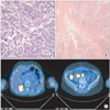
The menopausal 56 year old female patient (parity, 2-0-0-2) without specific medical history and history of hormone replacement visited the outpatient clinic for vaginal bleeding on August 2004. She was diagnosed with a cervical myoma 2 years ago at a local clinic. Since there was no change in the size of the myoma and bleeding, regular follow-up was conducted at the local clinic. Papanicolaou test (PAP) smears has remained normal for the recent 2 years. On speculum exam, polyps were not observed but an approximately 9 cm sized mass occupying the vagina was observed. Since it was difficult to find the cervical os due to the mass, the endometrium was difficult to evaluate properly. Endometrial thickness on ultrasound was approximately 3 mm and no specific findings were observed. Because postmenopausal vaginal bleeding continued for approximately 3 months, and high vascularity of the irregular contoured-mass was observed on Doppler ultrasound, total abdominal hysterectomy and bilateral salpingo-oophorectomy was performed due to the possibility of malignancy on August 27, 2004. After incising the tumor along the middle border of the anterior uterine wall, a huge well circumscribed solid mass was found fully filling the whole lower uterus. The mass was slightly friable with bleeding and necrosis. An ill-defined scattered necrotic solid mass was observed along the whole myometrial layer. According to the pathology report (Fig. 1A), it was consistent with an uterine carcinosarcoma (with heterologous components) with negative resection margin. Extensive lymphatic and vascular invasion were observed on the tumor. Metastasis to both ovaries was additionally diagnosed by the pathology (Fig. 1B) despite the absence of no gross lesions at the time of surgery. On immunohistochemical staining, cytokeratin was positive while vimentin, CK20, CD10, epithelial membrane antigen was negative.
To identify the metastatic loci, abdominopelvic magetic resonance imaging (MRI), positron emission tomographycomputed tomography (PET-CT) and tumor markers (CA-125 and CA 19-9) were conducted 4 weeks after the operation. On MRI, multiple metastatic lymph nodes were observed in the pelvic cavity. Two metastatic nodules on the right lobe of the liver were observed. Furthermore, additional peritoneal seeding nodules were observed between the right lobe of the liver and upper pole of the right kidney, and a soft-tissue mass was observed on the vaginal stump. On PET (Fig. 1C), abnormal fluorodeoxyglucose (FDG) uptake increase was confirmed on the aortocaval, mesenteric, bilateral iliac chain, left obturator lymph node, hepatic nodules, and peritoneal seeding nodules. The CA-125 was 50.0 U/mL. With the impression of an uterine carcinosarcoma stage IVB, we started palliative chemotherapy for 6 cycles from September 18, 2004 to January first, 2005; 5,000 mg/m2 of ifosfamide (with mesna) and 50 mg/m2 of cisplatin. No adjuvant radiotherapy was conducted. During the 6 cycles of chemotherapy, no serious side effects were found and the Eastern Cooperative Oncology Group (ECOG) performance scale was 0-1. The tumor marker (CA-125) normalized after 1 cycle of chemotherapy. Four weeks after completion of chemotherapy, MRI and PET-CT scans were followed-up. The lesions mentioned above were almost diminished over 80% (partial response by Response Evaluation Criteria In Solid Tumors [RECIST] criteria). Thereafter, the progress was observed every 3 months for the first year and every 6 months thereafter. No tumor recurrence, distant metastasis and meaningful lymph node enlargement has been reported on the image studies. Recent PAP smears as well as the tumor marker are all normal as of present (June, 2010). Her general condition is very tolerable with excellent ECOG performance scale (0-1).
DISCUSSION
The rarity of this tumor has limited large epidemiologic studies to report clinical behavior and outcome. Current evidence would suggest that carcinosarcomas of the uterus undergo full surgical staging and resection of any gross metastatic disease. Lymph node dissection has not been shown to be therapeutic.
Adjuvant chemotherapy may be beneficial for advanced stage. A phase II trial of ifosfamide and mesna in patients with advanced or recurrent carcinosarcomas was conducted by the Gynecologic Oncologic Group. Ifosfamide was unusually effective with a total response rate of 32.2% (complete response, 17.9%; partial response, 14.3%) [2]. The results of a randomized study comparing ifosfamide alone versus ifosfamide and paclitaxel in 179 patients with advanced uterine carcinosarcomas reported a response rate of 29% with ifosfamide alone and 45% in combination. The median progression-free and overall survival for ifosfamide compared with the combination was 3.6 vs. 5.8 months, and 8.4 vs. 13.5 months, respectively [3]. The results of a relatively large randomized clinical trial comparing whole-abdominal radiation (WAR) with three cycles of cisplatin, ifosfamide, and mesna (CIM) in 201 patients with stages I to IV uterine carcinosarcomas were reported in 2007. The estimated crude probability of recurring within 5 years was 58% for WAR and 52% for CIM. Adjusting for stage and age, the recurrence rate was 21% lower for CIM patients than for WAI patients (relative hazard [RH], 0.789; 95% confidence interval [CI], 0.530 to 1.176; p=0.245; 2-tail test). The estimated death rate was 29% lower among the CIM group (RH, 0.712; 95% CI, 0.484 to 1.048; p=0.085; 2-tail test). There were no statistically significant advantages in the recurrence rate or survival for adjuvant CIM over WAI. However, the observed differences favored the use of combination chemotherapy [4].
The benefit of adjuvant radiotherapy is unclear regarding overall survival so far. Some studies have revealed the advantage of radiotherapy for disease specific survival in early-stage tumors as well as local control in advanced-stage tumors [5].
This is a case report for stage IVB carcinosarcoma of the uterus with long-term survival over 5 years. Only chemotherapy was performed as adjuvant therapy. Since the data and clinical trial related to prognosis and adjuvant therapy is extremely rare, close follow-up of the clinical course of patient seems to be critical from the clinical point of view.
 XML Download