

 PDF
PDF ePub
ePub Citation
 Print
Print


INTRODUCTION
Choledochal cyst disease (CCD) is a pathologic condition characterized by congenital dilatation of the biliary system, including the common, intrahepatic, and intrapancreatic bile ducts. It is common in Asian patients, especially in females and infants. Due to recent advances in diagnostic imaging techniques, CCD can be diagnosed at any age, from the antenatal period to adult life.123456 CCD diagnosis in adult age has been made more frequently over time than before. Surgery in adult patients has been performed to resolve symptoms and to prevent or treat CCD-associated malignancies. There are several points of concern regarding surgical treatment such as surgical complications, remnant cystic lesions, malignant changes and minimal-incision approach.
Laparoscopic or robotic resection of choledochal cyst in adult patients has been occasionally reported, but its long-term outcomes especially with respect to anastomotic stenosis and incomplete cyst resection appear to have not yet been fully assessed.6789 Thus, until now, open surgery has been regarded as the standard procedure for resection of CCD in adult patients. Open surgery has a definite disadvantage of skin incision scar from operative wound, which can cause psychological and emotional stress especially in young female patients. Therefore, this study focused on the cosmetic aspect of two skin incisions for resection of CCD in young female patients.
Go to : 
MATERIALS AND METHODS
This study included 11 adult female patients with CCD who were less than 40 years of age (age range: 18-38 years). They underwent primary open surgery for CCD by a single surgeon (SH) within 2 years between January 2014 and December 2015. Patients who had undergone prior upper abdominal surgery such as CCD operation in childhood were excluded. In addition, patients who underwent a concurrent surgical procedure such as combined hepatectomy for intrahepatic lesions were also excluded. CCDs were classified according to the Todani modification of the original Alonso-Lej classification as type I in 8 patients and type IVa in 3 patients.10 Thus, all 11 study patients had naïve CCD, which was treated by mini-laparotomy. Detailed surgical procedures have been described previously.1 All patients recovered uneventfully after surgery and were regularly followed up to date; therefore, this study focused on cosmetic selection of skin incision and postoperative scar management. This study was approved by the Institutional Review Board of the Asan Medical Center.
In young female patients, a mini-laparotomy incision of around 9 cm in length is usually performed. The right subcostal incision is preferred because it is placed oblique to the transverse skin creases on the upper abdomen. Another surgical option is to perform an upper midline incision, but it crosses the transverse skin creases. The advantages and disadvantages of these two skin incisions were compared in this study.
Go to :
RESULTS
During the first year of the study period, all five patients underwent mini-laparotomy via the right subcostal incision. In contrast, during the second year, 6 patients underwent mini-laparotomy via either the right subcostal incision (n=3) or the upper midline incision (n=3).
In 9 patients with the right subcostal incision, the mean length of skin incision was 10 cm (range: 8-12 cm), which was dependent on the depth of the subcutaneous fat and location of the right costal cartilages. After skin marking over the location of bilateral costal cartilages and xiphoid process, the skin incision was made about 2 cm away from the right costal cartilage margins (Fig. 1A). The uppermost segment of the right rectus muscle was transected. The incision was extended by using two retractors, one retractor each from the right side and left side. No additional incision was performed because it might result in malalignment of the extended skin incision line, leading to disadvantage in wound cosmetics. Many surgical gauze sponges (5-8 sheets) were placed in the right subphrenic area to displace the liver towards the caudal side, so that the hepatic hilum would be beneath the operative wound. After completing the surgical procedure, the operative wound was repaired with interrupted sutures in two layers (peritoneum, transversalis and internal oblique muscle; and external oblique muscle and fascia) using absorbable suture materials. A Jackson-Pratt drain measuring 3.2 mm in outer diameter was inserted under the interrupted sutures on Scarpa's fascia. The skin was closed with interrupted subcuticular monofilament sutures along with topical application of adhesive sterile strips. None of the patients developed wound problem. These patients were advised to apply a silicone gel sheet (Cica-Care, Smith & Nephew, London, UK) from 6 weeks after surgery until 1 year. At the 1 year follow-up, a slight bulge on the skin scar was observed in 3 patients (37.5%) and the remaining 5 patients showed a flat skin scar. None of the patients showed noticeable atrophy of the ipsilateral rectus muscle. None of the patients complained of skin discomfort or paresthesia over the denervated skin area.
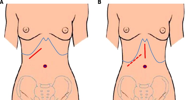 | Fig. 1Selection of incision for mini-laparotomy in female young adult patients with choledochal cyst. In patients with a relatively wide epigastrium (A), a right subcostal incision is more appropriate. In contrast, an upper midline incision appears to be more suitable for patients with a relatively narrow epigastrium (B).
|
An upper midline incision was made in 3 selected patients with a narrow epigastric area between the costal cartilages due to a slim body dimension (Fig. 1B). The length of skin incision uniformly measured as 9 cm. The incision was extended by using two retractors, one retractor each from the right side and left side (Fig. 2A). No additional incision extension was performed. Many surgical gauze sponges (7-10 sheets) were placed in the right lateral area to shift the liver towards the midline, so that the hepatic hilum was placed under the operative wound. After completing the surgical procedure, the operative wound was repaired with interrupted sutures in two layers (peritoneum and posterior sheath of the rectus muscles; anterior sheath of the rectus muscles) using absorbable suture materials. A Jackson-Pratt drain measuring 3.2 mm in outer diameter was inserted under the interrupted sutures on Scarpa's fascia. The skin was closed with interrupted subcuticular monofilament sutures and both ends of skin incision were additionally repaired with 5-0 Nylon to reinforce skin approximation (Fig. 2B). Finally, adhesive sterile strips were attached. None of the patients developed wound problem. They were also advised to apply a silicone gel sheet. At the 6 month to 1 year follow-up, a slight bulge on the skin scar was observed in 1 patient (33.3%) and the other 2 patients showed a flat skin scar.
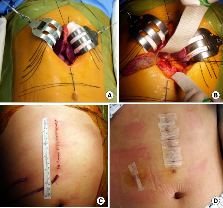 | Fig. 2Operative photographs of upper midline mini-laparotomy in a 32-year-old female patient. (A) After skin marking of the bilateral subcostal margins, a 9.5 cm-long upper midline incision is made. (B) Hepatic hilum is displaced toward the midline after gauze packing at the right lateral side. (C) The wound is repaired meticulously and then adhesive tapes are applied. (D) The small skin stitches and wound drain are removed at 5 days after surgery.
|
Go to :
DISCUSSION
After resection of choledochal cyst in adult patients, anastomotic stenosis of hepaticojejunostomy is the most common and most serious complication unless the patients do not have accompanying malignancy. We have previously reported that the late liver-related complications such as intrahepatic stone formation, recurrent cholangitis and overt anastomotic stricture occurred in at least 16 of the 102 patients (15.7%) with CCD type I and 15 of the 72 patients (20.8%) with CCD type IVa.1
The anastomotic stricture may be closely associated with technical factors, although its risk factors were reported as CCD type IVa, large cyst size, short symptom duration and high grade infiltration of inflammatory cells.11 All portions of choledochal cysts should be removed, but in practice, residual proximal cyst walls may be left to facilitate biliary anastomosis.112 The proximal end of the cyst extending from the confluence of the hepatic duct to the junction of the common duct is often difficult to define clearly. Therefore, we used a customized procedure including the initial incision-opening the cyst wall and then identifying the luminal appearance grossly to determine the proximal transection line. Although this technique occasionally resulted in some cyst remnants in the hepatic ducts, it effectively reduced the risk of intractable anastomotic stricture. The size of the anastomosis and the blood supply to the bile duct stump also seem to be important factors related to the occurrence of anastomotic stricture. If there is a stenotic portion around the hilar bile ducts, it should be widened through individualized ductoplasty. If the hepatic duct openings appear to be relatively small or hypoplastic, a wider anastomosis must be beneficial, after leaving some cyst wall remnant.
Such an individualized approach requires a considerable experience with CCD surgery and biliary reconstruction. Secure biliary reconstruction in CCD surgery is often demanding in adult patients; therefore, a higher incidence of surgical complications is anticipated after laparoscopic or robotic resection than after open surgery, unlike in pediatric patients.6789 Considering that we experienced a transferred case requiring reoperation due to intractable anastomotic stricture after robotic surgery as well as we observed unreported surgical complications following laparoscopic approach, the indications for minimal-incision approach should be prudently selected as well as the surgical technique should be further refined.
As shown in the results of this study, we recognized that the cosmetic effect of both right subcostal and upper midline mini-laparotomy incisions may be similar. We have performed the upper midline incision more than 1,000 times for various hepatobiliary operations, but we have rarely performed it in young female patients except for living liver donors.13 Based on the preliminary results of this study, the upper midline approach may be permissible for CCD surgery from the standpoint of wound cosmetics. After placing several surgical gauze sponges in the right lateral area to shift the liver towards the midline, the hepatic hilar structures are readily exposed. If the right subcostal incision had been used in our 3 selected patients with upper midline incision, the incision in these patients might have been elongated and its right lower end might have been lowered to the umbilical level. Regarding the relaxed skin tension line, subcostal incision is usually 30-40° oblique and upper midline incision is perpendicular, thus the latter is more disadvantageous. In contrast, it is reasonable to consider preservation of the rectus muscle function, especially in young patients with vigorous physical activity. Thus, regarding the rectus muscle function, the upper midline incision is more advantageous.
We think that the indications for upper midline incision include a narrow epigastrium with slender body dimension and special conditions requiring full rectus muscle function due to occupation-specific physical activity. In such patients, selection of the upper midline incision would easily facilitate CCD surgery as well as help in wound cosmetics.
To ensure that the skin wound is cosmetically sound, two points should be emphasized. First, for upper midline incision, this incision should be placed exactly at the midline, thus resulting in complete symmetry. Based on the anatomical characteristics and the typical fat deposition pattern, the abdominal area can be considered to have 10 aesthetic subunits including the abdominal region and adjacent areas that significantly affect the aesthetic appearance of the midsection (Fig. 3).14 The upper midline incision is located at the upper midline aesthetic subunit which overlies the midline of the abdomen extending from the xyphoid sternum to the umbilicus. Since these aesthetic subunits give aesthetic silhouette to the body, incisions should be placed to show symmetry. Second, meticulous time-consuming surgical techniques are mandatory.151617 It is important to incise the skin sharply with a knife with low-power electrocauterization because thermal burns involving the epidermis and dermis lead to delayed formation of hypertrophic scar. During surgery, excessive skin tension from wound traction should be avoided because it also induces ischemia of the skin wound edge, which also leads to hypertrophic scar. A slightly longer incision would be cosmetically better than a very tightly retracted small-in-size skin incision. We usually spend 1 hour for the repair of mini-laparotomy incision measuring approximately 9-10 cm in young female patients. It includes two interrupted layer-by-layer abdominal wall closures, wound drain insertion, Scarpa's fascia repair, interrupted subcuticular sutures, reinforcement of edge skin sutures, and adhesive tape application.
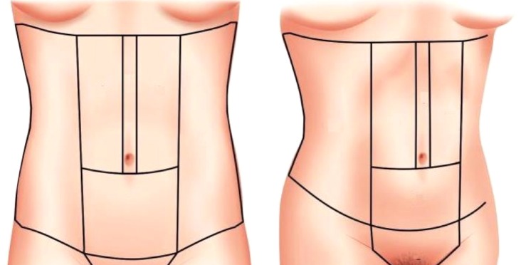 | Fig. 3Aesthetic subunits of the abdominal region and adjacent areas. The upper midline incision is located at the upper midline aesthetic subunit which overlies the midline of the abdomen.14
|
After surgery, we also suggest application of a silicone gel sheet up to 1 year, but more than a half of our patients did not comply with the application, probably due to inconvenience and ineffectiveness.17181920 For patients showing low compliance to silicone gel sheet, it would be recommended to use silicone gel since 3 months after surgery. Self-drying silicone gel is appealing because it is effective, no fixation is required and it is invisible when dry.21
In conclusion, the results of this preliminary study support the claim that cosmetic effect of the upper midline incision for CCD surgery appears to be non-inferior to that of the right subcostal incision if the incision is placed accurately and repaired very meticulously.
Go to :
 XML Download