Journal List > Korean J Hepatobiliary Pancreat Surg > v.20(3) > 1040677

Kim, Yoon, In, and Kim: Efficacy evaluation of SurgiGuard® in partially hepatectomized pigs

Abstract

Backgrounds/Aims

This study evaluated the hemostatic effects of a novel oxidized regenerated cellulose, SurgiGuard®, during liver surgery, using a reproducible and clinically relevant animal model.

Methods

Fifteen mini-pigs underwent left partial hepatectomy. They were randomized to treatment of the resected surface with SurgiGuard® (Group C [test], n=5), Surgicel® (Group B [reference], n=5), or nothing (Group A [control], n=5). Blood loss was measured 5, 7 and 9 min after resection. Time to hemostasis was recorded. Mini-pigs were necropsied 4 or 6 weeks postoperatively to evaluate toxicity changes and material dissolution.

Results

The median resected liver weight was 2.13 g (2.02-2.20) in control group, 2.04 g (2.01-2.13) in reference group, and 2.01 g (1.99-2.12) in test group (p=0.024). Median total blood loss was 57.18 g (52.02-59.54) in control group, 32.52 g (27.66-35.10) in reference group, and 35.52 g (25.70-38.71) in test group (p=0.008). Blood loss at 0-5 minutes and 7-9 minutes was significantly different between groups (p=0.009 and p=0.006, respectively). At necropsy, no hematomas, granulomas, or adhesions were noted in any group. Histopathological analysis revealed no changes suggesting toxicity related to SurgiGuard®.

Conclusions

SurgiGuard® is as effective as Surgicel® in achieving hemostasis after porcine partial liver resection.

INTRODUCTION

Postoperative morbidity and mortality after hepatectomy are adversely affected by blood loss and blood transfusion.1 There are several hemostatic methods to control bleeding. Mechanical techniques include manual pressure and ligation. Although these techniques are the oldest and most flexible method, they can be labor-intensive and add time to the operation.2 Thermal methods, such as electrocauterization, laser cauterization, or argon beam, can also be useful methods. However, these methods create necrotic tissue, which increases the rate of infection and may lead to impaired healing. Furthermore, these conventional techniques and methods are sometimes difficult to apply because of difficulty in accessing the areas of bleeding.3 Topical hemostatic agents may be useful in such situations. Currently, several hemostatic agents are commercially available. Broadly, these agents are one of two types: passive or active. Active agents, such as thrombin, fibrin sealants, and hemostatic patches, provide biologically active components of the coagulation cascade. In contrast, passive agents, such as oxidized regenerated cellulose, gelatin sponges, and collagen pads and sponges, cause the activation of the coagulation cascade.4
Among the passive hemostatic agents, oxidized regenerated cellulose (ORC) has been in use for several decades. ORC contributes to hemostatic action by absorption of blood, surface interaction with platelets and proteins, and coagulation cascade activation.5 Since ORC was first reported in 1943, several commercial products have been used.6 Surgicel® was approved by the United States Food and Drug Administration (FDA; http://www.fda.gov/) in 1960 for control of capillary, venous, and small arterial hemorrhage when standard surgical techniques are ineffective or impractical. ORCs are frequently used in hepatopancreatobiliary surgery, especially liver resections.7
A novel ORC system, SurgiGuard® (Samyang Biopharmaceuticals Corp.), has received approval from the Korean FDA (product license no. 47, 30/09/2014 KFDA). The present study was performed to evaluate the hemostatic effects of SurgiGuard® in liver surgery using a reproducible and clinically relevant animal model.

MATERIALS AND METHODS

Material

SurgiGuard® is a type of ORC. Because, this agent has similar chemical structure, it can be used like Surgicel®. SurgiGuard® is available as a woven patch, i.e., SurgiGuard-fabric® (Fig. 1). Like Surgicel®, SurgiGuard® is designed to assist in the control of capillary, venous, and small arterial hemorrhage when standard surgical techniques are ineffective or impractical.

Methods

This study was conducted in accordance with the Korea Food and Drug Administration notification No. 2012-61 'Good Laboratory Practice' (Aug 24, 2012) and Organization for Economic Co-operation and Development Principles of Good Laboratory Practice (1997) in consultation with the sponsor and was approved by our Institutional Animal Care and Use Committee (Approval No. IACU 12-KE-054).
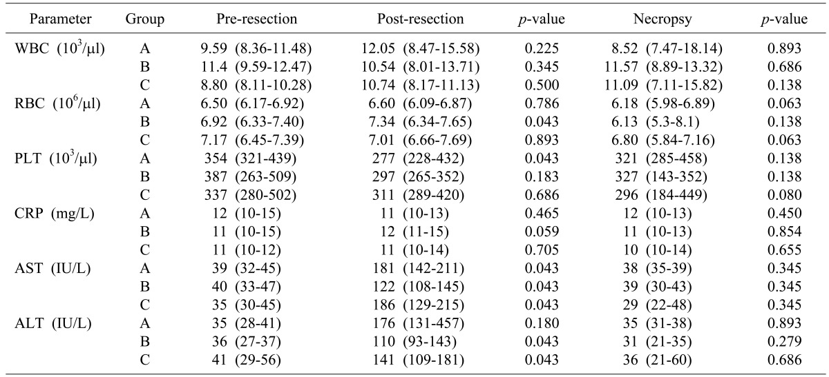
Fifteen mini-pigs (35±5 kg) (Medikinetics mini-pig supplies and services) were randomly allocated into 3 groups: SurgiGuard® (Group C [test], n=5), Surgicel® (Group B [reference], n=5), or none (Group A [control], n=5). Before surgery, all mini-pigs were weighed and blood samples were taken to determine the complete blood cell count (CBC), including white blood cell (WBC), red blood cell (RBC), and platelet (PLT) counts; C-reactive protein (CRP); and liver function tests (LFT), including alanine aminotransferase (ALT) and aspartate aminotransferase (AST). Anesthesia was induced with zoletil and rompun, and the hair in the surgical region (upper abdomen) was removed. Endotracheal intubation was performed and anesthesia was maintained by isoflurane inhalation. The animals were monitored during the procedure by recording the pulse rate and oxygen saturation.
The surgical region was disinfected with povidone iodine and alcohol, followed by the opening of the upper abdominal cavity for liver exposure. Similar to human liver resection, the hepatoduodenal ligament was dissected, and the left lobe was identified for wedge resection. Parenchymal resection of the liver was performed using the Kelly clamp-crushing technique. If the number of bleeding blood vessels was >1, all bleeding blood vessels were closed with forceps except for one blood vessel. However, if the number of bleeding blood vessels was <1, blood vessels were incised to generate one bleeding vessel. Subsequently, ORC of either Surgicel® or SurgiGuard® was applied to the resection margin. The resected liver was weighed.
Blood loss was measured at the time of exposing the resection margin after the confirmation of hemostasis. Blood loss was measured for 5 minutes after the resection. If bleeding was not stopped after 5 minutes, this was regarded as a failure of 1st hemostasis, and blood loss was measured twice at every 2 minutes (i.e., 7 minutes and 9 minutes after resection). The control group received no topical treatment at the resection margin after liver resection. For the 1st hemostasis in the control group, reference group and test group, cotton gauze sheets were applied to the resection margin immediately after resection. Five minutes later, one sheet was applied if 1st hemostasis had not been achieved; this was repeated 2 minutes later if complete hemostasis had not been achieved at the 2nd measurement (at 7 minutes after resection). If complete hemostasis was not achieved after the 3rd measurement (at 9 minutes after resection), a mechanical or thermal method was performed to stop the bleeding.
Detailed measurement of blood loss was performed as follows. In the control group, one sheet of a sterilized waterproof surgical drape was placed below the region of resection, and blood was absorbed by sterilized gauze immediately after the liver resection. In the reference group and test group, two sheets of sterilized waterproof surgical drapes were placed below the region of resection. Surgicel® and SurgiGuard® were applied and one sheet of surgical drapes was removed at the same time. The blood in the surgical field was absorbed by sterilized gauze. The wet gauzes were weighed, and the blood loss was calculated as the difference between the weight of the wet gauze and the premeasured weight of the dry gauze (Fig. 2). After measurement of the blood loss, the surgical region was arranged to avoid adhesion with the resection margin of the liver. The muscle and skin were then sutured. The time of every procedure of the operation was recorded.
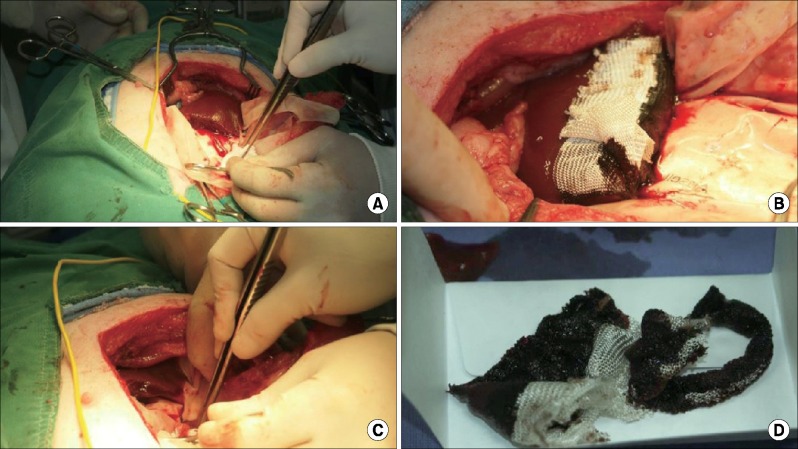
After surgery, the animals were permitted food and water as normal upon recovery from anesthesia. They were subsequently monitored once daily. If any abnormality was found, the type and date of occurrence and severity of signs were recorded for each abnormality. All animals were weighed once weekly throughout the experimental period. One week after the operation, a blood sample was taken to measure the same parameters determined preoperatively.
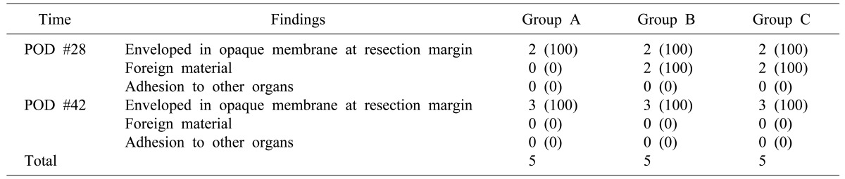
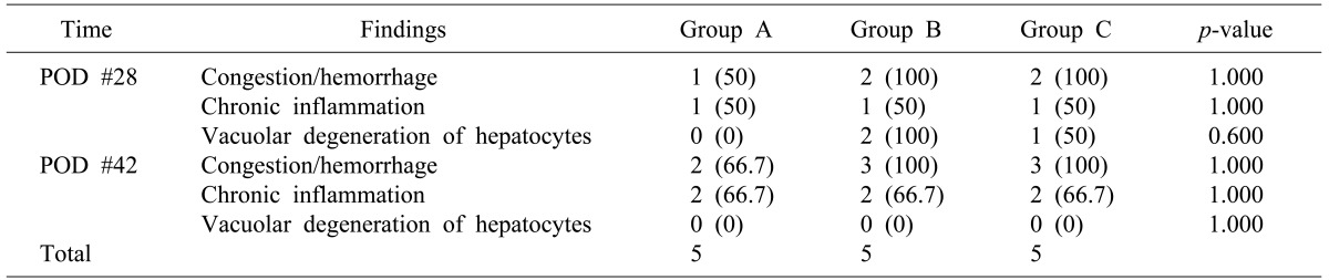
The animals in each study group (control, reference, and test) were randomly divided into 2 subgroups for necropsy. Two mini-pigs were allocated to 1st necropsy group and the other 3 mini-pigs were allocated to 2nd necropsy group. Necropsy was performed 4 weeks after the operation in the 1st necropsy group and 6 weeks after surgery in 2nd necropsy group. All mini-pigs were fasted before their necropsy for at least 12 hours. Before anesthesia, another set of blood samples was obtained from each mini-pig. The animals were deeply anesthetized with zoletil and rompun and euthanized by exsanguination from the carotid artery. The resected livers were examined grossly. After recording the results, the livers, including resection margins, were fixed in 10% neutral formalin solution. They were then embedded in paraffin, and microsections with a thickness of 4-5 µm were made from the blocks. Hematoxylin & Eosin - stained slides were prepared, and the specimens were examined with an optical microscope.

Statistical analysis

Differences between the 3 groups were evaluated by the Kruskal-Wallis test. If significant differences were identified, post-hoc analysis was conducted using the Mann-Whitney test adding Bonferroni's method for correction of type I error to perform pair-wise comparisons between groups. Changes in pre- and post-operative laboratory parameters were evaluated using the Wilcoxon signed-rank test. The criterion for statistical significance was p-value<0.05 and adjusted p value by Bonferroni's method was p<0.017. The commercial statistical program, SPSS 20.1 software, was used for the analyses. The data were presented using nonparametric method, except when indicated otherwise.

RESULTS

Baseline porcine characteristics

The baseline characteristics of the 15 mini-pigs who underwent left hepatectomy are shown in Table 1. Their median body weight was 33.2 kg in the control group, 35.7 kg in the reference group and 34.8 kg in the test group, showing significant difference (overall p=0.003, control vs. reference: p=0.008, reference vs. test: p=0.008, test vs. control: p=0.032). The preoperative CBC, CRP, and LFT values were not significantly different among the three groups.

Hemostasis during liver resection

The only significant difference in the median weight of the resected liver was observed between control group (2.13 g) and reference group (2.01 g) (p=0.024 for overall comparison and p=0.016 for control vs. reference). There were no significant differences between test group and control group (p=0.095) or between reference group and test group (p=0.151). Blood loss after the application of SurgiGuard® (median 35.52 g) and Surgicel® (median 32.52 g) was significantly lower than blood loss in the control group (median 57.18 g) (p=0.008 for the overall comparison, p=0.008 for control vs. reference, and p=0.008 for test vs. control). However, blood loss was not significantly different between the test group and reference group (p=0.548) (Fig. 3). During subgroup analysis according to time periods, blood loss at 0-5 minutes and 7-9 minutes was significantly different between groups at both 0-5 minutes and 7-9 minutes. Median blood loss at 0-5 minutes was as follows: control group 44.15 g; reference group 25.69 g; and test group 24.50 g (p=0.009). At 7-9 minutes, the median blood loss was as follows: control group 4.14 g; reference group 3.00 g; and test group 2.01 g (p=0.006) (Table 2).

Changes in laboratory parameters after liver resection

Comparing preoperative laboratory parameters, including CBC and CRP, to postoperative laboratory parameters on postoperative day #7, there were no significant differences between the groups. By contrast, changes in LFT exhibited significant differences. The median AST level increased significantly from preoperatively to postoperatively in all groups: control group 39 IU/L vs. 181 IU/L (p=0.043); reference group 40 IU/L vs. 122 IU/L (p=0.043); and test group 35 IU/L vs. 186 IU/L (p=0.043). The median ALT level increased significantly from preoperatively to postoperatively in only reference and test group: control group 35 IU/L vs. 176 IU/L (p=0.180; reference group 36 IU/L vs. 110 IU/L (p=0.043); and test group: 41 IU/L vs. 141 IU/L (p=0.043). However, the LFT values returned to the normal range by the necropsy date (Table 3).

Necropsy and histopathological findings

During necropsy, no hematomas, granulomas, or adhesions were observed in any group. In reference and test group, foreign body material noted in the postoperative day #28 pigs were not observed in the postoperative day #42 mini-pigs (Table 4). Histopathological analysis revealed no changes suggesting toxicity related to the hemostatic agents in either reference or test group (Table 5).

DISCUSSION

Several hemostatic agents can overcome the unsatisfactory hemostatic effects of woven cotton gauze. Because the ideal hemostatic material should have ample hemostatic action, minimal tissue reactivity, low cost, in vivo biodegradability, and lack of antigenicity, analyses of the clinical benefits and risks of such materials should be performed by considering these qualities as standards.8
Since ORC was first described in 1943, numerous studies have shown the hemostatic effects of ORC, including when used for hepatectomy.91011 In addition to its application in laparotomy or laparoscopic surgery, ORC is now also used for hemostasis during endoscopic procedures.12 The results of the current study were similar to those previously reported. Although the median weights of the resected liver differed between groups, statistically significant differences were only demonstrated between the control and reference groups. Therefore, this factor did not influence the blood loss comparisons between the test and reference groups. Moreover, because the weight of the resected liver does not always represent the extent of the resected surface margin, the correlation between the resected liver weight and the amount of blood loss may be of minor importance. The time required to achieve hemostasis is an important determinant of the amount of blood loss. In our study, two cases of complete hemostasis within 5-7 minutes occurred in the test as well as the reference group, and there were significant differences in blood loss between the control group and treated groups at 7-9 minutes. These data suggested that the use of the ORC agents promoted time-saving during liver resection.
Because the presence of a foreign body increases the susceptibility to infection, most local hemostatic agents may increase infection and should not be used in an infected wound.1314 However, ORC is resistant to infection because its acidic pH is fatal to bacteria.15 In the current study, laboratory parameters that suggest the presence of an infection, such as WBC and CRP, showed no significant preoperative to postoperative change. In addition, abscess or other infectious changes were absent at necropsy. This result, therefore, suggests that SurgiGuard® was not toxic to the host cells.
Healing quality is also important after the implantation of any medical material or device, and minimum inflammation without a strong foreign body reaction or inhibition of the healing process is desirable. The anti-adhesive effectiveness of ORC is previously reported.1617 To improve upon this characteristic, a novel agent, Interceed®, was developed and used for various types of surgery, especially gynecologic operations.1819 Although microscopic examination revealed chronic inflammatory changes at the liver surface in the current study, no adhesions were observed around the liver on gross examination during necropsy.
ORC reportedly dissolves promptly in various sites without toxicity, with complete dissolution by 6 weeks.620 Migrated ORC is occasionally mistaken for abscess formation or leads to severe complications; hence, complete dissolution without migration is important.2122 In our current study, foreign body materials were found attached to the liver at 4 weeks, but none were observed during the 6-week necropsy examinations; in addition, there was no significant difference in LFT at the time of the necropsy examinations. Although changes in LFT exhibited significant differences on postoperative day #7, the changes were possibly due to transient effects of liver resection and not from ORC toxicity. This result suggested that SurgiGuard® has a suitable capacity for dissolving in a reasonable time without toxicity and for adhering only to surfaces to which it is applied, but not to other organs.
This study has the major limitation of the small size of the experimental groups, which potentially influenced the evaluation of statistically significant outcomes. The difficulty of creating equivalent conditions is another limitation of this study. Although there were no significant differences in PLT counts preoperatively between groups, and the resected liver weights were relatively similar between groups, this does not necessarily guarantee that each mini-pig had similar hemostatic status or resected liver margin area. Moreover, compared to other organs such as the spleen and kidney, the liver has variable spontaneous clotting times and rates of hemorrhage, which leads to difficulties in obtaining precise measurement values.8 Nevertheless, SurgiGuard® exhibited significant hemostatic effects compared to the control group, which were equivalent to the effects exhibited by the reference agent. Furthermore, no toxic changes and adhesions to other organs were observed at the resection margin during the entire experimental period in mini-pigs who were treated with SurgiGuard®.
In conclusion, the current study suggested that the novel ORC, SurgiGuard®, could be as effective as Surgicel® in achieving hemostasis after porcine liver resection. To overcome the limitations of this study, future studies should be performed to provide more data regarding systemic environment such as similar hemostatic condition and obtaining similar resection margin.

ACKNOWLEDGEMENTS

This study was supported by a research grant of Samyang Biopharmaceuticals Corp (SYM-1151).

References

1. Wei AC, Tung-Ping Poon R, Fan ST, Wong J. Risk factors for perioperative morbidity and mortality after extended hepatectomy for hepatocellular carcinoma. Br J Surg. 2003; 90:33–41. PMID: 12520572.
crossref
2. Uranüs S, Mischinger HJ, Pfeifer J, Kronberger L Jr, Rabl H, Werkgartner G, et al. Hemostatic methods for the management of spleen and liver injuries. World J Surg. 1996; 20:1107–1111. PMID: 8798373.
crossref
3. Seyednejad H, Imani M, Jamieson T, Seifalian AM. Topical haemostatic agents. Br J Surg. 2008; 95:1197–1225. PMID: 18763249.
crossref
4. Muench TR, Kong W, Harmon AM. The performance of a hemostatic agent based on oxidized regenerated cellulose--polyglactin 910 composite in a liver defect model in immunocompetent and athymic rats. Biomaterials. 2010; 31:3649–3656. PMID: 20144845.
5. Kanko M, Liman T, Topcu S. A low-cost and simple method to stop intraoperative leakage-type bleeding: use of the vancomycin-oxidized regenerated cellulose (ORC) sandwich. J Invest Surg. 2006; 19:323–327. PMID: 16966211.
crossref
6. Frantz VK. bsorbable cotton, paper and gauze: (oxidized cellulose). Ann Surg. 1943; 118:116–126. PMID: 17858245.
7. Simo KA, Hanna EM, Imagawa DK, Iannitti DA. Hemostatic agents in hepatobiliary and pancreas surgery: a review of the literature and critical evaluation of a novel carrier-bound fibrin sealant (TachoSil). ISRN Surg. 2012; 2012:729086. PMID: 23029624.
crossref
8. Chvapil M, Owen JA, DeYoung DW. A standardized animal model for evaluation of hemostatic effectiveness of various materials. J Trauma. 1983; 23:1042–1047. PMID: 6655749.
crossref
9. Blair SD, Backhouse CM, Harper R, Matthews J, McCollum CN. Comparison of absorbable materials for surgical haemostasis. Br J Surg. 1988; 75:969–971. PMID: 3219544.
crossref
10. Frantz VK, Clarke HT, Lattes R. Hemostasis with absorbable gauze (oxidized cellulose). Ann Surg. 1944; 120:181–198. PMID: 17858482.
11. Wright GF, Tyson RR. Oxidized regenerated cellulose for hepatic hemostasis. Arch Surg. 1963; 87:669–672. PMID: 14056251.
crossref
12. Velázquez-Aviña J, Mönkemüller K, Sakai P, Sulbaran M, Chávez-Vargas C, Montalvo Javé E, et al. Hemostatic effect of oxidized regenerated cellulose in an experimental gastric mucosal resection model. Endoscopy. 2014; 46:878–882. PMID: 25225961.
crossref
13. Scher KS, Coil JA Jr. Effects of oxidized cellulose and microfibrillar collagen on infection. Surgery. 1982; 91:301–304. PMID: 7058512.
14. Tomizawa Y. Clinical benefits and risk analysis of topical hemostats: a review. J Artif Organs. 2005; 8:137–142. PMID: 16235029.
crossref
15. Dineen P. The effect of oxidized regenerated cellulose on experimental intravascular infection. Surgery. 1977; 82:576–579. PMID: 918847.
16. Larsson B, Nisell H, Granberg I. Surgicel--an absorbable hemostatic material--in prevention of peritoneal adhesions in rats. Acta Chir Scand. 1978; 144:375–378. PMID: 735674.
17. Raftery AT. Absorbable haemostatic materials and intraperitoneal adhesion formation. Br J Surg. 1980; 67:57–58. PMID: 7357246.
crossref
18. Prevention of postsurgical adhesions by INTERCEED(TC7), an absorbable adhesion barrier: a prospective randomized multicenter clinical study. INTERCEED(TC7) Adhesion Barrier Study Group. Fertil Steril. 1989; 51:933–938. PMID: 2524407.
19. Ahmad G, O'Flynn H, Hindocha A, Watson A. Barrier agents for adhesion prevention after gynaecological surgery. Cochrane Database Syst Rev. 2015; (4):CD000475. PMID: 25924805.
crossref
20. Katti DS, Lakshmi S, Langer R, Laurencin CT. Toxicity, biodegradation and elimination of polyanhydrides. Adv Drug Deliv Rev. 2002; 54:933–961. PMID: 12384316.
crossref
21. Wada E, Yonenobu K, Ebara S, Kuwahara O, Ono K. Epidural migration of hemostatic agents as a cause of postthoracotomy paraplegia. Report of two cases. J Neurosurg. 1993; 78:658–660. PMID: 8450341.
22. Behbehani S, Tulandi T. Oxidized regenerated cellulose imitating pelvic abscess. Obstet Gynecol. 2013; 121(2 Pt 2 Suppl 1):447–449. PMID: 23344404.
Fig. 1

SurgiGuard-fabric® is a woven patch type of oxidized regenerated cellulose.

kjhbps-20-102-g001
Fig. 2

Experimental procedures: (A) After wedge resection, (B) Application of hemostatic material, (C) Blood absorption by sterilized gauze and (D) Measurement of blood loss.

kjhbps-20-102-g002
Fig. 3

Total blood loss according to hemostatic agent. (A) Control group, (B) Reference group and (C) Test group.

kjhbps-20-102-g002
Table 1

Basic porcine characteristics

kjhbps-20-102-i001

Data are median (range). Group A: control group; Group B: reference group; Group C: test group. *A vs. B: p=0.032, B vs. C: p=0.008, C vs. A: p=0.008. ALT, alanine aminotransferase; AST, aspartate aminotransferase; CBC, complete blood cell count; CRP, C-reactive protein; PLT, platelets; RBC, red blood cells; WBC, white blood cells

Table 2

Resection characteristics: weight of resected liver and blood loss

kjhbps-20-102-i002

Data are median (range). Group A: control group; Group B: reference group; Group C: test group. *A vs. B: p=0.095, B vs. C: p=0.151, C vs. A: p=0.016. A vs. B: p=0.008, B vs. C: p=0.690, C vs. A: p=0.008. A vs. B: p=0.008, B vs. C: p=0.222, C vs. A: p=0.008. #A vs. B: p=0.008, B vs. C: p=0.548, C vs. A: p=0.008

Table 3

Changes between preoperative and postoperative laboratory parameters

kjhbps-20-102-i003

Data are median (range). Group A: control group; Group B: reference group; Group C: test group. ALT, alanine aminotransferase; AST, aspartate aminotransferase; CBC, complete blood cell count; CRP, C-reactive protein; PLT, platelets; pre, preoperative; post, postoperative; RBC, red blood cells; WBC, white blood cells

Table 4

The results of the mini-pig necropsies

kjhbps-20-102-i004

Data are number (%). Group A: control group; Group B: reference group; Group C: test group. POD, postoperative day

Table 5

Histopathological findings of the resected livers

kjhbps-20-102-i005

Data are number (%). Group A: control group; Group B: reference group; Group C: test group. POD, postoperative day

TOOLS
Similar articles