

 PDF
PDF ePub
ePub Citation
 Print
Print


INTRODUCTION
Accompanying the development of novel diagnostic instruments, such as endoscopic ultrasonography or magnetic resonance imaging, asymptomatic cystic tumors of the pancreas have been identified and diagnosed with increasing frequency.123
Intraductal papillary mucinous neoplasm (IPMN) of the pancreas involves the main duct (MD), and/or side branch ducts (BD), and can be classified histopathologically as one of the three following types: main duct IPMN (MD-IPMN), branch duct IPMN (BD-IPMN), and a mixed duct type that is clinicopathologically included in the category of MD-IPMN. Various authors have reported that MD-IPMN has a higher potential of causing malignant or invasive disease, compared to BD-IPMN, and any invasive carcinoma arising in MD-IPMN is more aggressive. In MD-IPMN versus BD-IPMN, their mean frequencies of malignancy are 61.6% vs. 25.5% and those of invasiveness are 43.1% vs. 17.7%, respectively.12345678910111213 Classifying these morphological subtypes of ductal involvement is important for deciding upon a treatment strategy.
International consensus guidelines for the management of IPMN were established in 2006, and revised in 2012.1213 In the 2012 guidelines, surgical resection for MD-IPMN was strongly recommended for all surgically fit patients, with the exception of cases having a main pancreatic duct (MPD) dilation of 5-9 mm; in this instance, the treatment strategies for BD-IPMN are more complicated, and it is these cases for which the concepts of 'high-risk stigmata of malignancy' and 'worrisome features' were adopted. 'High-risk stigmata of malignancy', in BD-IPMN, includes obstructive jaundice in a patient with a cystic lesion of the head of the pancreas, an enhancing solid component within the cyst, and an MPD size of ≥10 mm. 'Worrisome features' include a cyst size of ≥3 cm, thickened/enhancing cyst walls, an MPD size of 5-9 mm, a non-enhancing mural nodule, and an abrupt change in the caliber of the pancreatic duct with distal pancreatic atrophy.13
In this study, we retrospectively reviewed the pathologically confirmed BD-IPMN in patients who underwent surgical resection. The clinicopathological characteristics of BD-IPMN were investigated, by associating the malignant and invasive tendencies with the levels of the total bilirubin, the MPD diameter, the tumor size, and the presence of mural nodules that were classified as 'high-risk stigmata' and/or 'worrisome features,' and we assessed the correlation of these parameters with patient survival and recurrence patterns.
MATERIALS AND METHODS
Among the consecutive patients who underwent pancreatic resection at the Asan Medical Center, between March 1997 and December 2013, 699 patients were pathologically diagnosed as IPMN. The two categories of BD- or MD-mixed type IPMN, and the four pathologic degrees of low- (LGD), intermediate- (IMGD), and high-grade dysplasia (HGD), as well as invasive intraductal papillary mucinous carcinoma (IPMC/invIPMC), were described in the pathological reports. The degree of dysplasia was classified according to the WHO classification, 4th edition,14 and the IPMNs were divided into both non-invasive (LGD, IMGD, and HGD) and invIPMC groups,9 and benign (LGD and IMGD) and malignant (HGD and invIPMC) groups. Among these 699 patients, 337 (48.2%) were proven to be BD-IPMNs, which were defined as being IPMN that did not microscopically involve the MPD. In this study, 324 patients with BD-IPMN were included, and their medical records were reviewed retrospectively; 13 cases were lost during follow-up.
To obtain accurate diagnoses, all patients preoperatively underwent computed tomography (CT) scans, and most also underwent at least one of the following procedures: magnetic resonance cholangiopancreatography (MRCP), endoscopic ultrasonography (EUS), or endoscopic retrograde cholangiopancreatography (ERCP). Although we confirmed and enrolled patients who had pathologically confirmed BD-IPMN, other data (including tumor location, tumor size, the presence of mural nodules, and the MPD diameter) were collected from preoperative imaging studies to investigate the significance of the features of the disease at the time of diagnosis.
At our institution, the surveillance intervals are every 3-6 months for invasive carcinoma and 6-12 months for noninvasive IPMN. All patients underwent CT scans, and some also underwent transcutaneous ultrasonography at each visit. Tumor recurrence and patient survival data were obtained from patient medical records.
The chi-square test or Fisher's exact test were used to compare categorical variables, and Student's t-test was used to compare continuous variables in univariate analyses. An actuarial survival analysis, and comparisons, were performed using the Kaplan-Meier method and the log-rank test. To identify the independent predictive factors, a logistic regression model was employed. A p-value <0.05 was considered to indicate a statistically significant difference. All statistical analyses were performed using SPSS software (version 18; SPSS, Inc, Chicago, IL, USA).
RESULTS
Demographic features and univariate analysis associated with malignant and/or invasive IPMN
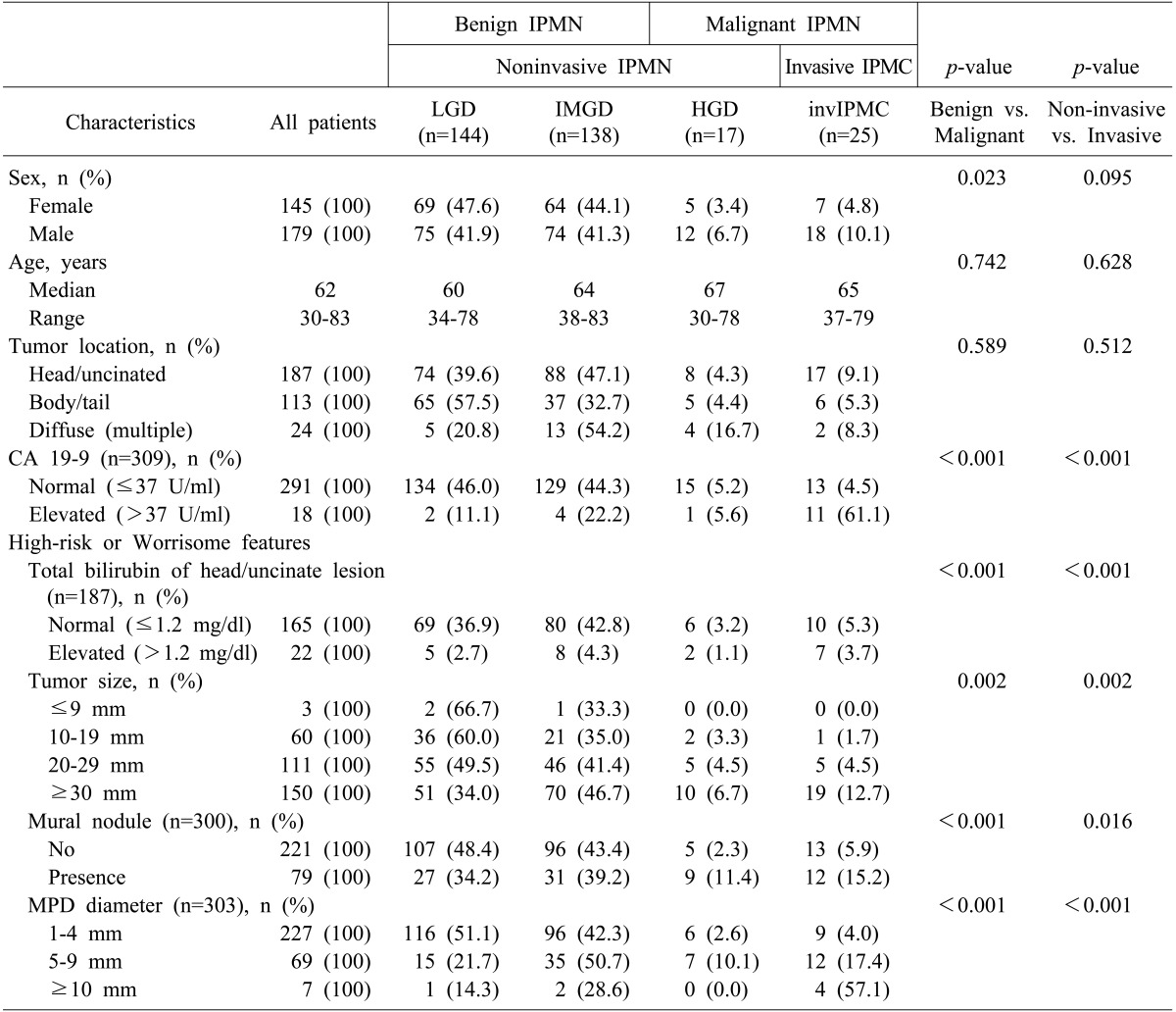
Among the 324 patients with BD-IPMN, there were LGD in 144 (44.4%), IMGD in 138 (42.6%), HGD in 17 (5.3%), and invIPMC in 25 (7.7%). Additionally, 282 patients had benign IPMN (LGD and IMGD), and 42 had malignant IPMN (HGD and invIPMC). They could also be categorized as non-invasive IPMN (LGD, IMGD, and HGD) in 299, and invasive IPMC in 25 cases (Table 1).
Table 1 also shows the univariate analyses that detected the correlations between the clinical characteristics and the malignant and invasive diseases. Male patients were more likely to have malignant IPMN (p=0.023), but there was no significant difference in the non-invasive and invasive subgroups according to the sex (p=0.095). The median ages were higher in HGD and invasive carcinoma (67 and 65, respectively) than in benign IPMN, although this difference was not statistically significant. The location of the tumor was not associated with malignancy or invasiveness (p=0.589 and p=0.512, respectively). Elevated levels of CA19-9 (>37 U/ml) were associated with both malignant and invasive disease (for both, p<0.001). All four factors that were included in the high-risk stigmata and/or worrisome features of the 2012 international consensus were significantly correlated with both malignant and invasive diseases. These factors included the total bilirubin of the head/uncinate process lesion (for both, p<0.001), the tumor size (for both, p=0.002), the presence of a mural nodule (p<0.001 or p=0.016, respectively), and the MPD diameter (for both, p<0.001).
Patient survival
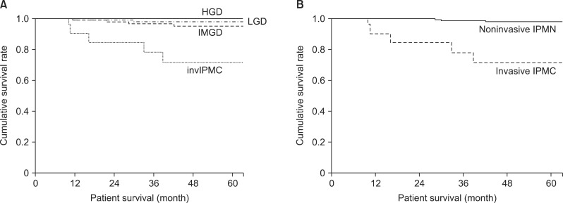
Patient survival curves are shown in Fig. 1A and 1B. The 5-year survival rates were 98.1% in LGD, 95.3% in IMGD, 100% in HGD, and 71.8% in invIPMC cases (Fig. 1A). Although HGD was included in malignant IPMN, the three survival curves for LGD, IMGD, and HGD were not significantly different. We did detect a significant difference between noninvasive IPMN and invasive IPMC (p<0.001; Fig. 1B).
Multivariate analysis of factors associated with malignant or invasive IPMN
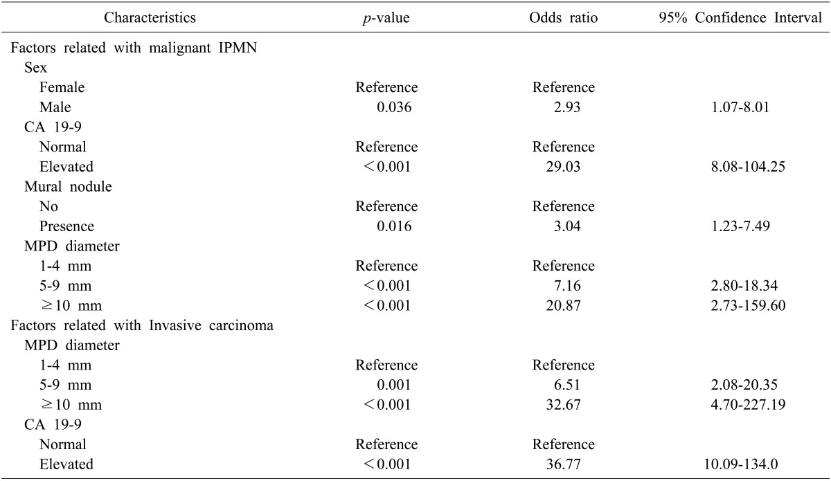
Among the factors associated with malignant IPMN described in Table 1, we identified four statistically significant factors, via a multivariate analysis (Table 2), that included the following: male sex (odds ratio: OR 2.928), the presence of a mural nodule (OR 3.093), the dilated MPD diameter (OR 7.158 in 5-9 mm and 20.872 in more than 10 mm), and an elevated level of CA19-9 (OR 29.019). The Multivariate analysis identified two factors as being predictive of invasive carcinoma (Table 2) that included the following: a dilated MPD diameter (OR 6.512 in 5-9 mm and 32.674 in more than 10 mm) and an elevated level of CA19-9 (OR 36.774).
Disease recurrence following surgical resection
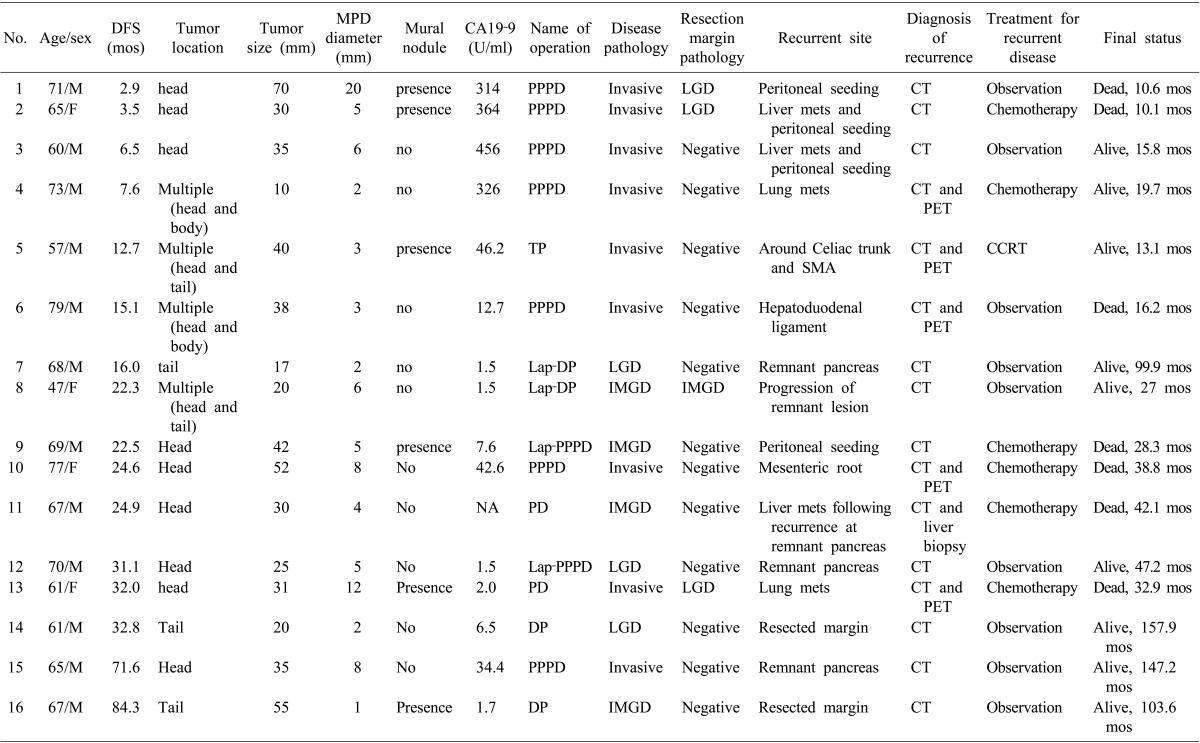
There were 16 patients who experienced disease recurrence following surgical resection; their clinicopathological characteristics are described in Table 3. The affected patients included 9 invasive carcinoma, 4 IMGD, and 3 LGD cases at the initial diagnosis. Of the 7 patients with initially noninvasive IPMN, 5 had recurrences or a progression of disease in the remnant pancreas (No. 7, 8, 12, 14, and 16; Table 3). 2 of the initially noninvasive IPMN patients experienced carcinomatous recurrences (peritoneal seeding and liver metastasis) at 22.5 and 24.9 months following surgical resection, and died as a consequence of disease progression (No. 9 and 11; Table 3).
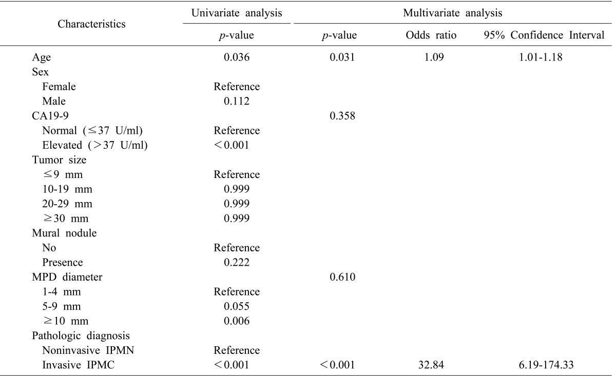
Old age, an elevated level of CA19-9 (>37 U/ml), a dilated MPD diameter (≥10 mm), and invasive pathology were associated with disease recurrence via a univariate analysis (p=0.036, p<0.001, p=0.006, and p<0.001, respectively; Table 4). According to the multivariate analysis, old age (OR 1.092) and invasive IPMC (OR 32.840) patients were independently significant, in regard to disease recurrence following surgical resection.
DISCUSSION
IPMN is a disease entity that has a wide range of histological subtypes, and a natural history that follows an adenoma-to-carcinoma sequence. Knowledge of the natural history and disease traits of each subtype of IPMN is important for devising treatment strategies that avail patients of optimal treatment opportunities. International consensus guidelines for the management of IPMN were generated to assist in devising treatment strategies, by the working group of the International Association of Pancreatology. The 2012 guidelines adopted new concepts, 'high-risk stigmata of malignancy' and 'worrisome features,' so we stratified patients into these two categories.13 The recommendation of these guidelines was to let the stratified patients follow different therapeutic flows. In this retrospective single-institutional study, we investigated preoperative clinical predictors of malignant and invasive diseases of BD-IPMN of the pancreas, and validated the flows recommended by the 2012 international consensus guidelines. To the best of our knowledge, this is the largest series- to date- of patients with BD-IPMNs that were resected and confirmed pathologically.
The prevalence of malignant IPMN was reported to range from 10% to 32% in patients with BD-IPMN.5101516 In the series presented here, malignant IPMN (HGD and invIPMC) was 13.0%, and invasive carcinoma (invIPMC) was 7.7% of the entire resected BD-IPMN cohort. Therefore, the proportion of malignant or invasive diseases of BD-IPMN would be reduced if non-resected non-invasive BD-IPMNs under observation had been included.
Our survival analysis showed that the 5-year survival rate of non-invasive IPMN was 98.2%. Although HGD was included in the malignant category, its survival curve resembled that of benign IPMNs. The postoperative survival rate of HGD of BD-IPMN was already verified by a previous study.2 However, Winner et al.17 showed that carcinoma in situ (HGD) of IPMN showed a higher recurrence rate after a postoperative period of 5 years. Tamura et al also demonstrated that HGD of MD-IPMN showed a high recurrence rate and poor survival, when long-term surveillance of more than 5 years was carried out.11 Therefore, although HGD shows favorable survival outcomes for the initial 5 years after surgical resection, it should be followed as a malignant disease using long-term surveillance.
The strategic factors that were considered to be 'high risk stigmata' and/or 'worrisome features', from the 2012 international consensus guidelines, were validated in this study. Although we did not investigate the presence of a thickened cystic wall or cytological pathology, additional factors that were associated with malignant and/or invasive disease were identified. We also revealed other prognostic factors, including sex and CA19-9, which were correlated with malignant and/or invasive IPMN in the multivariate analysis. Pedrazzoli et al.18 reported that positron emission tomography was more accurate than the guidelines, in distinguishing benign from malignant IPMN. Additionally, more predictive factors will likely be identified in future studies. When more prognostic and diagnostic data are accumulated, these guidelines should then be revised. Therefore, various trials, including those with retrospective and prospective designs, should be repeated to identify more accurate factors. The present algorithm for management is quite complicated for application in an outpatient clinical setting, and it only focuses on the morphological features, without regard to patient factors or biomarkers that are capable of reflecting the propensity of the disease.
Previously, patients with invasive IPMC were reported to be more likely to recur with a probability of hazard ratio of 5.2, but the original pathology did not predict disease severity on recurrence.17 Here, we observed recurrences in 16 (4.9%) of the 324 patients with resected BD-IPMN, including 36% of the invasive IPMC and 2.5% of the non-invasive IPMN cases. We determined that the initial disease pathology was the most powerful predictor of recurrence (OR 32.84: 95% CI 6.19-174.33), and that the initial pathology was related to the recurrence pattern (or severity) following resection. There were 8 of 9 patients with initially invasive IPMN who had distant metastases. or peritoneal seeding, while 5 of 7 patients with originally non-invasive IPMN had recurrences on the remnant pancreas or resected margin (Table 3). The latter 5 patients have remained alive and are under periods of regular observation that have ranged from 27.0 to 157.9 months. We experienced 2 cases of malignant recurrences of IMGD in the two year follow-up period after surgical resection (No. 9 and 11 in Table 3). Those cases initially had a single lesion in the pancreas, and the resected margins were clear from disease. Therefore, there must still be undisclosed biological characteristics of IPMN, and molecular or large scale clinical studies to prevent the deaths of patients with initially benign disease should be considered.
In conclusion, in a high-volume single-institutional study, we investigated the clinicopathological characteristics of BD-IPMN of the pancreas. Although the current treatment guidelines for BD-IPMN reflect malignant and invasive features appropriately, they are confined to the morphological characteristics of the disease. Patient factors and biological features should be considered when devising optimal therapeutic or surveillance strategies.
 XML Download