

 PDF
PDF ePub
ePub Citation
 Print
Print


Abstract
Leonardo da Vinci is remembered as the greatest genius of the Renaissance. He left outstanding achievements as an artist, scientist and inventor, and contributes up to today's science. He ranks the best in a variety of fields, such as botany, mathematics, geology, astronomy, geometry and optics. It has well known that Leonardo is an artist, scientist, inventor and philosopher. And he was a great anatomist that dissected dead bodies and animals directly and left many anatomical drawings.
He took an interest in anatomy from the point of view of the artist, which is why the human body structure and function to know the sakes were “ignorant of the anatomy should not be upset.” Over time, he became interested in the structure and function of the body, even get the human body in a difficult environment; he dissected many the human bodies directly. His scientific inquiry and infatuation made him as an advanced pioneer for more than 100 years, and got enough level to surpass the artistry. Leonardo left about 1,800 anatomical figures of the muscular, skeletal, vascular, nervous and urogenital system, and they are also very scientific and high artistic achievements. The aim of this article is to take a look at Leonardo da Vinci's anatomical achievements and thoughts. In addition, the goal is to knowledge today's anatomists about Leonardo da Vinci's astonishing achievements as a great pioneer in anatomy.
Go to : 
References
1. Jose AM. Anatomy and Leonardo da Vinci. Yale J Biol Med. 2001; 74:185–95.
2. Shoja MM, Agutter PS, Loukas M, Benninger B, Shokouhi G, Namdar H, et al. Leonardo da Vinci's studies of the heart. Int J Cardiol. 2013; 167:1126–33.

3. O'Malley CD, Saunders JB, de CM. Leonardo on the Human Body. New York: Dover Publication, Inc.;1983. p. 17,. p. 27–30.
4. Leonardo da Vinci. [cited 2016 May 2] Available from:. https://en.wikipedia.org/wiki/Leonardo_da_Vinci.
5. Dunn PM. Leonardo Da Vinci (1452–1519) and reproductive anatomy. Arch Dis Child Fetal Neonatal Ed. 1997; 77:249–51.
6. Keele KD. Three early masters of experimental medicine-Erasistratus, Galen and Leonardo da Vinci. Proc R Soc Med. 1961; 54:577–88.
7. Song CH. Historical figures in anatomy. Seoul, Republic of Korea: Jeongseok Publisher.;2015. p. 81–83. 172–173.
8. Persaud TVN. Early history of human anatomy. Springfield. Illinois: Charles C Thomas Publisher LTD;1984. p. 45–46.
9. Singer C. A short history of anatomy and physiology from the Greeks to Harvey. New York: Dover Publications, Inc.;1957. p. 28–29. 90, 92.
10. Persaud TVN. A history of human anatomy. The post-Vesalian era. Springfield. Illinois: Charles C Thomas Publisher LTD;1997. p. 3–5. 69.
11. Janson HW. History of art. New York: Harry N. Abrams, Inc.;1977. p. 76.
12. Persaud TVN, Loukas M, Tubbs RS. A history of human anatomy. 2nd Edt., Springfield. Illinois: Charles C Thomas Publisher LTD;2014. p. 60–65.
13. Lester T (author)/Oh SG (translator). da Vinci, Draw a the vitruvian man. Seoul, Republic of Korea: Roots and leaves;2014. p. 84.
14. Keele KD. Leonardo da Vinci, and the movement of the heart. Proc R Soc Med. 1951; 44:209–13.
15. Jones R. Leonardo da Vinci: anatomist. Br J Gen Pract. 2012; 62:319.
16. Sooke A. Leonardo da Vinci: Anatomy of an artist, [cited 2016 May 2] Available from:. http://www.telegraph.co.uk/culture/art/leonardo-da-vinci/10202124/Leonardo-da-Vinci-Anatomy-of-an-artist.html.
17. Ogden MS. The Galenic works cited in Guy de Chauliac's Chirurgia magna. J Hist Med Allied Sci. 1973; 28:24–33.
18. Johannes N, Frank Z. Leonardo da Vinci, 1452–1519, The complete paintings and drawings. Köln. London, Taschen: Benedikt Taschen Verlag;2003. p. 254 and p.p. 400–479.
19. Keele KD. Leonardo da Vinci's ‘Anatomia Naturale'. The inaugural John F. Fulton lecture. Yale University School of Medicine. November 3, 1978, Yale J Biol Med. 1979; 52:369–409.
Go to :
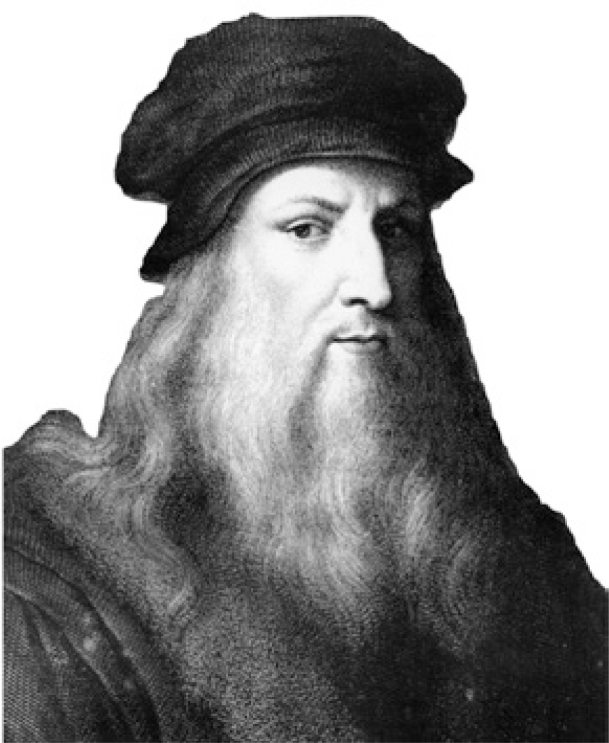 | Fig. 1.Leonardo da Vinci (1452∼1519). Cited from: Song CH. Historical figures in anatomy. Seoul, Republic of Korea: Jeongseok Publisher. 2015. p. 81. |
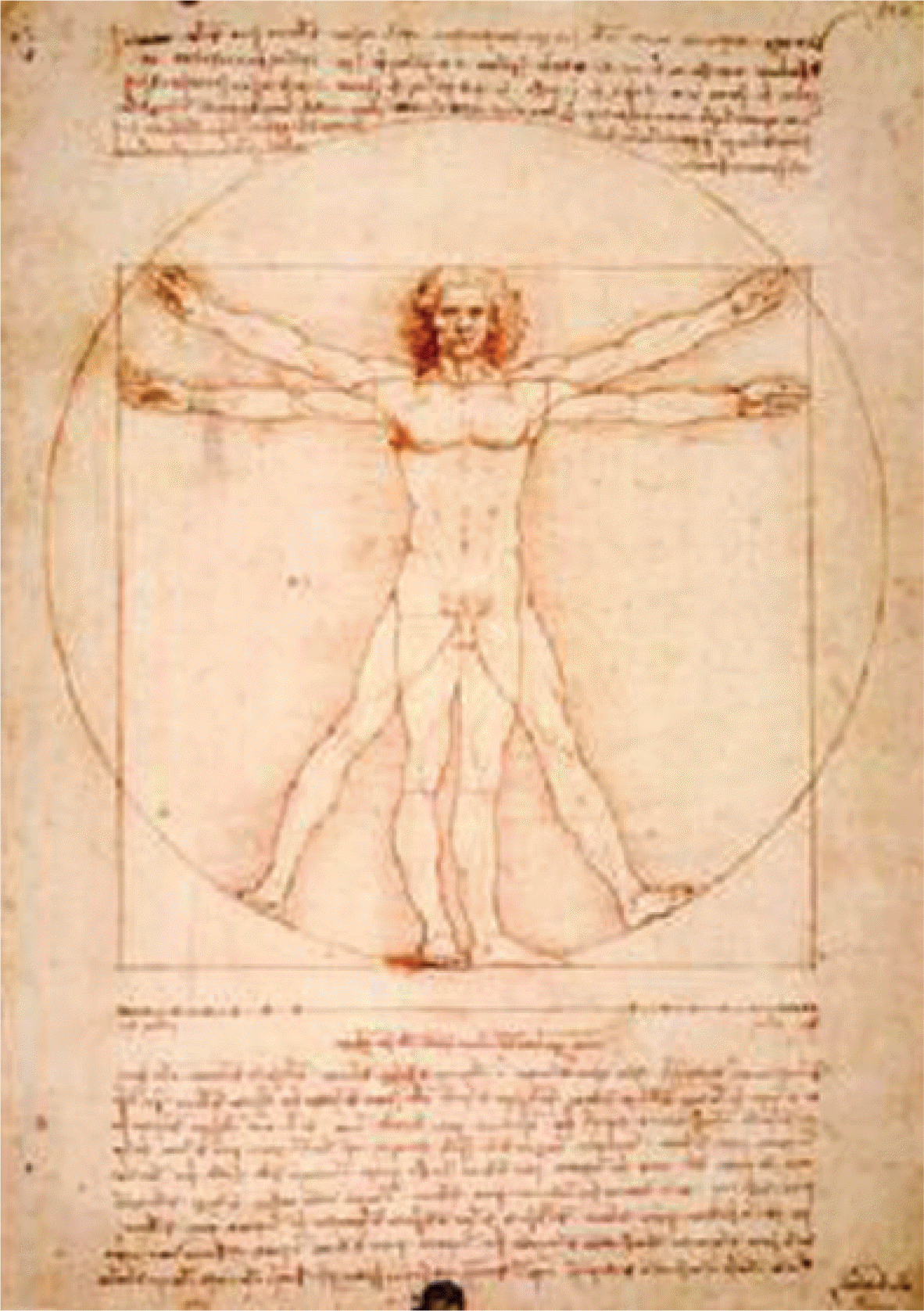 | Fig. 2.The vitruvian manour of Leonardo da Vinci (1490). Cited from: https://en.wikipedia.org/wiki/Leonardo_da_Vinci. |
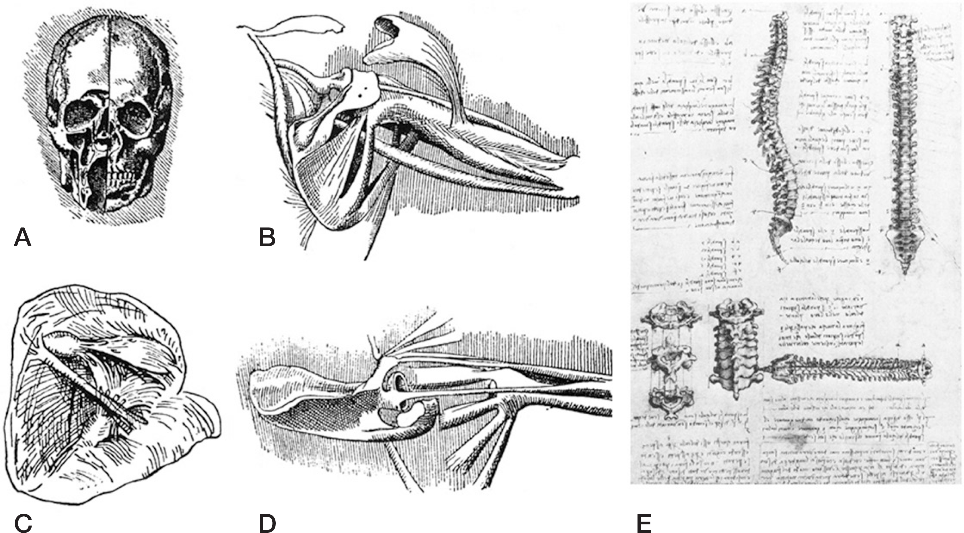 | Fig. 3.Anatomical sketches of Leonardo da Vinci. A. Dissection of the skull showing maxillary sinus, frontal sinus, and nasal fossa. B and D. Dissection of the shoulder. C. The right ventricle of heart laid open showing the tricuspid valve and the intra-ventricular moderate band. E. Drawings of the articulated vertebral column (viewed from the lateral, anterior, and posterior aspects). A∼D: Cited from Singer C. A short history of anatomy and physiology from the Greeks to Harvey. New York: Dover Publications, Inc.; 1957. p. 89. E: Cited from Persaud TVN, Loukas M, Tubbs RS. A history of human anatomy. 2nd Edt., Springfield, Illinois: Charles C Thomas Publisher LTD; 2014. p. 68. |
 | Fig. 4.The drawings of muscles, nerves, arteries and bones by Leonardo da Vinci. A. Series of drawings showing the clavicle, superficial and deep facial muscles, arm and forearm muscles, the ulnar and median nerves, and the ulnar artery (superficial palmar arterial arch). B. Drawings of the muscles of the shoulder region, arm, and forearm. The bones of the ankle region and foot are shown below on the right. A & B: Cited from Persaud TVN, Loukas M, Tubbs RS. A history of human anatomy. 2nd Edt., Springfield, Illinois: Charles C Thomas Publisher LTD; 2014. p. 63. |
 | Fig. 5.The drawings of heart by Leonardo da Vinci. Sketches showing the distribution of the coronary vessels, viewed from different aspects. On the lower right-hand corner are the pulmonary and tricuspid valves seen from above. Cited from Persaud TVN, Lou-kas M, Tubbs RS. A history of human anatomy. 2nd Edt., Springfield, Illinois: Charles C Thomas Publisher LTD; 2014. p. 64. |
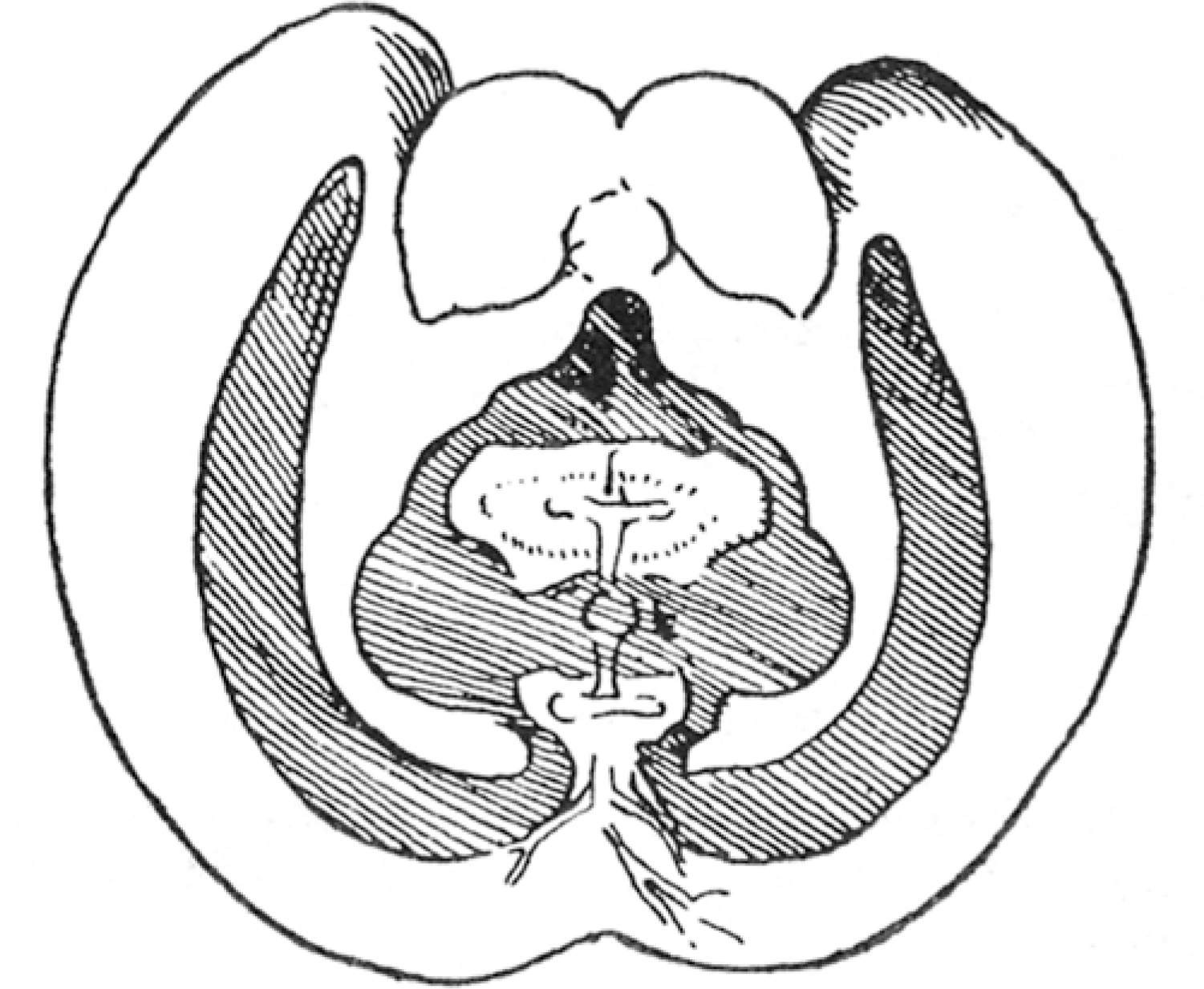 | Fig. 6.Wax cast of ventricles of the brain as portrayed by Leonardo da Vinci. Cited from Singer C. A short history of anatomy and physiology from the Greeks to Harvey. New York: Dover Publications, Inc.; 1957. p. 91. |
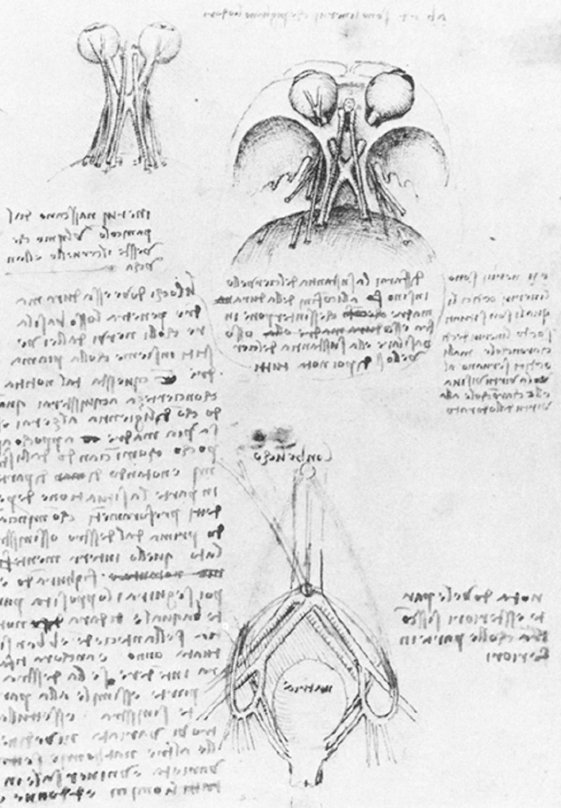 | Fig. 7.The sketches above show the eyeballs, optic nerve and chiasm, and several cranial nerves. Below is a drawing of the uterus with paired vessels supplying it from either side. Cited from Persaud TVN, Loukas M, Tubbs RS. A history of human anatomy. 2nd Edt., Springfield, Illinois: Charles C Thomas Publisher LTD; 2014. p. 67. |
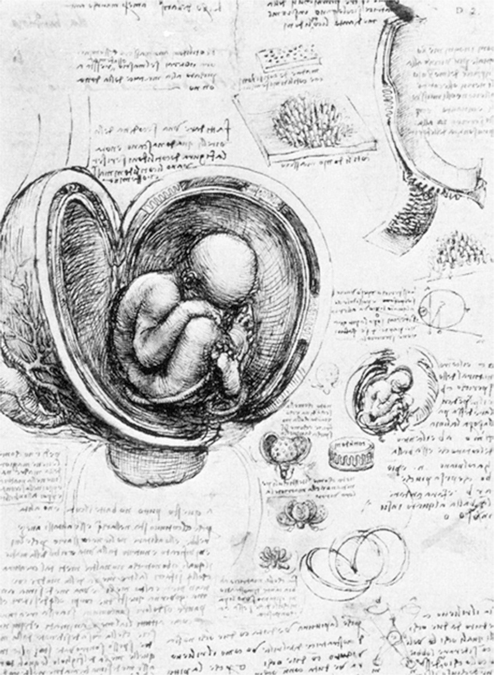 | Fig. 8.The pregnant uterus bisected to show the fetus in its natural position. The placenta, however, is that of a cow. The sketches on the right and below reveal the different layers of fetal membranes. In one of the small diagrams, the fetus can be seen through the transparent amnion following removal of the chorion and uterine wall. Cited from Persaud TVN, Loukas M, Tubbs RS. A history of human anatomy. 2nd Edt., Springfield, Illinois: Charles C Thomas Publisher LTD; 2014. p. 66. |
 | Fig. 9.The principal organs and vascular and urino-genital systems of a woman. This elaborate and impressive drawing is undoubtedly an earlier work of Leonardo da Vinci. There are many inaccuracies and the anatomical representations suggest that the drawing was based on the dissection of animals. An attempt has been made to consolidate the structure of the female anatomy with Galenic concepts of function. Cited from Persaud TVN, Loukas M, Tubbs RS. A history of human anatomy. 2nd Edt., Springfield, Illinois: Charles C Thomas Publisher LTD; 2014. p. 67. |
 XML Download