

 PDF
PDF ePub
ePub Citation
 Print
Print


Abstract
Corpus striatum is subcortical nuclei composed of caudate nucleus and putamen. It has been considered to be associated with motor control and learning. Dysfunction of the striatum is related to Huntington's disease, Tourette's syndrome, obsessive-compulsive disorder and schizophrenia. Nevertheless, standard Korean striatum volume was not set yet. Here, we report the striatum volume in healthy Korean youths.
The subjects were composed of 57 youths (male, 28; female, 29). The MRI study was undertaken after a brief history taking and neurological examination. The DICOM files were imported into V-Works program. Volume models of the intracranial cavity, whole brain, caudate nucleus, and putamen were made and their volumes were calculated by the program.
The average caudate volume was 7.23±1.18 cm3 in male group and 6.23±0.96 cm3 in female group. The average volume of putamen was 7.19±1.25 cm3 in male group and 6.38±0.86 cm3 in female group. Interestingly the right caudate volume is significantly larger in both group, although there is no difference in putamen volume.
This study reports Korean corpus striatum volume in healthy volunteers. These results would provide an important standard reference for further study.
Go to : 
References
1. Dupuis JH, McGavarn CA, Raz N, Briggs SD, Acker JD. Aging of the cerebellar hemispheres and vermis observed in IN VIVO. Soc Neurosci. Abstr. 1995; 21:1536 (614.4).
2. DeLisi LE, Sakuma M, Tew W, Kushner M, Hoff AL, Grimson R. Schizophrenia as a chronic active brain process: a study of progressive brain structural change subsequent to the onset of schizophrenia. Psychiatry Res. 1997; 74:129–40.

3. Matsumae M, Kikinis R, Morocz IA, Lorenzo AV, Sandor T, Albert MS, et al. Age-related changes in intracranial compartment volumes in normal adults assessed by magnetic resonance imaging. J Neurosurg. 1996; 84:982–91.
4. Lenroot RK, Giedd JN. Brain development in children and adolescents: Insights from anatomical magnetic resonance imaging. Neurosci and Biobehav Rev. 2006; 30:718–29.
5. Holttum JR, Minshew NJ, Sanders RS, Phillips NE. Magnetic resonance imaging of the posterior fossa in autism. Biol Psychiatry. 1992; 32:1091–101.
6. Piven J, Saliba K, Bailey J, Arndt S. An MRI study of autism: the cerebellum revisited. Neurology. 1997; 49:546–51.
7. van Eijndhoven P, van Wingen G, van Oijen K, Rijpkema M, Goraj B, Jan Verkes R, et al. Amygdala volume marks the acute state in the early course of depression. Biol Psychiatry. 2009; 65:812–8.
8. Sun J, Maller JJ, Guo L, Fitzgerald PB. Superior temporal gyrus volume change in schizophrenia: a review on region of interest volumetric studies. Brain Res Rev. 2009; 61:14–32.
9. Reiss AL, Abrams MT, Singer H, Ross J, Denckla. Brain development, gender and IQ in children-a volumetric imaging study. Brain. 1996; 119:1763–74.
10. Coffey CE, Lucke JF, Saxton JA, Ratcliff G, Unitas LJ, Billig B, et al. Sex differences in brain aging: a quantitative magnetic resonance imaging study. Arch Neurol. 1998; 55:169–79.
11. Nagai F. Investigation of Korean brain sulcus. Seoul Medical Education Institution Bulletin. 1933; 3:15–29. Korean.
12. Shibata I. Brain weight of the Korean. Am J Phys Anthropol. 1936; 22:27–35.
13. Lee Y, Ku KH, Yu CH. A Statistical Study on the weight of the brain Korean. Chosun Medical Report. 1947; 1:1–71. Korean.
14. Wee DY. Morphological Study on the Korean infants'brain sulcus. J Cathlic Med College. 1960; 4:49–106. Korean.
15. Lee MB, Park TS, Choi IY, Park YH. Statistics on the Korean brain weight. The New Medical Journal. 1963; 6:771–5. Korean.
16. Hong KE, Ock SM, Kang MH, Kim CE, Bae JN, Lim MK, et al. The segmented regional volumes of the cerebrum and cerebellum in boys with Tourette syndrome. J Korean Med Sci. 2002; 17:530–6.
17. Choi JS, Kang DH, Kim JJ, Ha TH, Lee JM, Youn T, et al. Left anterior subregion of orbitofrontal cortex volume reduction and impaired organizational strategies in obsessive-compulsive disorder. J Psychiatr Res. 2004; 38:193–9.
18. Rhyu IJ, Cho TH, Lee NJ, Kim H, Suh YS. Study of the Normal Cerebellar Volume Estimated by Magnetic Resonance Imaging (MRI) in Korean. Kor J Anat. 1997; 30:575–9. Korean.
19. Koh I, Lee MS, Lee NJ, Park KW, Kim KH, Kim H, et al. Body size effect on brain volume in Korean youth. Neuroreport. 2005; 16:2029–32.
20. Rhyu IJ, Cho TH, Lee NJ, Uhm CS, Kim H, Suh YS. Magnetic resonance image-based cerebellar volumetry in healthy Korean adults. Neurosci Lett. 1999; 270:149–52.
21. Chung SC, Tack GR, Yi JH, Lee B, Choi MH, Lee BY, et al. Effects of gender, age, and body parameters on the ventricular volume of Korean people. Neurosci Lett. 2006; 395:155–8.
22. Lee BY, Sohn JH, Choi MH, Lee SJ, Kim HS, Yang JW, et al. A volumetric study of the corpus callosum in 20s and 40s Korean people. Brain Struct Funct. 2009; 213:463–7.
23. Lee MS. Korean standard brain volume based on 3 dimensional MRI volumetry. Doctoral dissertation, Korea University Seoul Korea. 2004.
24. Anatasi G, Cutroneo G, Tomasello F, Lucerna S, VitetTa A, Bramanti P, et al. In vivo basal ganglia volumetry through application of NURBS models to MR images. Neuroradiology. 2006; 48:338–45.
25. Jang JH, Kwon JS, Jang DP, Moon WJ, Lee JM, Ha TH, et al. A proton MRSI study of brain N-acetylaspartate level after 12 weeks of citalopram treatment in drug-naive patients with obsessive-compulsive disorder. Am J Psychiatry. 2006; 163:1202–7.
26. Harris GJ, Pearlson GD, Peyser CE, Aylward EH, Roberts J, Barta PE, et al. Putamen volume reduction on magnetic resonance imaging exceeds caudate changes in mild Huntington's disease. Annals of Neurology. 1992; 31:69–75.
27. Aylward EH, Brandt J, Codori AM, Mangus RS, Barta PE, Harris GJ. Reduced basal ganglia volume associated with the gene for Huntington's disease in asymptomatic at-risk persons. Neurology. 1994; 44:823–8.
28. Hyde TM, Stacey ME, Coppola R, Handel SF, Rickler KC, Weinberger DR. Cerebral morphometric abnormalities in Tourette's syndrome A quantitative MRI study of monozygotic twins. Neurology. 1995; 45:1176–82.
29. Filipek PA, Richelme C, Kennedy DN, Caviness VS Jr. The young adult human brain: an MRI-based morphometric analysis. Cereb Cortex. 1994; 4:344–60.
30. Ostby Y, Tamnes CK, Fjell AM, Westlye LT, Due-T⊘nnes-sen P, Walhovd KB. Heterogeneity in subcortical brain development: A structural magnetic resonance imaging study of brain maturation from 8 to 30 years. J Neurosci. 2009; 29:11772–82.
31. Yamashita K, Yoshiura T, Hiwatashi A, Noguchi T, Togao O, Takayama Y, et al. Volumetric Asymmetry and Differential Aging Effect of the Human Caudate Nucleus in Normal Individuals: A Prospective MR Imaging Study. J Neuroimaging. 2011; 21:34–7.
32. Goldstein JM, Seidman LJ, Horton NJ, Makris N, Kennedy DN, Caviness VS Jr, et al. Normal sexual dimorphism of the adult human brain assessed by in vivo magnetic resonance imaging. Cereb Cortex. 2001; 11:490–97.
33. Hynd GW, Hern KL, Novey ES, Eliopulos D, Marshall R, Gonzalez JJ, et al. Attention deficit-hyperactivity disorder and asymmetry of the caudate nucleus. J Child Neurol. 1993; 8:339–47.
34. Mataró M, Garcia-Sánchez C, Junqué C, Estévez-González A, Pujol J. Magnetic resonance imaging measurement of the caudate nucleus in adolescents with attention-deficit hyperactivity disorder and its relationship with neuropsychological and behavioral measures. Arch Neurol. 1997; 54:963–8.
35. Gunning-Dixon FM, Head D, McQuain J, Acker JD, Raz N. Differential aging of the human striatum: a prospective MR imaging study. AJNR. 1998; 19:1501–7.
36. Peterson B, Riddle MA, Cohen DJ, Katz LD, Smith JC, Hardin MT, et al. Reduced basal ganglia volumes in Tourette's syndrome using three-dimensional reconstruction techniques from magnetic resonance images. Neurology. 1993; 43:941–9.
37. Castellanos FX, Giedd JN, Eckburg P, Marsh WL, Vaituzis AC, Kaysen D, et al. Quantitative morphology of the caudate nucleus in attention deficit hyperactivity disorder. Am J Psychiatry. 1994; 151:1791–6.
38. Castellanos FX, Giedd JN, Marsh WL, Hamburger SD, Vaituzis AC, Dickstein DP, et al. Quantitative brain magnetic resonance imaging in attention-deficit hyperactivity disorder. Arch Gen Psychiatry. 1996; 53:607–16.
39. Castelo JM, Courtney MG, Melrose RJ, Stern CE. Putamen hypertrophy in nondemented patients with human immunodeficiency virus infection and cognitive compromise. Arch Neurol. 2007; 64:1275–80.
40. Ifthikharuddin SF, Shrier DA, Numaguchi Y, Tang X, Ning R, Shibata DK, et al. MR volumetric analysis of the human basal ganglia: normative data. Acad Radiol. 2000; 7:627–34.
41. Giedd JN, Snell JW, Lange N, Rajapakse JC, Casey BJ, Kozuch PL, et al. Quantitative magnetic resonance imaging of human brain development: ages 4–18. Cereb Cortex. 1996; 6:551–60.
42. Giedd JN. Structural magnetic resonance imaging of the adolescent brain. Ann N Y Acad Sci. 2004; 1021:77–85.
43. Savic I. Asymmetry of cerebral gray and white matter and structural volumes in relation to sex hormones and chromosomes. Frontiers in Neuroscience. 2014; 8:329.
Go to :
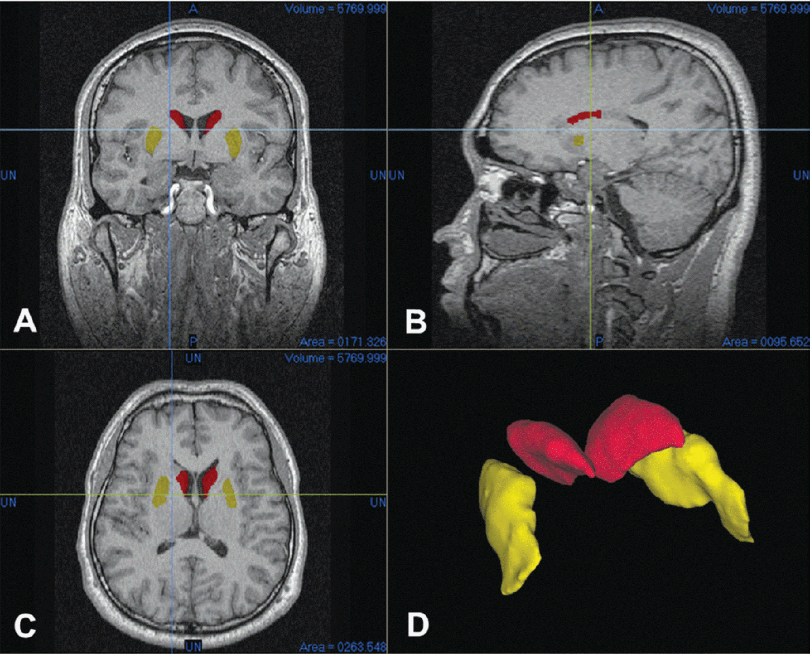 | Fig. 1.Screen capture of segmentation works and 3-D modeling in V-works program. The segmented region was confirmed from coronal (A), sagittal (B), and horizontal plane (C). The 3-D volume rendered models of cuaduate (red) and putamen (yellow) appeared on (D). |
Table 1.
Caudate volume about male and female.
Table 2.
Putamen volume about male and female.
Table 3.
Left and right asymmetry of caudate nucleus volume. Caudate nucleus volume shows that the right caudate is larger than the left regardless of male and female. (∗) means statistical significance under p-value<0.05.
Table 4.
Left and Right asymmetry of Putamen volume. The Putamen in both male and female does not show volume difference between the right and left side.
 XML Download