Abstract
The purpose of this study is to enable children and adolescents to experience anatomy and clinics. For the purpose, the ways to use the anatomy educational resources (comics, 3-dimensional images, and 2-dimensional images) and diagnostic tools (stethoscope, sphygmomanometer, pen light, and reflex hammer) were described in a guide book. Following the guide book, students experienced anatomy and clinics in a course of the science museum. They learned anatomy with the comics, then did virtual dissection with the 3-dimensional and 2-dimensional images. Sequentially, with the diagnostic tools, they listened to heart sound, measured blood pressure, and performed light reflex and knee jerk. Through this study, we have found that anatomy and clinics should be experienced pleasantly. The com-plimentary guide book is expected to be further improved in future, so as to achieve better experience at home, science museum, and school.
References
1. Hwang SB, Chung MS, Park JS. Anatomy cartoon for common people. Korean J Anat. 2005; 38:433–41.
2. Jang HG, Chung MS, Chae KS, Lee ST, Park HS, Lee SH, et al. Multimedia data for common people to learn anatomy easily. J Lifestyle Med. 2012; 2:47–55.
3. Shin DS, Chung MS, Park JS, Park HS, Lee S, Moon YL, et al. Portable document format file showing the surface models of cadaver whole body. J Korean Med Sci. 2012; 27:849–56.

4. Shin DS, Chung MS, Park HS, Park JS, Hwang SB. Browsing software of the Visible Korean data used for teaching sectional anatomy. Anat Sci Educ. 2011; 4:327–32.

5. Korean Association of Anatomists. Anatomical terminology. 6th ed.Anyang: Academya;2014.
6. Song JJ, Lee HC, Yoo PK. The effect of science cartoon reading on the levels of interest in science, the academic achievements and the scientific attitudes of elementary students. Elem Sci Educ. 2013; 32:581–92.
7. Salend SJ. Creating inclusive classrooms: effective and re-flective practices. 7th ed.New York City: Pearson;2011.
8. Han MJ, Yang CH, Noh TH. An analysis of teaching strategies of science teacher's teaching in science museum. J Korean Assoc Sci Educ. 2014; 34:559–69.

9. Oh CS, Kim KJ, Chung E, Choi HJ. Digital report in an anatomy laboratory: a new method for team-based dissection, reporting, and evaluation. Surg Radiol Anat. 2015; 37:293–8.

10. Chung BS, Chung MS. Free manual of the cadaver dissection modifiable by other anatomists. Anat Sci Int. 2015; 90:201–2.
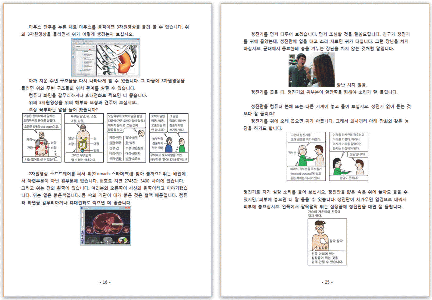
Fig. 2.
Anatomy tools for the experience. Easy anatomy (left), PDF file of 3-dimensional images (center), and browsing software of 2-di-mensional images (right).

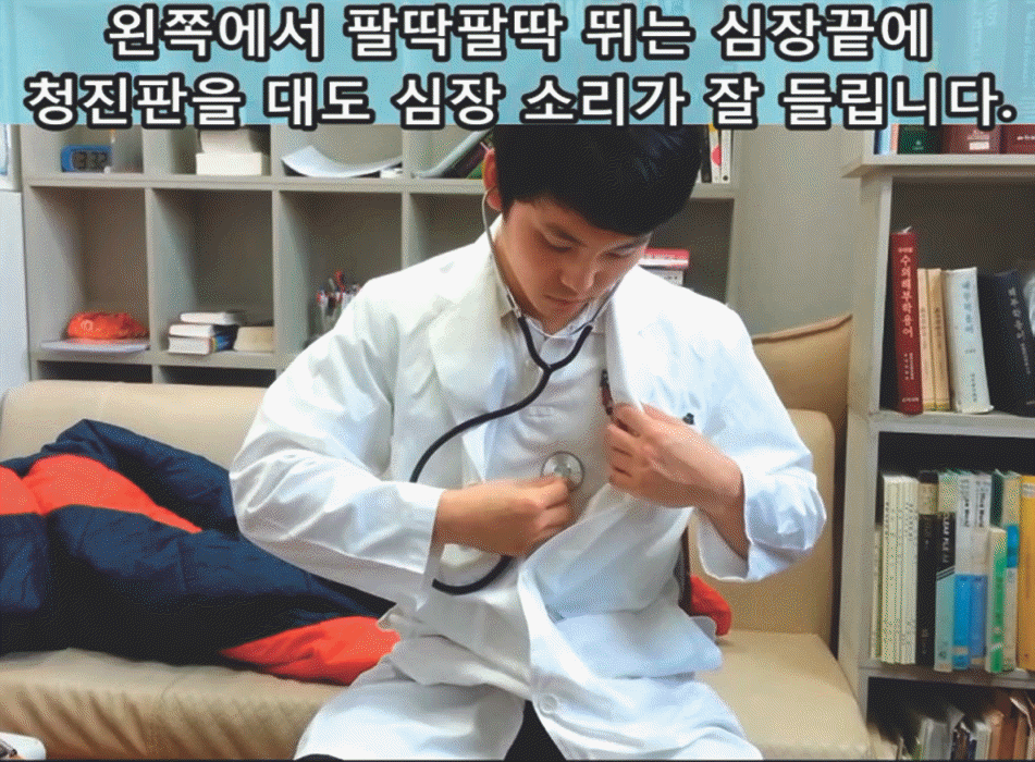
Fig. 3.
Video guide to explain how to examine heart sound using stethoscope. The Korean subtitle at the top delivers instruction for each step of experience.

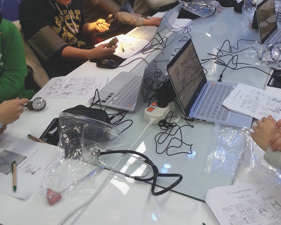
Fig. 4.
Students reading the anatomy comics and using the stetho-scope in the course, held in Gwangju National Science Museum, Korea.

Table 1.
Correlation of the anatomy and clinical experiences with the requisite tools.
| Anatomy tools | Clinical tools | ||||||
|---|---|---|---|---|---|---|---|
| Comics | 3-dimensional images | 2-dimensional images | Stethoscope, Sphygmo- manometer | Pen light | Reflex hammer | ||
| Anatomy | Visceral organs1 | O | O | O | |||
| experience | Muscles2 | O | O | ||||
| Heart sound | O | O | O | ||||
| Clinical experience | Blood pressure | O | O | O | |||
| Light reflex | O | O | |||||
| experience | Oral inspection | O | O | ||||
| Knee jerk | O | O | O | ||||
Table 2.
Remarks from the high school students, who attended the anatomy and clinical experiences, held in Gwangju National Science Museum, Korea.


 PDF
PDF ePub
ePub Citation
 Print
Print


 XML Download