

 PDF
PDF ePub
ePub Citation
 Print
Print


INTRODUCTION
Tuberculosis (TB) remains one of the major public health problems causing significant morbidity and mortality worldwide. The most commonly involved site of TB is the respiratory tract, which results in pulmonary tuberculosis (PTB). Additionally, it can affect any sites of the body including lymph nodes, pleura, the gastrointestinal tract, and the genitourinary tract, which results in extrapulmonary tuberculosis (EPTB). The incidence and prevalence rate of TB and EPTB vary from country to country. According to the notification rate of TB, there were reported cases of 36,305 TB (74.3 cases per 100,000 people) and 8,129 EPTB (16.6 cases per 100,000) in Korea in 2010 (http://www.knta.or.kr/) [1]. The reported proportion of EPTB among all TB in Korea from 2004 to 2010 was 11% (3,556/31,503), 15% (5,171/35,269), 14% (5,044/35,361), 14% (5,005/34,710), 17% (5,813/34,157), 19% (6,923/35,845), and 22% (8,129/36,305) during successive years [1]. However, considering lower voluntary reporting, diagnostic difficulty of EPTB, and missed cases, it is likely that actual proportion of EPTB was much higher than what was reported.
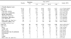
Studies have been conducted on EPTB epidemiology according to the region and the country. The proportion of EPTB in the United States (USA) was 18.7% (47,293/253,299) during 1993-2006 [2]. The most commonly affected sites of EPTB in the USA were lymph nodes (40%), pleura (20%), and bone and/or joint (11%) [2]. Another epidemiologic study showed that the proportion of EPTB in England and Wales increased from 48% in 1999 to 53% in 2006 [3]. A study in Turkey demonstrated that EPTB prevalence during 2001-2007 was 26% (103/397) of total TB, and the most common types of EPTB were genitourinary TB (27%) and meningeal TB (19%) [4]. Except for the last study performed in Turkey, several prior studies regarding EPTB have been performed using cases diagnosed clinically based on histological, radiological, and microbiological evidence (Table 1) [2,4-13]. Only some of these clinically-diagnosed (CD) cases were confirmed by microbiological culture. So far, there have been few studies regarding microbiologically-confirmed EPTB.
The aim of the present study was to investigate the characteristics of EPTB at a tertiary hospital in Seoul, Korea. In addition, the data of culture-confirmed (CC) EPTB were compared with those of CD EPTB.
MATERIALS AND METHODS
1. Study design and study subjects
This study was performed using the data obtained at Samsung Medical Center, which is the second largest medical center located in Seoul, Korea, from January 1, 1995 to December 31, 2010. Samsung Medical Center is a tertiary care center with 2,005 beds and about 7,500 outpatients per year in Seoul, which has a population of about 10,039,000 (http://kostat.go.kr/portal/korea/index.action) [14]. For this study, the CC cases were defined as TB confirmed via the mycobacterial culture method, which is the gold standard for the diagnosis of TB. In contrast, the CD cases were defined as TB via the clinical judgment based on clinical, radiological, histological, as well as microbiological methods. By definition, the CD cases included the CC cases in the current study. We retrospectively reviewed the data regarding both cases with clinical diagnosis of TB and culture-positive TB. All the study subjects were non-duplicated during the study period, and the age was selected at the time of diagnosis of TB. The cases of EPTB with concurrent PTB were excluded, while the cases of EPTB involved simultaneously in the various sites were all included according to the sites. The subjects were categorized according to the type of culture-positive specimen or clinically-affected sites. The cases of EPTB were defined as TB diagnosed in any site other than the respiratory tract.
2. Culture of Mycobacterium tuberculosis
The culture of Mycobacterium from a clinical specimen was done in Ogawa medium using conventional solid egg-based methods. The automated broth-based system, BACTEC MGIT 960 system (Becton Dickinson, Franklin Lakes, NJ, USA), was used, along with traditional solid-based methods since February 2009. The final culture interpretations were done after 8 weeks (in case of the solid-based method) or 6 weeks (in case of the broth-based method) of incubation at 37 degree Celsius. In addition to the culture, Ziehl-Neelsen staining was also performed to find acid-fast bacilli (AFB). The differentiation of Mycobacterium tuberculosis (MTB) from Nontuberculous Mycobacteria (NTM) was based on the presence of cord formation with AFB staining, an MPT-64 antigen test (SD, Yongin, Korea), a Gen-Probe MTD test (Gen-Probe, Inc., San Diego, CA, USA), or Multiplex-PCR (M&D, Wonju, Korea). Specimens of stool and homogenized tissue were treated before incubation with 4%, and 1% NaOH-N-acetyl-L-cysteine (NaOH-NALC), respectively. All other specimens were treated before incubation with 2% NaOH-NALC except for the sterile sample.
3. Statistical analysis
Data were expressed as mean±SD. We compared the categorical data, such as age and gender, according to the type of specimen or involved sites using the chi-square test. P values less than 0.05 were considered statistically significant. All analyses were performed with the PASW version 19.0 (SPSS Inc., Chicago, IL, USA).
RESULTS
1. Clinically-diagnosed cases
Clinical diagnosis of TB consisted of 30,118 cases (78%) of PTB and 8,608 cases (22%) of EPTB during the study period (Table 2). In addition, the proportions of EPTB per year during 16 years ranged from 19-22% with a CV of 2.4%. Of the 8,608 EPTB cases, the most commonly involved types were tuberculous lymphadenitis (N=2,655, 31%), followed by pleural TB (N=2,147, 25%), abdominal TB (gastrointestinal tract and peritoneum; N=1,146, 13%), skeletal TB (N=737, 9%), genitourinary TB (N=504, 6%), CNS TB (N=494, 6%), others (N=548, 6%) and miliary TB (N=377, 4%) (Table 2).
The mean ages of patients with EPTB and PTB were 43 years and 47 years, respectively. The male to female (M/F) ratio was 0.88 (4,020/4,588) in EPTB, while it was 1.31 (17,058/13,060) in PTB. The distribution of both age and gender between PTB and EPTB cases were significantly different (P<0.001 for both, Table 2). In addition, pleural TB and CNS tuberculosis were seen more in males than females (The M/F ratios in pleural TB and CNS tuberculosis were 1.6 and 1.2, respectively). While tuberculous lymphadenitis and miliary tuberculosis were more frequent in females than males (The M/F ratios of tuberculous lymphadenitis and miliary tuberculosis were 0.5 and 0.9, respectively)
2. Culture-confirmed cases
The PTB and EPTB among the culture-positive cases were composed of 5,504 (88%) and 745 (12%) cases, respectively (Table 2). In addition, the proportions of EPTB per year during 16 years ranged from 5.3-21% with a CV of 3.4%. The distribution of EPTB in 745 culture-positive specimens were in the pleura (N=197, 26%), lymph nodes (N=148, 20%), bone and joint (N=118, 16%), gastrointestinal tract (N=80, 11%), peritoneum (N=57, 7%), genitourinary area (N=77, 10%), the CNS (N=51, 7%), cardiac region (N=9, 1%), and others (N=8, 1%) (Table 2).
The mean ages of patients with EPTB and PTB were 49 years and 50 years, respectively. The M/F ratio was 0.99 (370/375) in EPTB, while it was 1.45 (3,253/2,251) in PTB. The age and gender distributions were significantly different between PTB and EPTB (P<0.001 for both, Table 2). In addition, pleura and cardiac involvement were more common in males (The M/F ratio in pleura and cardiac EPTB were 2.1 and 2.0, respectively, Table 2). Lymph node and CNS involvement was observed more often in females (The M/F ratio was 0.5 for both, Table 2).
DISCUSSION
Although a national surveillance program has continuously decreased the incidence of PTB, the incidence of EPTB has significantly increased in Korea since 2007 [1]. These contrasting increases of EPTB were also previously reported in the U.S., in addition to the Caucasian population [2,6]. According to the National Tuberculosis Surveillance System from 50 states in the U.S., the proportion of EPTB was reported to be increased from 15.7% in 1993 to 21.0% in 2006 [2] That study revealed an association between EPTB and female gender, nonwhite race, foreign birth, and a disproportionately slower decrease in EPTB prevalence, compared with the decrease in PTB cases [2]. In addition, the Caucasian population also showed a proportional increase of EPTB, along with a decreased incidence of TB [6]. The causes of the proportional increase of EPTB in the Caucasian population were reported to be an increase of life expectancy and the female-predominant distribution, in addition to the decline of BCG-vaccinated patients. Based on these previous studies, the epidemiology of EPTB might be different from those of PTB. For effective TB control in Korea, an epidemiologic study of EPTB in the Korean population is also needed. However, little studies regarding EPTB have been conducted in Korea [13]. Herein, we investigated the proportion and distribution of EPTB in a large Korean population. In addition, we were the first to compare the cases of CC EPTB to those of CD EPTB in Korean patients.
The current study revealed that the overall proportions of EPTB cases during the 16 years examined were 22% and 12% among the CD and CC cases, respectively. The CC cases consisted of only culture-positive cases of all EPTB; thus, the number of actual CC cases might be underestimated. Indeed, the diagnosis of EPTB may be difficult due to nonspecific clinical manifestations, lower sensitivity of AFB staining and mycobacterial culture, and difficulties in obtaining direct specimens from the extrapulmonary sites compared to those of PTB. Among a total of 745 CC cases of EPTB, the pleura, lymph nodes, and gastrointestinal tract with peritoneum were the most commonly affected sites in the Korean population. In the same manner, tuberculous lymphadenitis, pleural TB, and abdominal TB were the most common types among 8,608 cases with clinical diagnoses of EPTB. There was no difference in distributions of EPTB in both CC and CD cases in the current study. However, according to previous studies, the results analyzed using the cases with clinical diagnosis of EPTB were significantly different from the results evaluated using culture-proven EPTB (Table 1) [4,9]. The one epidemiologic study revealed that EPTB was common in early adulthood (mean age 23 years) and lymph nodes was the most common sites in the Turkish city of Kocaeli during 1996-2000 (Table 1) [9]. However, the other study demonstrated that genitourinary TB and meningeal TB were most common two types in Malatya, Turkey, during 2001-2007 (Table 1) [4]. The former study was performed using the cases with diagnoses based on the clinical evidence of EPTB, while the latter study was done using only culture-confirmed cases of EPTB [4,9]. Indeed, the epidemiologic results might be different according to study design, as well as country, region, city, and time, such as the results from two different studies in Turkey. The current study firstly demonstrated that there was no difference in the distributions of EPTB when the cases of CC were compared to those of CD in Korean population during the same 16 years.
Interestingly, the proportions of EPTB per year were much more variable in CC cases than CD cases (The coefficient of variation from CC cases was 3.4%, while that from CD cases was 2.4%). There were some possible reasons for the variability in proportions of EPTB in the study duration. First, the national program for control of TB has been focused on PTB rather than EPTB, which results in the contrasting increase of EPTB. Second, the EPTB detection rate might be increased due to the improvement of diagnostic tests, including introduction of the broth-based culture method and interferon-gamma releasing assay, as well as the genotype-based assay. In the present study, the broth-based culture system was introduced in February 2009. Indeed, the proportion of culture-positive cases of EPTB was 12% (99/835) in 2007-2008, increasing to 20% (183/932) in 2009-2010 (P<0.001). Also, the proportions of CD cases of EPTB were 23% (1,027/4,449) and 25% (933/3716) in 2007-2008 and 2009-2010, respectively (P=0.0352).
The present study showed that the female gender and younger age were more prevalent in cases with EPTB compared with PTB cases. Pleural TB was more commonly found in males, while tuberculous lymphadenitis was more commonly observed in females in both CC and CD cases. However, there was a discrepancy in the M/F ratio of CNS TB between CC cases and CD cases (The M/F ratios of CNS TB were 1.19 and 0.55 in CD and CC cases, respectively). The previous study in Malatya, Turkey, showed that meningeal and bone and/or joint TB were commonly found in males, while lymphatic, genitourinary, and peritoneal TB were observed more in females [4].
In conclusion, the proportion and number of EPTB cases in Korea were increased during the study period, which was more significant in CC cases during 2007-2010. The epidemiologic characteristics of EPTB were different from those of PTB as well as EPTB from other countries. The most common types of EPTB were tuberculous lymphadenitis, pleural TB, and abdominal TB. Further study would be needed to find the causes of the increase in EPTB and to investigate the differences of radiologic and histologic findings among the cases.

 XML Download