

 PDF
PDF ePub
ePub Citation
 Print
Print


INTRODUCTION
Traditionally, the diagnosis of infective endocarditis (IE) is mainly based on the clinical suspicion, conventional culture method, and echocardiography. Etiological diagnosis by culture method is very important to provide adequate treatment and favorable clinical outcome. However, blood culture negative infective endocarditis (CNE) has been reported with a frequency of 2.5-31%, which is a diagnostic dilemma and results in delayed treatments and high mortality [1,2]. The most common cause of CNE is antibiotic administration preceding blood culture, which occurs in 45-60% of CNE [3-5]. Fastidious and slow-growing organism is also reported as another cause of CNE. In addition, other factors such as right sided endocarditis and cases with permanent pacemaker have been reported for the causes of CNE [2].
The HACEK group (Haemophilus spp., Aggregatibacter actinomycetemcomitans gen. nov., comb. nov., Cardiobacterium hominis, Eikenella corrodens, Kingella kingae) is classified as one of the fastidious organism causing CNE [1,6]. Haemophilus parainfluenzae as one of the HACEK group constitutes part of normal flora of the oropharyngeal cavity. Thus, risk factors such as dental procedure, nasopharyngeal infection, tongue piercing, and the use of tongue scrapers seem to be needed for the development of IE by Haemophilus parainfluenzae [7-9]. Although it is a fastidious organism that requires V factor for growth, the mean incubation time for growth of HACEK group is 3-5 days except Actinobacillus actinomycetemcomitans, which requires up to 30 days for growth [10,11].
Herein, we present a case of CNE due to Haemophilus parainfluenzae, which was identified only via sequencing of direct tissue specimen in a healthy boy without definitive predisposing factors.
CASE REPORT
A 17-year-old healthy boy was referred from other hospital with fever of unknown origin and weight loss (5 kg/month) for 1 month. He complained that fever was highly spiking and responded to antipyretics. Chest tightness, dyspnea, and cough were added as new symptoms 1-2 days earlier than admission. His medical history was not remarkable except the present illness. On admission to hospital, he had blood pressure of 93/51 mmHg, heart rate of 123 beats per min, temperature of 39.2℃, and oxygen saturation of 97%. Physical examination revealed Grade III pansystolic murmur at lower left sternal border and mild splenomegaly. Chest X-ray showed that peribronchial marking in both lungs was increased with normal cardiac size, which was suggestive of viral pneumonia. Oxygen and empirical antibiotics such as ampicillin-sulbactam and azithromycin were administrated. However, worsening of chest discomfort and bilateral pulmonary edema were developed along with persistent fever on day 2 after admission. A transthoracic echocardiography (TTE) showed severe mitral regurgitation (MR), chordal rupture of anterior mitral valve leaflet (AMVL), and left atrium dilation. IE was suspected and antibiotics were exchanged into combination of ampicillin, nafcillin, and gentamicin. Subsequent transesophageal echocardiography (TEE) showed linear mobile mass like lesion on AMVL, which indicates the vegetation. The sizes of the vegetation were 0.65×0.26 cm and 0.62×0.55 cm.
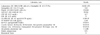
Gram stain and 3 sets of cultures using the blood obtained on admission day showed no growth of microorganism even after 14 days of incubation in Bact/Alert 3D automated microbial detection system (bioMerieux, Inc, Durham, NC, USA). One day after that, 1 set of blood culture also was negative for the growth of microorganism. Other laboratory findings were described in Table 1. Delayed mitral valvuloplasty with artificial chordae formation was performed to save native valve on day 14 after admission. Ruptured chordaes were removed and fibrous tissue on tip of separated chordae was collected to perform the Gram stain and culture. Histologic sections of removed mitral valve demonstrated fibromyxoid valvulopathy and fibrin deposition with dystrophic calcification, which is suggestive of IE. However, no microorganisms were identified using Gram stain of mitral valve. In addition, Gram stain and culture of the chordae tissue showed no microorganism. We did not prepare wet smear in this case.
To identify the causative agent, genomic DNA was extracted from the direct chordae tissue using the MagNa Pure LC Total Nucleic Acid Isolation Kit (Roche, Mannheim, Germany). Approximately 500 bp of 16S ribosomal RNA (rRNA) gene was amplified in an ABI 9700 Thermal Cycler (Applied Biosystems, Foster City, CA, USA). The polymerase chain reaction consisted of 1 cycle at 94℃ for 5 min, followed by a 32 cylce amplification (94℃ for 30 sec, 60℃ for 30 sec, and 72℃ for 30 sec) and a final extension step at 72℃ for 7 min. Standard 16S rRNA primer pairs were following sequences according to CLSI guideline; Forward primer, 4F:5'-TTG GAG AGT TTG ATC CTG GCT C-3' and reverse primer, 534R: 5'- TAC CGC GGC TGC TGG CAC-3' and forward primer 27F: 5'-AGA GTT TGA TCM TGG CTC AG-3' and reverse primer 801R: 5'-CGGC GTG GAC TTC CAG GGT ATC T-3' [11]. Direct sequencing was performed using the BigDye Terminator Cycle Sequencing Kit 3.1 (Applied Biosystems) on ABI prism 3730 Genetic analyzer (Applied Biosystems). Sequence analysis showed that the 16S rRNA sequences of the isolate had 100% homology (548/548 bp) with those of Haemophilus parainfluenzae (GenBank accession number, FJ939586.1).
The patient had no more fever since the day 3 after admission. Antibiotic treatment was done for 6 weeks and the patient had a full recovery without any complication. Follow-up echocardiography after 2 months showed minimal MR.
DISCUSSION
Fastidious organisms as causative organism constitute 5-15% of IE and 50% of CNE [5]. CNE caused by fastidious organisms can be diagnosed mainly by serologic testing and molecular techniques. There are several organisms in this category; intracellular organisms (Coxiella burnetii and Chlamydia spp.), slow-growing bacteria (Bartonella spp., Brucella spp. and Legionella spp.), and nutritionally deficient bacteria (Abiotrophia spp. Mycoplasma spp. and Haemophilus spp.) [5]. Our patient was also all negative for serologic tests of antibodies responding to Coxiella burnetii, Brucella, Bartonella, and Legionella pneumophilia. When traditional microbiological and serologic diagnosis was not definitive, molecular method is the most powerful tool for etiologic diagnosis of CNE. According to one study, molecular approach showed a sensitivity of 72% and specificity of 100%, while the culture-based approach only showed a sensitivity of 26% and a specificity of 62% [12]. There are some situations when sequencing of the 16S rRNA gene is useful in case of; 1) antibiotic treatment preceding blood culture, 2) fastidious pathogen that is difficult to grow by conventional culture method, 3) some pathogen such as Coxiella burnetii and Brucella spp. that give a significant biohazard.
Fewer than 80 cases of H. parainfluenzae endocarditis including 4 cases in Korea have been reported in the literature [13]. Typical clinical manifestations of H. parainfluenzae endocarditis are subacute endocarditis, predisposing factors of dental procedure, nasopharyngeal infection, tongue piercing, and preexisting cardiac abnormalities in the literature [11,14,15]. Also, septic embolization is well known complication of H. parainfluenzae endocarditis, because which they have a propensity to form friable and large vegetations [11]. Our patient showed only prolonged fever and heart murmur without any other physical findings such as Roth's spots, and Janeway's lesions. In addition, he had no definite predisposing conditions except for probable respiratory infection, which was supported by the increased Mycoplasma pneumoniae IgG titer on admission. It was not certain that Mycoplasma pneumoniae infection was occurred prior to H. parainfluenzae infection. Conventional culture method failed to identify the causative organism in spite of long incubation time of 2 weeks. Considering the HACEK organisms could be recovered within 3 days in improved blood culture system, negative results of culture in our case might be due to previous antibiotic treated in other hospital during unknown period. If antibiotics were administrated for short period, result of blood culture turned negative into positive. However, longer courses of incurative antibiotics lead to negative results of blood culture for several weeks [1].
In summary, we report the case of H. parainfluenzae endocarditis diagnosed via only molecular technique with culture negative tissue specimen. Early awareness of causative organism leading to CNE is very important because empirical antibiotic therapy might not be effective and delayed diagnosis could result in destruction of cardiac structure.
 XML Download