Abstract
Background
Bacteremia is a life-threatening infection, and prognosis is highly dependent on early recognition and treatment with appropriate antimicrobial agents. We investigated the diagnostic performance of serum procalcitonin (PCT) for differentiation between contaminants and true pathogens in blood cultures.
Methods
Serum PCT, C-reactive protein (CRP) and blood culture were performed for 473 patients between February 2008 and October 2008. We retrospectively reviewed the patients' clinical characteristics and laboratory results based on medical records.
Results
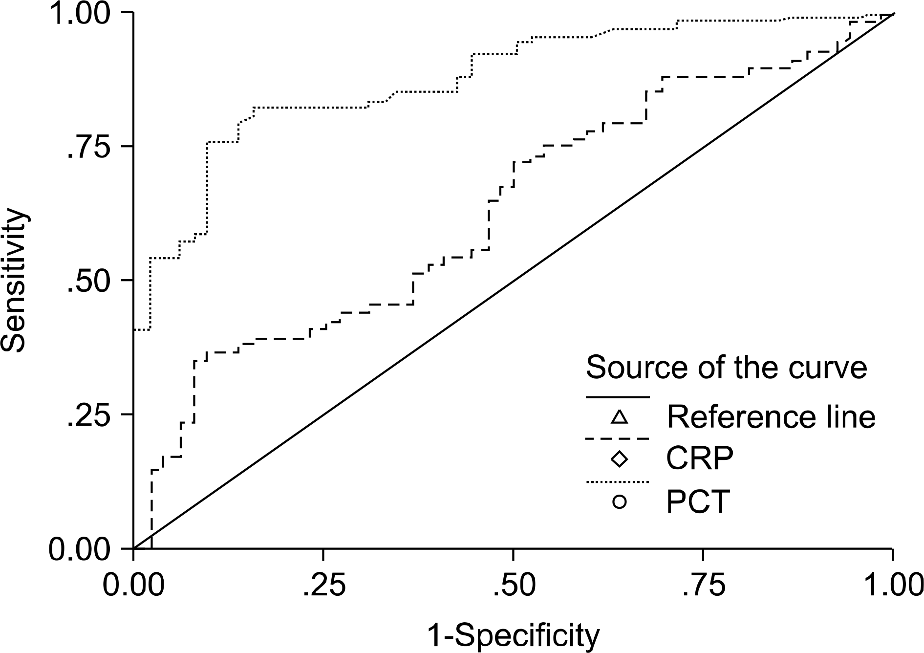
The mean concentration of PCT was significantly different between the two negative and positive blood culture groups (6.45 ng/mL vs 28.77 ng/ mL, P<0.001). Procalcitonin levels were found to be markedly higher in those with Gram-negative bacilli (mean±SD; 59.58±67.00 ng/mL) bacteremia than in those with Gram-positive cocci (mean±SD; 17.75±42.88 ng/mL) bacteremia (P<0.001). The areas under the receiver operating characteristic curves (95% confidence interval) for PCT and CRP were 0.880 (0.820∼ 0.940) and 0.637 (0.538∼0.736), respectively. The use of a PCT level of 2 ng/mL as a cutoff value yielded an 83.6% positive predictive value and a 77.4% negative predictive value for the detection of bacteremia pathogens.
REFERENCES
1. Angus DC, Linde-Zwirble WT, Lidicker J, Clermont G, Carcillo J, Pinsky MR. Epidemiology of severe sepsis in the United States: analysis of incidence, outcome, and associated costs of care. Crit Care Med. 2001; 29:1303–10.

2. Digiovine B, Chenoweth C, Watts C, Higgins M. The attributable mortality and costs of primary nosocomial bloodstream infections in the intensive care unit. Am J Respir Crit Care Med. 1999; 160:976–81.

3. Ibrahim EH, Sherman G, Ward S, Fraser VJ, Kollef MH. The influence of inadequate antimicrobial treatment of bloodstream infections on patient outcomes in the ICU setting. Chest. 2000; 118:146–55.

4. Charles PE, Ladoire S, Aho S, Quenot JP, Doise JM, Prin S, et al. Serum procalcitonin elevation in critically ill patients at the onset of bacteremia caused by either Gram negative or Gram positive bacteria. BMC Infect Dis. 2008; 8:38.

5. Christ-Crain M and Müller B. Procalcitonin in bacterial infections-hype, hope, more or less? Swiss Med Wkly. 2005; 135(31-32):451–60.
6. American College of Chest Physicians/Society of Critical Care Medicine Consensus Conference: definitions for sepsis and organ failure and guidelines for the use of innovative therapies in sepsis. Crit Care Med. 1992; 20:864–74.
7. Hur M, Moon HW, Yun YM, Kim KH, Kim HS, Lee KM. Comparison of diagnostic utility between procalcitonin and C-reactive protein for the patients with blood culture-positive sepsis. Korean J Lab Med. 2009; 29:529–35.

8. Weinstein MP, Murphy JR, Reller LB, Lichtenstein KA. The clinical significance of positive blood cultures: a comprehensive analysis of 500 episodes of bacteremia and fungemia in adults. II. Clinical observations, with special reference to factors influencing prognosis. Rev Infect Dis. 1983; 5:54–70.

9. Müller B, Schuetz P, Trampuz A. Circulating biomarkers as surrogates for bloodstream infections. Int J Antimicrob Agents. 2007; 30(Suppl 1):S16–23.

10. Nahm CH, Choi JW, Lee J. Delta neutrophil index in automated immature granulocyte counts for assessing disease severity of patients with sepsis. Ann Clin Lab Sci. 2008; 38:241–6.
Table 1.
C-reactive protein levels and positive rate of blood culture according to the five groups of procalcitonin concentrations
| Procalcitonin groups (ng/mL) | No. (%) | C-reactive protein (mg/dL) | No. (%) of positive blood culture | |
|---|---|---|---|---|
| Mean±SD | Range | |||
| I (<0.05) | 58 (12.3) | 3.58±4.10∗ | 0.01∼17.52 | 9 (15.5) |
| II (0.05∼0.49) | 122 (25.8) | 9.58±7.54† | 0.01∼31.26 | 22 (18.0) |
| III (0.5∼1.99) | 114 (24.1) | 13.28±10.10‡ | 0.03∼41.52 | 22 (19.3) |
| IV (2∼9.99) | 83 (17.5) | 16.17±10.25§ | 0.12∼46.20 | 21 (25.3) |
| V (≥10) | 96 (20.3) | 20.02±11.34 | 0.17∼50.80 | 46 (47.9) |
| Total | 473 (100) | 13.02±10.54 | 0.01∼50.80 | 120 (25.4) |
Table 2.
Procalcitonin and C-reactive protein levels according to blood culture and SIRS results
| Blood culture | No. (%) | Procalcitonin (ng/mL) | C-reactive protein (mg/dL) |
|---|---|---|---|
| Mean±SD (range) | Mean±SD (range) | ||
| Negative | 353 (74.6) | 6.45±17.61 (<0.05∼184.72) | 12.53±10.43 (0.01∼50.80) |
| SIRS + | 160 (33.8) | 6.99±19.04 (<0.05∼184.72) | 12.62±11.31 (0.01∼50.80) |
| SIRS − | 193 (40.8) | 6.01±16.36 (<0.05∼134.08) | 12.46±9.68 (0.01∼46.20) |
| Positive | 120 (25.4) | 28.77±52.54 (<0.05∼200.00)∗ | 14.45±10.73 (0.01∼49.37) |
| SIRS + | 61 (12.9) | 41.18±58.50 (<0.05∼200.00)† | 14.89±11.13 (0.33∼40.92) |
| SIRS − | 59 (12.5) | 15.95±42.34 (<0.05∼200.00) | 14.00±10.41 (0.01∼49.37) |
Table 3.
Procalcitonin and C-reactive protein levels according to organisms isolated from blood culture
Table 4.
Procalcitonin and C-reactive protein levels according to contaminants and pathogens in blood cultures
Table 5.
Diagnostic performance of procalcitonin according to cutoff values for discrimination of pathogen of bacteremia


 PDF
PDF ePub
ePub Citation
 Print
Print


 XML Download