Abstract
Balantidium coli is the only largest ciliated protozoon known to infect human and nonhuman primates. Balantidiasis is a zoonotic disease and is acquired by humans via fecal-oral contact between pigs and humans. The clinical manifestation includes mainly gastrointestinal symptoms; diarrhea and abdominal pain, but in rare cases extraintestinal spread to lungs has been reported. A few reports of B. coli were found in vaginal secretion, skin, gastric juice, and omentum, but there have been no previous isolated cases in the respiratory tract in Korea. We reported that the first case of pneumonia caused by B. coli in Korea in an immunocompetent 40-year-old woman who displayed symptoms of chest discomfort and cough, and was cured with metronidazole.
REFERENCES
1. Schuster FL and Ramirez-Avila L. Current world status of Balantidium coli. Clin Microbiol Rev. 2008; 21:626–38.
2. Esteban JG, Aguirre C, Angles R, Ash LR, Mas-Coma S. Balantidiasis in Aymara children from the northern Bolivian Altiplano. Am J Trop Med Hyg. 1998; 59:922–7.

3. Kim YP, Lee JS, Kahng JB, Park SD. Studies of new culture and staining methods for ciliata, Balantidium coli, found parasitized in a plantar ulcer of a leprosy patient. Korean J Dermatol. 1979; 17:319–27.
4. Keum KH, Suh MA, Park HJ, Lee KH, Lee GH, Choi EJ, et al. A case of Balantidium coli in the gastric juice of a neonate. Korean J Perinatol. 2008; 19:84–7.
5. Moon Y, Kim HS, Nahm CH, Choi JW. Balantidium coli in an asymptomatic patient: a case report. Korean J Lab Med. 2004; 24:234–6.
6. Choi YM, Kim KA, Park YC, Kang DY. Multiple intestinal perforation due to Balantidium coli. J Korean Surg Soc. 1974; 16:45–8.
7. Anargyrou K, Petrikkos GL, Suller MT, Skiada A, Siakantaris MP, Osuntoyinbo RT, et al. Pulmonary Balantidium coli infection in a leukemic patient. Am J Hematol. 2003; 73:180–3.
8. Vasilakopoulou A, Dimarongona K, Samakovli A, Papadimitris K, Avlami A. Balantidium coli pneumonia in an immunocompromised patient. Scand J Infect Dis. 2003; 35:144–6.

9. Sharma S and Harding G. Necrotizing lung infection caused by the protozoan Balantidium coli. Can J Infect Dis. 2003; 14:163–6.
10. Leuckart R. Ueber Paramecium coli Malmst. Arch Naturgesch. 1861; 27:81–6.
11. Schuster FL and Visvesvara GS. Amebae and ciliated protozoa as causal agents of waterborne zoonotic disease. Vet Parasitol. 2004; 126:91–120.
12. Ferry T, Bouhour D, De Monbrison F, Laurent F, Dumouchel-Champagne H, Picot S, et al. Severe peritonitis due to Balantidium coli acquired in France. Eur J Clin Microbiol Infect Dis. 2004; 23:393–5.

13. Dodd LG. Balantidium coli infestation as a cause of acute appendicitis. J Infect Dis. 1991; 163:1392.

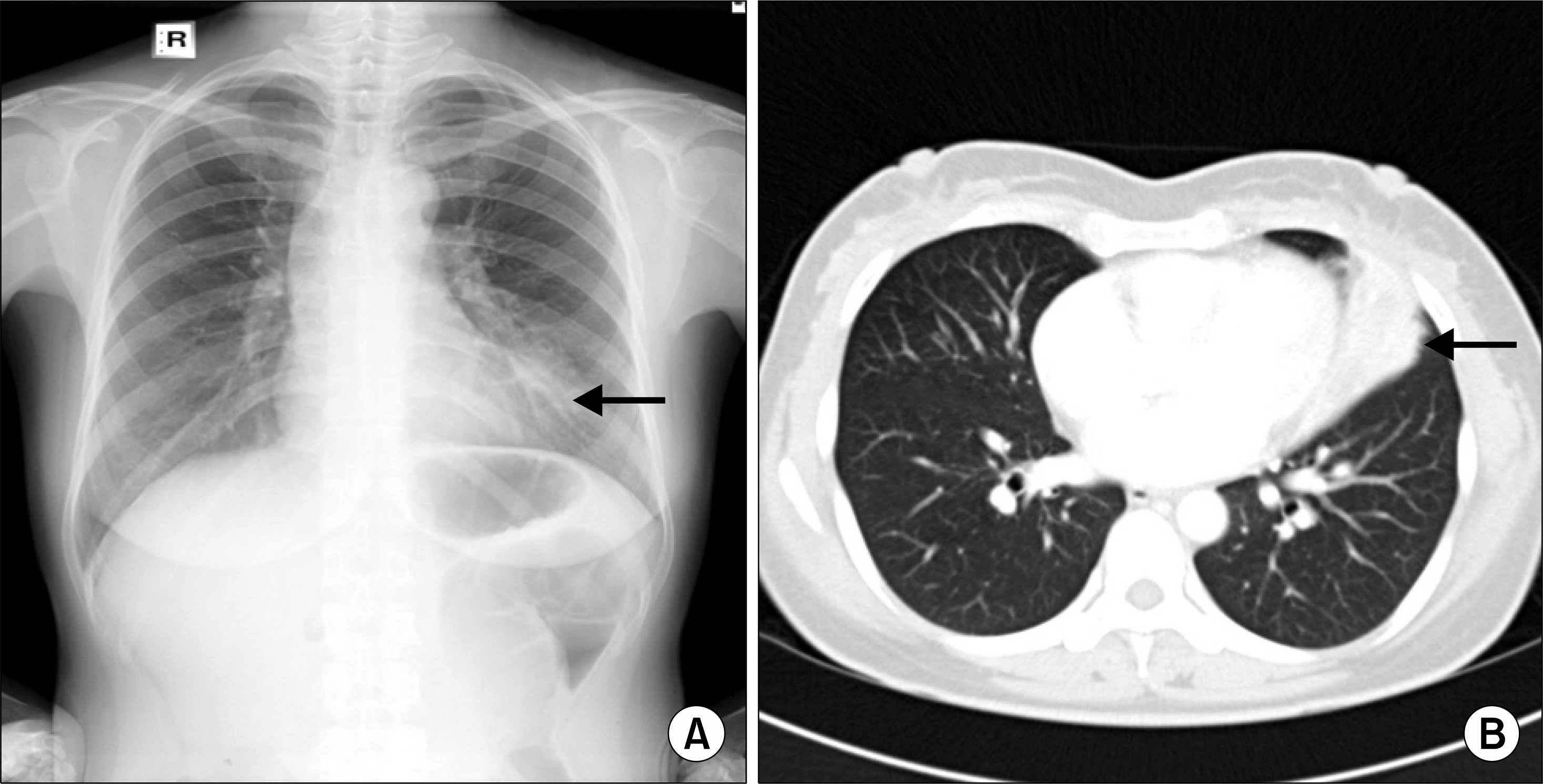
Fig. 1.
Radiologic finding of organizing pneumonia at left upper lung field. (A) Chest PA showed segmental distribution of air space consolidation at the left upper lung field. (B) Chest CT showed air space consolidation and atelectasis at lingular division of left upper lung.


 PDF
PDF ePub
ePub Citation
 Print
Print


 XML Download