

 PDF
PDF ePub
ePub Citation
 Print
Print


Abstract
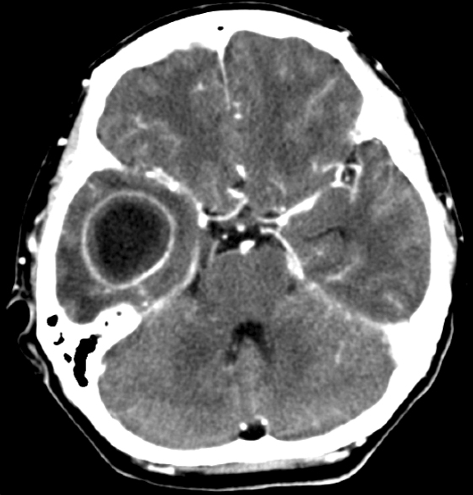
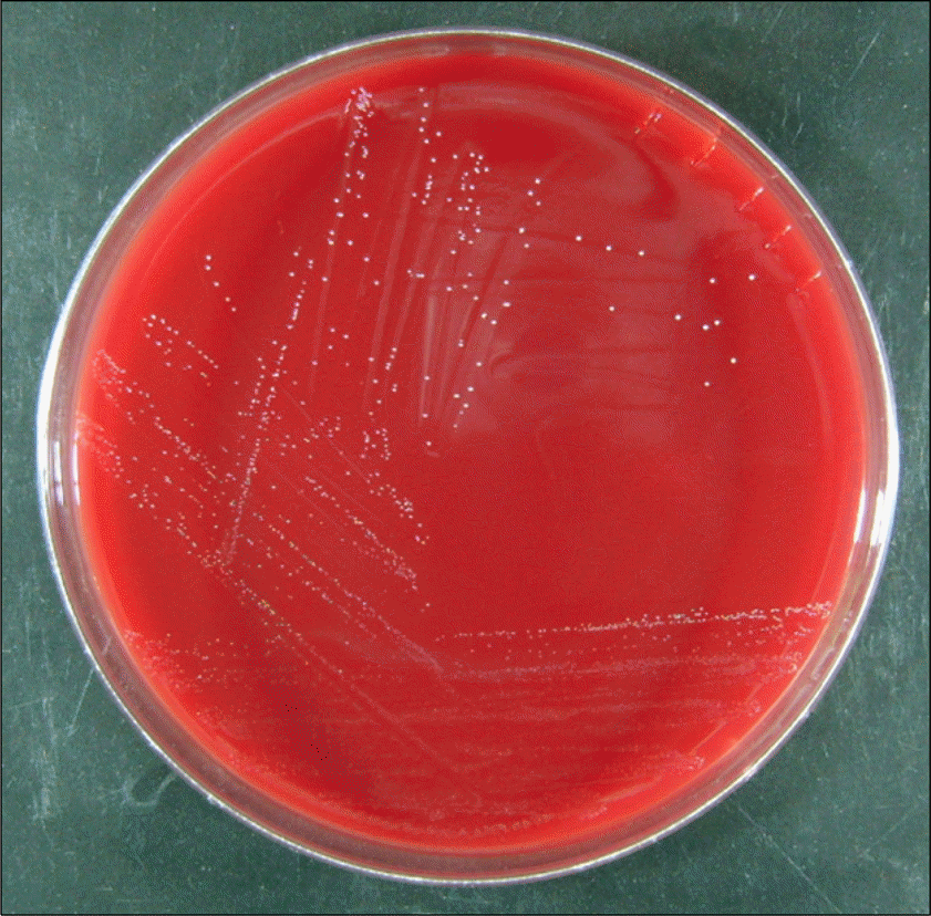
Parvimonas micra is a non-spore-forming anaerobic gram-positive coccus, widely distributed as normal flora in the skin, vagina and mucosa, and able to cause opportunistic infections, particularly endocarditis and brain abscess following dental manipulations. A 49-year-old woman was hospitalized due to fever and headache. She had been diagnosed with periodontitis at the beginning of fever. A brain abscess was noted in the right temporal lobe on the brain CT, and she was treated with ceftriaxone, isepamicin and metronidazole. In the next day, abscess was aspirated and drained by a surgical procedure. An organism was isolated from an anaerobic culture of the abscess aspirate, and was identified as P. micra by a commercial kit and 16S rRNA sequencing.
REFERENCES
1. Tindall BJ, Euzéby JP. Proposal of Parvimonas gen. nov. and Quatrionicoccus gen. nov. as replacements for the illegitimate, prokaryotic, generic names Micromonas Murdoch and Shah 2000 and Quadricoccus Maszenan et al. 2002, respectively. Int J Syst Evol Microbiol. 2006; 56:2711–3.
2. Murray PR, Baron EJ, et al. eds. Manual of Clinical Microbiology. 9th ed.Washington, D.C.: American Society for Microbiology;2007. p. 862–71.
3. Murdoch DA, Mitchelmore IJ, Tabaqchali S. Peptostreptococcus micros in polymicrobial abscesses. Lancet. 1988; 1:594.
4. Chung HY, Sung H, Lee MY, Yoon NS, Lee SG, Suh DJ, et al. A case of bacteremia by Atopobium rimae in a patient with liver cirrhosis. Korean J Lab Med. 2007; 27:351–4.
5. Hong YK, Ha YS, Huh CW, Song JU. Gas-forming brain abscess due to Peptostreptococcus. J Korean Neurosurg Soc. 1984; 13:761–4.
6. Atlas RM. Handbook of Microbiological Media. 1st ed.Boca Raton: CRC Press;1993. : 163.
7. Sharma R, Mohandas K, Cooke RP. Intracranial abscesses: changes in epidemiology and management over five decades in merseyside. Infection. 2009; 37:39–43.

8. Prasad KN, Mishra AM, Gupta D, Husain N, Husain M, Gupta RK. Analysis of microbial etiology and mortality in patients with brain abscess. J Infect. 2006; 53:221–7.
9. Corson MA, Postlethwaite KP, Seymour RA. Are dental infections a cause of brain abscess? Case report and review of the literature. Oral Dis. 2001; 7:61–5.
10. Li X, Tronstad L, Olsen I. Brain abscesses caused by oral infection. Endod Dent Traumatol. 1999; 15:95–101.
11. Tomás I, Alvarez M, Limeres J, Potel C, Medina J, Diz P. Prevalence, duration and aetiology of bacteraemia following dental extractions. Oral Dis. 2007; 13:56–62.
12. Lucas VS, Gafan G, Dewhurst S, Roberts GJ. Prevalence, intensity and nature of bacteraemia after toothbrushing. J Dent. 2008; 36:481–7.
13. Turng BF, Guthmiller JM, Minah GE, Falkler WA Jr. Development and evaluation of a selective and differential medium for the primary isolation of Peptostreptococcus micros. Oral Microbiol Immunol. 1996; 11:356–61.
 XML Download