Abstract
Background
In July 2007, three neonates in the neonatal intensive care unit (NICU) of Chosun University Hospital expired due to Escherichia coli sepsis. An E. coli outbreak was suspected.
Conclusion
The result of rep-PCR assay showed that the outbreak had originated from a single clone of E. coli. But we could not identify risk factors for the infection. The attack rate of E. coli in NICU returned to the basal level after implement of the in-fection control measures such as disinfection of NICU environment and equipments, thorough hand washing, and education of health care workers.
Methods
To investigate the outbreak, environmental cultures were taken from NICU. We performed repetitive extragenic palindromic (rep)-PCR to compare genotypes of the three isolates from the cases and one environmental strain of E. coli. A case-control study was done in order to identify risk factors for the infection.
Results
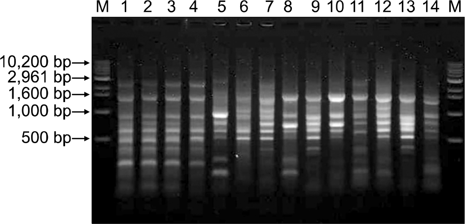
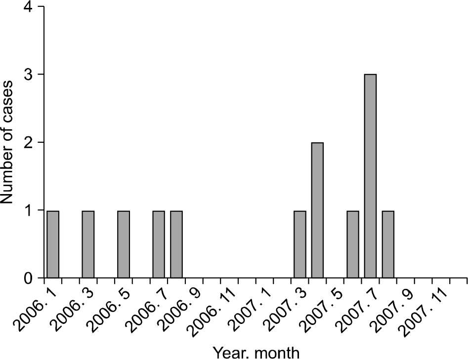
In July 2007, the attack rate of E. coli was 11.1%, which was higher than the basal rate. All the three E. coli isolates from the cases presented the same antimicrobial susceptibility pattern whereas other E. coli isolated from non-outbreak period presented different patterns. Among environmental cultures, only one specimen collected from the surface of a bathtub for neonates was culture positive for E. coli. Three strains of the cases and one environmental strain of E. coli showed the same rep-PCR pattern, while control strains showed different patterns. No statistically significant difference in risk factors was found between the case and control groups in the case-control study.
Go to : 
References
2. Vergnano S, Sharland M, Kazembe P, Mwansambo C, Heath PT. Neonatal sepsis: an international perspective. Arch Dis Child Fetal Neonatal Ed. 2005; 90:F220–4.

3. Gastmeier P, Loui A, Stamm-Balderjahn S, Hansen S, Zuschneid I, Sohr D, et al. Outbreaks in neonatal intensive care units? They are not like others. Am J Infec Control. 2007; 35:172–6.
4. Damjanovic V and van Saene HKF. Outbreaks of infection in neonatal intensive care units (NICU). J Hosp Infect. 1997; 35:237–42.
5. Taneja N, Das A, Raman Rao DSV, Jain N, Singh M, Sharma M. Nosocomial outbreak of diarrhoea by enterotoxigenic Escherichia coli among preterm neonates in a tertiary care hospital in India: pitfalls in healthcare. J Hosp Infect. 2003; 53:193–7.

6. Wong HC and Lin CH. Evaluation of typing of Vibriopa-rahaemolyticus by three PCR methods using specific primers. J Clin Microbiol. 2001; 39:4233–40.
7. Zaza S and Jarvis WR. Investigation of outbreaks. Mayhall CG, editor. Hospital Epidemiology and Infection Control. 1st ed.Baltimore: Williams & Wilkins;2006. p. 105–13.
8. Olive and Bean P. Principles and applications of methods for DNA-based typing of microbial organisms. J Clin Microbiol. 1999; 37:1661–9.

Go to : 
 | Fig. 1.Temporal distribution of admissions, discharges, and date of specimen sampling. The gray boxes indicate admission period of cases with septicemia due to E. coli outbreak strain infection. The ▼ marks indicate the date of isolation of E. coli from the cases. |
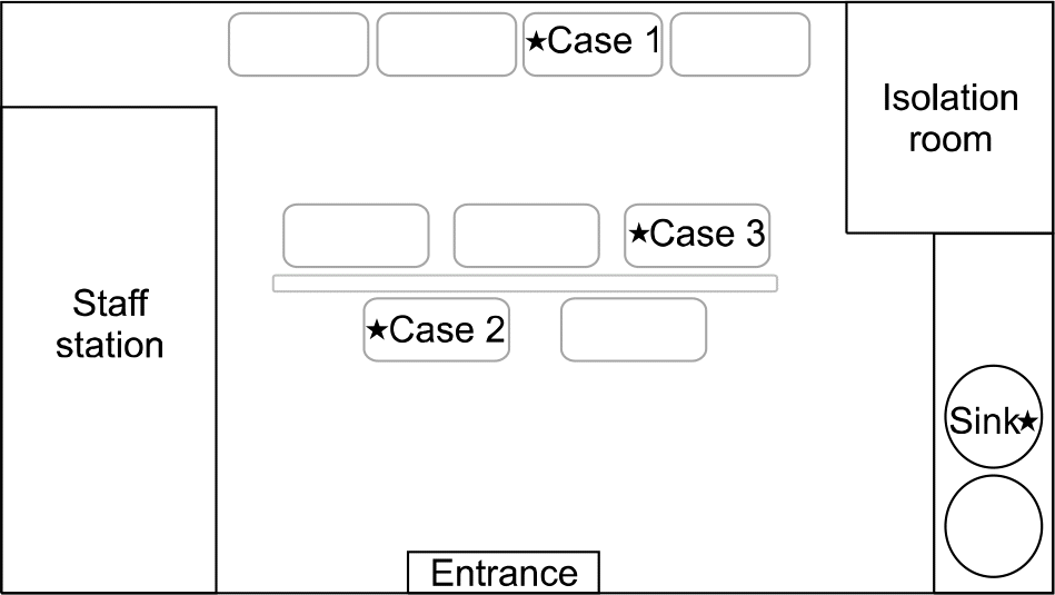
 | Fig. 3.The spot map of E. coli outbreak in NICU. The ★ marks indicate the location of incubators of involved cases and the environmental culture site where E. coli was isolated. |
 | Fig. 4.Rep-PCR patterns of E.coli isolated from patients, environmental culture, and non-outbreak control strains. Lane M, molecular weight marker; Lane 1, case 1; Lane 2, case 2; Lane 3, case 3; Lane 4, environmental culture strain, Lane 5 to 14, non-outbreak control strains. |
Table 1.
Clinical features of the case patients with septicemia due to E. coli outbreak strain infection
Table 2.
Antimicrobial susceptibility of E. coli isolated from NICU in 2007
Abbreviations: AMK, amikacin; AMC, amoxicillin-clavulanic acid; AMP, ampicillin; CFZ, cafazolin; FEP, cefepime; CTX, cefotaxime; FOX, cefoxitin; CAZ, caftazidime; ESBL, extended-spectrum beta-lactamase; CIP, ciprofloxacin; GEN, gentamicin; IPM, imipenem; PIP, piperacillin; TZP, piperacillin-tazobactam; TET, tetracycline; SXT, trimethoprim-sulfamethoxazole.
Table 3.
Univariate analysis of risk factors for infection with E. coli in NICU


 PDF
PDF ePub
ePub Citation
 Print
Print


 XML Download