

 PDF
PDF ePub
ePub Citation
 Print
Print


Abstract
Purpose
Iliosacral screw fixation is an effective and less invasive method that is used widely for the definitive treatment of unstable pelvic ring injuries. On the other hand, fixation failures after iliosacral screw fixation have been reported in vertically unstable pelvic ring injuries. This study examined the surgical outcomes of posterior pelvic fixation using S1 and S2 screws in vertically unstable pelvic ring injuries.
Materials and Methods
Between January 2011 and April 2016, 17 patients with vertically unstable pelvic ring injuries who met the minimum 1 year follow-up criteria were treated with internal fixation using posterior pelvic S1 and S2 screws. Their mean age was 43.9 years. According to the AO/OTA classification, 10 patients had C1, 6 had C2, and 1 had C3 injuries. Surgical treatments of single or multiple steps, where necessary, were performed by two surgeons. The clinical and radiologic outcomes were assessed retrospectively using radiographs and medical records.
Results
Overall, 16 patients had bone healing without screw loosening; however, one patient could not maintain anterior pelvic fixation because of an open fracture and deep infection in the anterior pelvic ring. Of five patients who complained of neurological symptoms after injury, three had partially recovered from their neurological deficit. At the last follow-up, the clinical outcomes according to the Majeed score were excellent in 5, good in 6, fair in 4, and poor in 2 patients. The postoperative radiologic outcomes by Matta and Tornetta's method were excellent in 5, good in 8, and fair in 4 patients. Malposition of the S2 screw was identified in one case. The mean time to union was 14.6 weeks after surgery.
Go to : 
References
1. Rommens PM, Hessmann MH. Staged reconstruction of pelvic ring disruption: differences in morbidity, mortality, radiologic results, and functional outcomes between B1, B2/B3, and C-type lesions. J Orthop Trauma. 16:92–98. 2002.

2. Kabak S, Halici M, Tuncel M, Avsarogullari L, Baktir A, Bas-turk M. Functional outcome of open reduction and internal fixation for completely unstable pelvic ring fractures (type C): a report of 40 cases. J Orthop Trauma. 17:555–562. 2003.
3. Lindahl J, Hirvensalo E, Böstman O, Santavirta S. Failure of reduction with an external fixator in the management of injuries of the pelvic ring. Long-term evaluation of 110 patients. J Bone Joint Surg Br. 81:955–962. 1999.
4. Miranda MA, Riemer BL, Butterfield SL, Burke CJ 3rd. Pelvic ring injuries. A long term functional outcome study. Clin Orthop Relat Res. 329:152–159. 1996.
5. Cole JD, Blum DA, Ansel LJ. Outcome after fixation of unstable posterior pelvic ring injuries. Clin Orthop Relat Res. 329:160–179. 1996.
6. Keating JF, Werier J, Blachut P, Broekhuyse H, Meek RN, O'Brien PJ. Early fixation of the vertically unstable pelvis: the role of iliosacral screw fixation of the posterior lesion. J Orthop Trauma. 13:107–113. 1999.
7. Tornetta P 3rd, Matta JM. Outcome of operatively treated unstable posterior pelvic ring disruptions. Clin Orthop Relat Res. 329:186–193. 1996.
8. Routt ML Jr, Kregor PJ, Simonian PT, Mayo KA. Early results of percutaneous iliosacral screws placed with the patient in the supine position. J Orthop Trauma. 9:207–214. 1995.
9. Gorczyca JT, Varga E, Woodside T, Hearn T, Powell J, Tile M. The strength of iliosacral lag screws and transiliac bars in the fixation of vertically unstable pelvic injuries with sacral fractures. Injury. 27:561–564. 1996.
10. Krappinger D, Larndorfer R, Struve P, Rosenberger R, Arora R, Blauth M. Minimally invasive transiliac plate osteosynthesis for type C injuries of the pelvic ring: a clinical and radiological follow-up. J Orthop Trauma. 21:595–602. 2007.
11. Sagi HC, Militano U, Caron T, Lindvall E. A comprehensive analysis with minimum 1-year follow-up of vertically unstable transforaminal sacral fractures treated with triangular osteosynthesis. J Orthop Trauma. 23:313–319. 2009.
12. Shuler TE, Boone DC, Gruen GS, Peitzman AB. Percutaneous iliosacral screw fixation: early treatment for unstable posterior pelvic ring disruptions. J Trauma. 38:453–458. 1995.
13. Griffin DR, Starr AJ, Reinert CM, Jones AL, Whitlock S. Vertically unstable pelvic fractures fixed with percutaneous iliosacral screws: does posterior injury pattern predict fixation failure? J Orthop Trauma. 17:399–405. 2003.
14. Kim JW, Oh CW, Oh JK, et al. The incidence of and factors affecting iliosacral screw loosening in pelvic ring injury. Arch Orthop Trauma Surg. 136:921–927. 2016.
15. Matta JM, Tornetta P 3rd. Internal fixation of unstable pelvic ring injuries. Clin Orthop Relat Res. 329:129–140. 1996.
16. Majeed SA. Grading the outcome of pelvic fractures. J Bone Joint Surg Br. 71:304–306. 1989.
17. Routt ML Jr, Simonian PT. Closed reduction and percutaneous skeletal fixation of sacral fractures. Clin Orthop Relat Res. 329:121–128. 1996.
18. Routt ML Jr, Nork SE, Mills WJ. Percutaneous fixation of pelvic ring disruptions. Clin Orthop Relat Res. 375:15–29. 2000.
19. Zhang L, Peng Y, Du C, Tang P. Biomechanical study of four kinds of percutaneous screw fixation in two types of unilateral sacroiliac joint dislocation: a finite element analysis. Injury. 45:2055–2059. 2014.
20. Gautier E, Bächler R, Heini PF, Nolte LP. Accuracy of computer-guided screw fixation of the sacroiliac joint. Clin Orthop Relat Res. 393:310–317. 2001.
21. Carlson DA, Scheid DK, Maar DC, Baele JR, Kaehr DM. Safe placement of S1 and S2 iliosacral screws: the “vestibule” concept. J Orthop Trauma. 14:264–269. 2000.
22. Hinsche AF, Giannoudis PV, Smith RM. Fluoroscopy-based multiplanar image guidance for insertion of sacroiliac screws. Clin Orthop Relat Res. 395:135–144. 2002.
23. Moed BR, Geer BL. S2 iliosacral screw fixation for disruptions of the posterior pelvic ring: a report of 49 cases. J Orthop Trauma. 20:378–383. 2006.
24. Osterhoff G, Ossendorf C, Wanner GA, Simmen HP, Werner CM. Percutaneous iliosacral screw fixation in S1 and S2 for posterior pelvic ring injuries: technique and perioperative complications. Arch Orthop Trauma Surg. 131:809–813. 2011.
25. Moon SW, Kim JW. Usefulness of intraoperative three-dimensional imaging in fracture surgery: a prospective study. J Orthop Sci. 19:125–131. 2014.
26. Zwingmann J, Konrad G, Kotter E, Südkamp NP, Oberst M. Computer-navigated iliosacral screw insertion reduces malposition rate and radiation exposure. Clin Orthop Relat Res. 467:1833–1838. 2009.
27. Zwingmann J, Konrad G, Mehlhorn AT, Südkamp NP, Oberst M. Percutaneous iliosacral screw insertion: malpositioning and revision rate of screws with regards to application technique (navigated vs. conventional). J Trauma. 69:1501–1506. 2010.
28. Lucas JF, Routt ML Jr, Eastman JG. A useful preoperative planning technique for transiliac-trassacral screws. J Orthop Trauma. 31:e25–e31. 2017.
29. Kim JW, Bae DH, Kim JH, Kim YC. Neurologic injury within pelvic ring injuries. J Korean Fract Soc. 27:17–22. 2014.
30. Sagi HC, Ordway NR, DiPasquale T. Biomechanical analysis of fixation for vertically unstable sacroiliac dislocations with iliosacral screws and symphyseal plating. J Orthop Trauma. 18:138–143. 2004.
Go to :
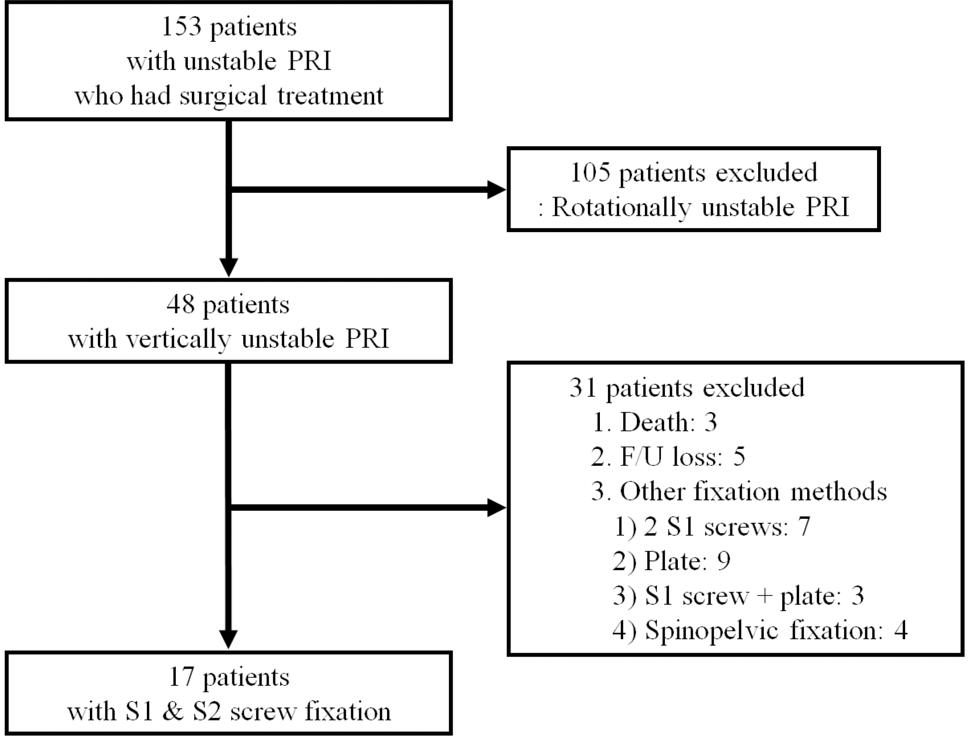 | Fig. 1.Flow chart of the enrollment of patients treated with posterior pelvic fixation using S1 and S2 screws for vertically unstable pelvic ring injuries. PRI: pelvic ring injury, F/U: follow-up. |
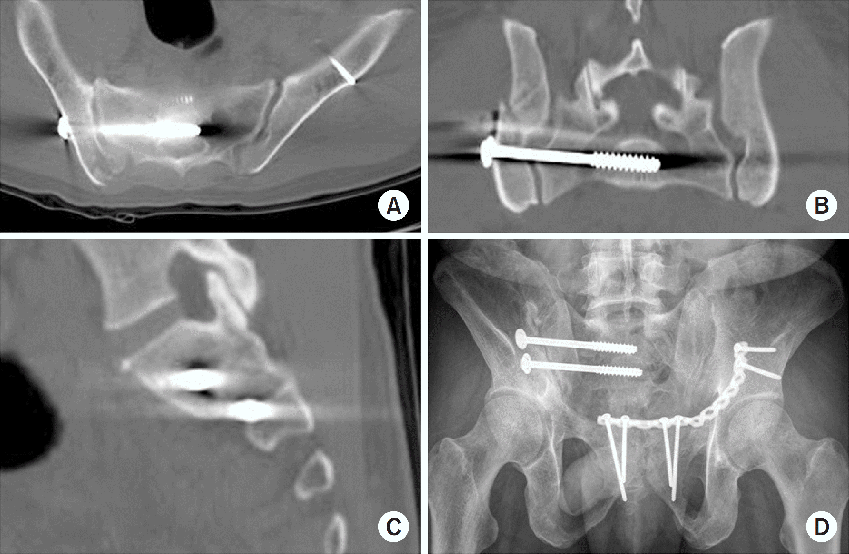 | Fig. 2.Malposition of the S2 screw in the S1 foramen was identified in the axial (A), coronal (B), and sagittal (C) images of postoperative computed tomography. (D) Postoperative radiograph of the pelvic outlet view. |
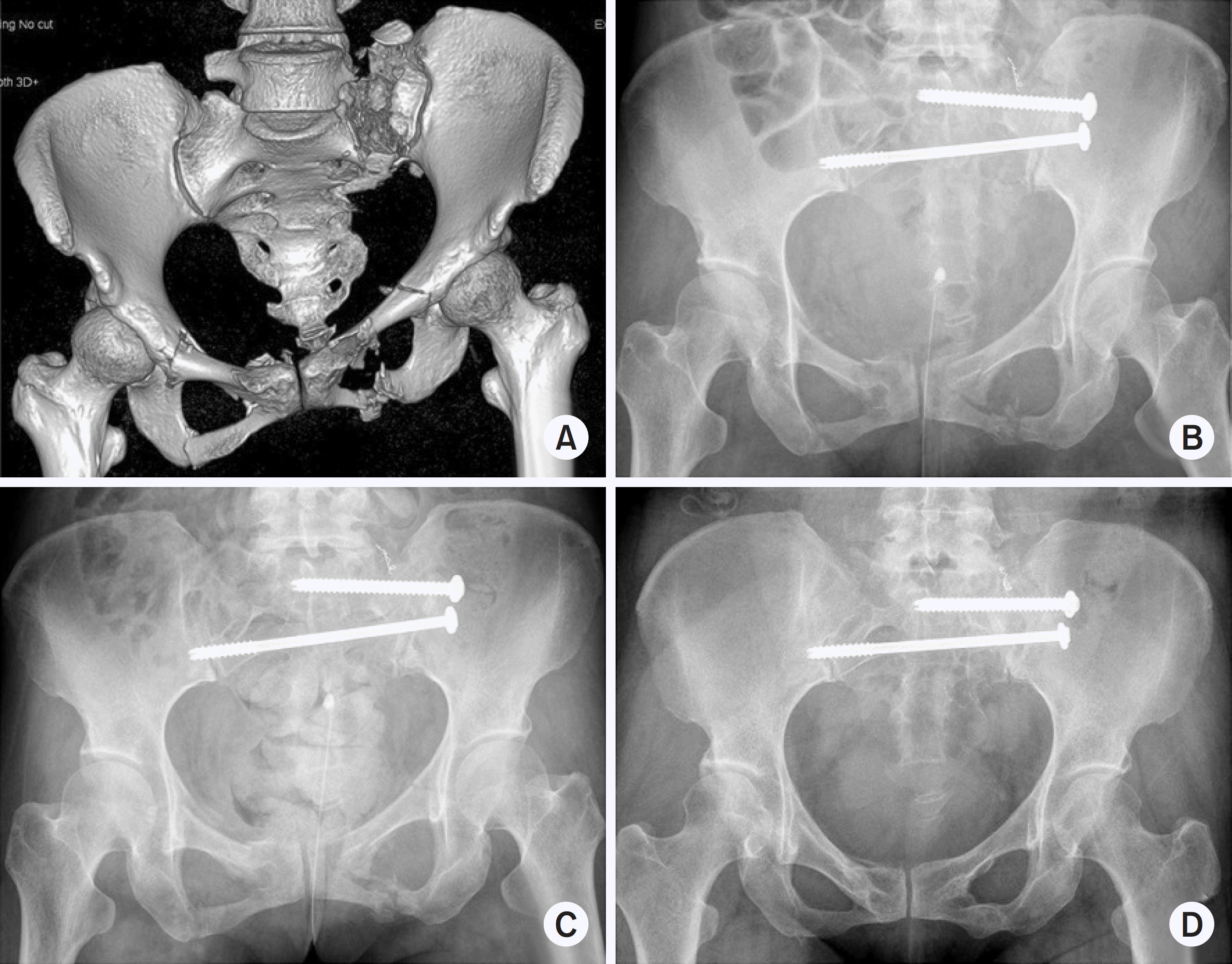 | Fig. 3.(A) A 35-year-old female patient fell from a height and sustained a type C pelvic ring injury. (B) Posterior pelvic stabilization using S1 and S2 screws was performed 1 day after the injury. No further procedure was performed for the anterior pelvic ring because acceptable reduction of the fractures in the pubic rami was identified after posterior fixation. (C) No displacement occurred, and callus formation was observed at the 4-week follow-up. (D) The radiograph obtained at the 1-year follow-up showed complete bony union without complications. |
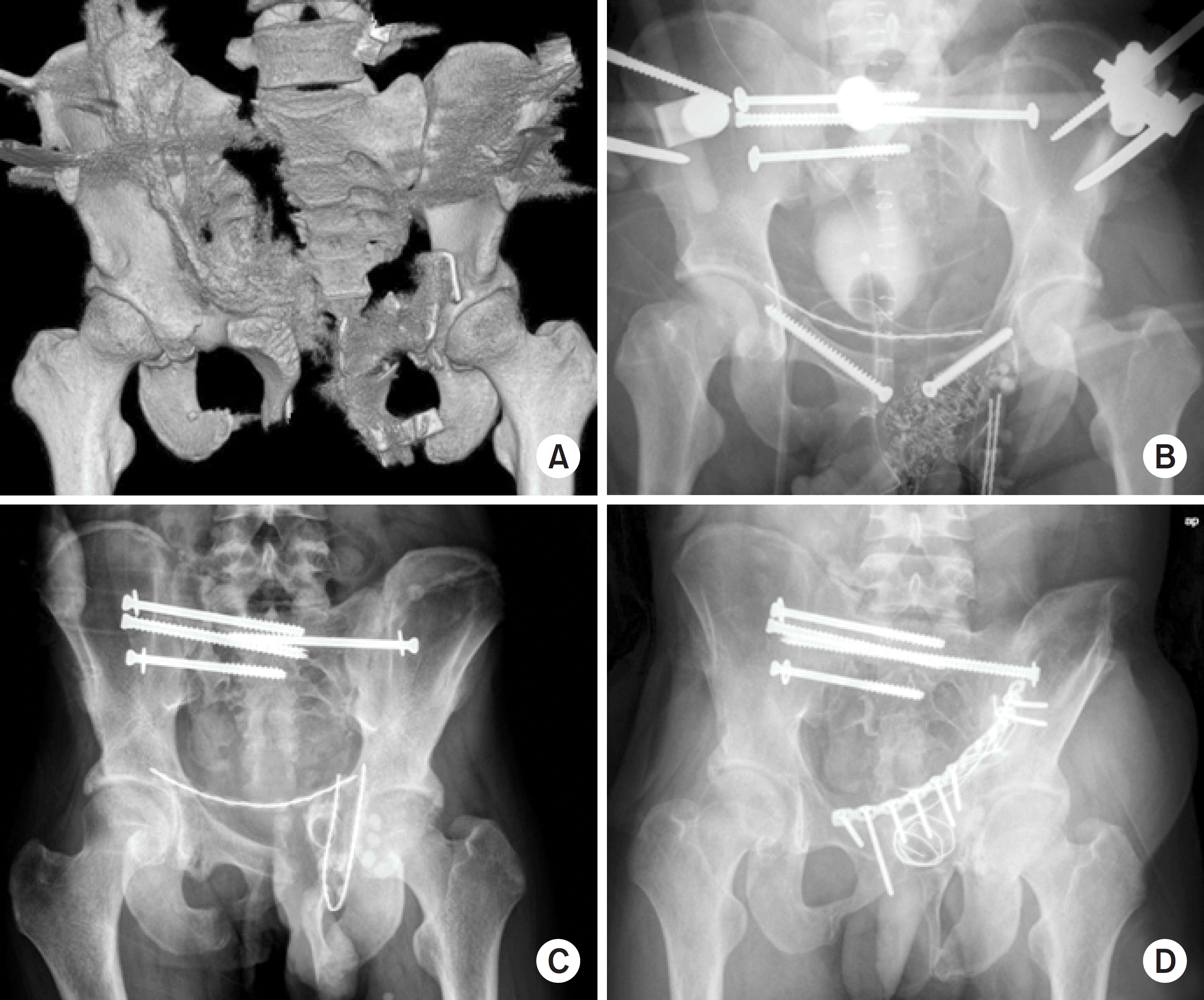 | Fig. 4.(A) Emergent external fixation and pelvic packing were performed in a 17-year-old male patient with a vertically unstable (type C) pelvic ring injury with an open wound. (B) Posterior pelvic fixation using S1 and S2 iliosacral screws and anterior fixation with rami screws were performed 2 days after the initial injury. (C) Rami screws and the external fixator were removed, and debridement and the insertion of antibiotic-impregnated cement beads were conducted because of a deep infection 4 weeks after the initial surgery. Sacroiliac screw loosening was identified. (D) Anterior pelvic plating using the modified Stoppa approach and posterior pelvic screw exchange were performed after the infection had subsided completely. |
Table 1.
Patient Demographics
AO/OTA: Arbeitsgemeinschaft fü r Osteosynthesefragen/Orthopaedic Trauma Association, MVA: motor vehicle accident, M: other musculoskeletal injury, U: urological injury, T: thoracic injury, A: abdominal injury, H: head injury, N: neurological lesion, R: case needed initial resuscitation, Lt.: right, Rt.: right, SIJ: sacroiliac joint, EF: external fixator, ISS: iliosacral screw, TSTIS: transsacral-transiliac screw.
 XML Download