

 PDF
PDF ePub
ePub Citation
 Print
Print


Abstract
Purpose
This study was to assess the morphological changes of the pronator quadratus (PQ) muscle using an ultrasonography in the volar locking plate fixation group and in the percutaneous K-wire fixation group for distal radius fracture, and to evaluate the impact on clinical outcomes.
Materials and Methods
Fifty-four patients who received surgical treatment for distal radius fracture were enrolled in this study. They were divided into two groups according to treatment modality: Group 1 included 34 patients who underwent internal fixation with volar locking plate and Group 2 included 20 patients with percutaneous K-wire fixation. Thickness of the PQ muscle was measured using an ultrasonography at the final follow-up. We evaluated the outcomes using the Mayo wrist score, wrist range of motion, and grip strength at the final follow-up.
Results
Compared with the uninjured side, thickness of the PQ muscle showed 31.9% of mean atrophy in Group 1 and 11.4% in Group 2. The atrophy of PQ muscle was severe in Group 1 (p=0.01). However, there was no significant difference in the mean Mayo wrist score between the two groups (83.1±10.9 in Group 1 and 80.2±8.9 in Group 2, p=0.28), except a mild limitation of pronation in Group 1.
Figures and Tables
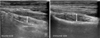 | Fig. 1Ultrasound examination in longitudinal view shows a significant decrease (40%) in the thickness of pronator quadratus muscle (arrows) at postoperative 21 months of volar locking plate.
|
Table 1
Demographic Characteristics
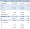
Values are presented as median (range), number (%), or mean±standard deviation. Group 1: patients of distal radius fractures treated with volar locking plate, Group 2: patients of distal radius fractures treated with percutaneous K-wire, BMI: body mass index, BMD: bone mineral density, AO: arbeitsgemeinschaft für osteosyntheses.
![]()
Table 2
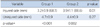
Thickness of the Pronator Quadratus Muscle at the Final Follow-Up
| Variable | Group 1 | Group 2 | p-value |
|---|---|---|---|
| Injured side (mm) | 3.2±0.9 (68.1) | 3.9±1.1 (88.6) | 0.01 |
| Uninjured side (mm) | 4.7±0.9 | 4.4±0.9 | 0.27 |
| p-value* | <0.001 | 0.002 |
![]()
Table 3
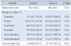
Clinical Outcomes at the Final Follow-Up
![]()
References
1. Drobetz H, Kutscha-Lissberg E. Osteosynthesis of distal radial fractures with a volar locking screw plate system. Int Orthop. 2003; 27:1–6.

2. Knox J, Ambrose H, McCallister W, Trumble T. Percutaneous pins versus volar plates for unstable distal radius fractures: a biomechanic study using a cadaver model. J Hand Surg Am. 2007; 32:813–817.
3. Lee KH. Volar plating of distal radius fractures. J Korean Fract Soc. 2008; 21:325–333.
4. Orbay JL, Badia A, Indriago IR, et al. The extended flexor carpi radialis approach: a new perspective for the distal radius fracture. Tech Hand Up Extrem Surg. 2001; 5:204–211.
5. Ahsan ZS, Yao J. The importance of pronator quadratus repair in the treatment of distal radius fractures with volar plating. Hand (N Y). 2012; 7:276–280.
6. Arora R, Lutz M, Hennerbichler A, Krappinger D, Espen D, Gabl M. Complications following internal fixation of unstable distal radius fracture with a palmar locking-plate. J Orthop Trauma. 2007; 21:316–322.
7. Hershman SH, Immerman I, Bechtel C, Lekic N, Paksima N, Egol KA. The effects of pronator quadratus repair on outcomes after volar plating of distal radius fractures. J Orthop Trauma. 2013; 27:130–133.
8. Sofka CM. Ultrasound of the hand and wrist. Ultrasound Q. 2014; 30:184–192.
9. Amadio PC, Berquist TH, Smith DK, Ilstrup DM, Cooney WP 3rd, Linscheid RL. Scaphoid malunion. J Hand Surg Am. 1989; 14:679–687.
10. Stuart PR. Pronator quadratus revisited. J Hand Surg Br. 1996; 21:714–722.
11. Gordon KD, Dunning CE, Johnson JA, King GJ. Influence of the pronator quadratus and supinator muscle load on DRUJ stability. J Hand Surg Am. 2003; 28:943–950.
12. McConkey MO, Schwab TD, Travlos A, Oxland TR, Goetz T. Quantification of pronator quadratus contribution to isometric pronation torque of the forearm. J Hand Surg Am. 2009; 34:1612–1617.
13. Armangil M, Bezirgan U, Başarır K, Bilen G, Demirtaş M, Bilgin SS. The pronator quadratus muscle after plating of distal radius fractures: is the muscle still working? Eur J Orthop Surg Traumatol. 2014; 24:335–339.
14. Swigart CR, Badon MA, Bruegel VL, Dodds SD. Assessment of pronator quadratus repair integrity following volar plate fixation for distal radius fractures: a prospective clinical cohort study. J Hand Surg Am. 2012; 37:1868–1873.
 XML Download